Dry eyes causes and treatment in adults. Dryness in the eyes. Causes, treatment, drops for dry eyes, alternative methods of treatment. Best Inexpensive Moisturizers
(xerophthalmia) - a state of insufficient moisture of the surface of the cornea and conjunctiva due to a violation of the quality and quantity of the tear fluid and the instability of the tear film. Manifestations of dry eye syndrome are burning and pain, a feeling of sand in the eyes, tearing, photophobia, rapid fatigue during visual work, intolerance to dry and dusty air. Dry eye syndrome is diagnosed according to the results of biomicroscopy, Schirmer and Norn tests, fluorescein instillation test, tiascopy, osmometry, tear fluid crystallography, and cytological examination of a smear from the conjunctiva. As a treatment for dry eye syndrome, artificial tear preparations, obstruction of the lacrimal ducts, tarsorrhaphy, keratoplasty, transplantation of the salivary glands are indicated.

General information
Dry eye syndrome is a fairly common condition in ophthalmology, which is characterized by a lack of hydration of the surface of the cornea and conjunctiva of the eye and the development of signs of xerosis. Dry eye syndrome occurs in 9-18% of the population, more often in women (almost 70% of cases), the incidence of the disease increases significantly with age: up to 50 years - 12%, after 50 - 67%.
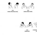
Normal anterior surface eyeball covered with a continuous thin (about 10 microns) tear film having a three-layer structure. The outer lipid layer - the oily secretion of the meibomian glands provides gliding upper eyelid on the surface of the eyeball and slows down the evaporation of the tear film. The aqueous layer with dissolved electrolytes and organic compounds flushes out foreign bodies from the eye, provides the cornea with nutrients and oxygen, and creates immune protection. The mucin layer is the mucous secretion of goblet and epithelial cells in direct contact with the cornea: it makes its surface even and smooth, binding the tear film to it and providing high quality vision.
Approximately every 10 seconds, the tear film breaks, initiating the blinking movement of the eyelids and the renewal of the tear fluid, restoring its integrity. Violation of the stability of the precorneal tear film leads to its frequent ruptures, dryness of the surface of the cornea and conjunctiva, and the development of dry eye syndrome.
Causes of dry eye syndrome
Dry eye syndrome results from insufficient amount and quality of tear fluid, as well as excessive evaporation of the precorneal tear film, which reduces its retention time or volume.
The reasons for the development of dry eye syndrome can be internal diseases and syndromes associated with a decrease in tear production: autoimmune (Sjögren's syndrome), diseases of the hematopoietic and reticuloendothelial systems (Felty's syndrome, malignant lymphoma), endocrine dysfunction (endocrine ophthalmopathy, menopause), kidney pathology, body exhaustion and infectious diseases, skin diseases(pemphigus), pregnancy.
Pathology of the organs of vision (chronic conjunctivitis, corneal and conjunctival scars, neuroparalytic keratitis, lagophthalmos, lacrimal gland dysfunction) and ophthalmic surgical interventions that destabilize the tear film (anterior radial keratotomy, corneal photoablation, keratoplasty, conjunctival plastic surgery, correction of ptosis) can lead to dry eye syndrome. ).
There are artifactual factors causing violation stability of the tear film - dry air from air conditioners and fan heaters, hard work with a PC, watching TV, errors in the selection and use of contact lenses, environmental problems.
Reduces tear production and causes dry eye syndrome Long-term use of ophthalmic medicines containing beta-blockers, anticholinergics, anesthetics; some systemic drugs (hormonal contraceptives, antihistamines, antihypertensives).
The appearance of dry eye syndrome is facilitated by too rare blinking movements, beriberi with impaired metabolism of fat-soluble vitamins, genetic predisposition, age after 40 years, belonging to the female sex. A decrease in the frequency of blinking movements may be due to a decrease in the sensitivity of the cornea of a functional or organic nature.
Classification of dry eye syndrome
According to the domestic classification, dry eye syndrome is distinguished by pathogenesis, which developed as a result of a decrease in the volume of tear fluid secretion, increased evaporation of the tear film, as well as their combined effects; according to etiology, syndromic dry eye, symptomatic, artefactial are distinguished.
Dry eye syndrome can be expressed in various clinical forms: recurrent macro- and microerosion of the cornea or conjunctiva of the eyeball; dry keratoconjunctivitis, filamentous keratitis.
According to the degree of severity, mild, moderate, severe and especially severe form dry eye syndrome.
Symptoms of dry eye syndrome
The clinical manifestations of dry eye syndrome are very diverse and are largely determined by the severity of the disease. The subjective symptoms of dry eye syndrome include a sensation foreign body(sand) in the conjunctival cavity, redness, burning and pain in the eyes; lacrimation, hypersensitivity to light, fatigue; blurred vision, pain when instilling eye drops.
Symptoms of dry eye syndrome are usually more pronounced in the evening, as well as when in a dry or polluted room, in the cold, wind, after prolonged or intense visual work.
Objective signs of dry eye syndrome are xerotic changes in the cornea and conjunctiva of varying severity (corneal-conjunctival xerosis). With mild corneal-conjunctival xerosis, a compensatory increase in tear production (hyperlacrimia) and an increase in the height of the lower lacrimal meniscus develop. With moderate xerosis, reflex lacrimation decreases, lacrimal menisci decrease or are completely absent, there is a feeling of “dryness” in the eyes, the swelling of the conjunctiva creeping onto the free edge of the lower eyelid and its displacement along with the adhering eyelid during blinking movements. Severe corneal-conjunctival xerosis is manifested by the following clinical forms: filamentous keratitis, dry keratoconjunctivitis and recurrent corneal erosion, occurring against the background of existing manifestations of dry eye syndrome.
With filamentous keratitis on the cornea, multiple epithelial growths are observed, manifestations of a moderately pronounced corneal syndrome without inflammatory changes in the conjunctiva.
With dry keratoconjunctivitis, pronounced corneal-conjunctival changes of an inflammatory-degenerative nature are noted: subepithelial opacities, dullness and roughness of the cornea, saucer-shaped epithelialized or non-epithelialized depressions on its surface, flaccid hyperemia, swelling and loss of luster of the conjunctiva of the eye, more pronounced adhesion of the eyeball to the conjunctiva of the eyelids .
With recurrent erosion of the cornea, surface microdefects of its epithelium periodically appear, which persist for up to 3-5 days or more, after their epithelization, prolonged discomfort is noted.
Particularly severe corneal-conjunctival xerosis usually develops with complete or partial non-closure of the palpebral fissure. Dry eye syndrome against the background of a pronounced lack of vitamin A is manifested by scaly metaplasia of the epithelium and keratinization of the conjunctiva.
Dry eye syndrome often co-occurs with blepharitis. Dry eye syndrome can lead to severe and irreversible xerotic changes and even corneal perforation.
Diagnosis of dry eye syndrome
Diagnostic examination of a patient with dry eye syndrome begins with the collection of complaints, assessment of anamnesis and clinical symptoms diseases, in order to identify pathognomonic and indirect signs of corneal-conjunctival xerosis.
During a physical examination for dry eye syndrome, an external examination is performed, during which the ophthalmologist determines the condition of the skin of the eyelids, the sufficiency of their closing, the nature and frequency of blinking movements. With biomicroscopy of the eye, the state of the tear film, cornea, conjunctiva of the eyeball and eyelids, and the height of the lacrimal meniscus are analyzed.
If dry eye syndrome is suspected, a fluorescein instillation test is performed using a staining solution, which makes it possible to determine the time of tear film rupture and to identify the presence of dry foci - areas of the cornea devoid of epithelium. With the help of special samples, the rate of formation of tear fluid is examined - the total tear production (Schirmer's test), the quality and rate of evaporation of the tear film (Norn's test). Non-invasive assessment of the strength of the precorneal tear film is performed using thiascopy (examination in polarized light) and measurement of the thickness of the lipid layer.
A complete ophthalmological examination for dry eye syndrome also includes laboratory research osmolarity and crystallography of the lacrimal fluid, cytological examination smear from the conjunctiva (including impression). In case of systemic or endocrine diseases in the anamnesis of a patient with dry eye syndrome, appropriate immunological and endocrinological studies are carried out.
Treatment of dry eye syndrome
Treatment of dry eye syndrome is focused on eliminating the etiological factors of xerosis; complete hydration ocular surface and increasing the stability of the precorneal tear film; cupping pathological changes cornea and conjunctiva and prevention of complications.
The most widely used in dry eye syndrome is regular instillation of artificial tear preparations (natural tears, gels with carbomer and dexpanthenol), which allow restoring a fairly stable tear film on the surface of the eyeball. In case of mild dry eye syndrome, low-viscosity preparations are prescribed, in moderate and severe forms - medium and high viscosity (gels), in especially severe cases of xerosis - low-viscosity preparations without preservatives.
Also, with dry eye syndrome, instillations of anti-inflammatory and immunotropic agents are indicated, in the presence of degenerative xerotic changes in the cornea - metabolic drugs. Additionally, antihistamines, mast cell membrane stabilizers, stabilizers of lysosomal membranes of macrophages are prescribed.
Surgical treatment of dry eye syndrome is carried out, if necessary, to limit the outflow and evaporation of native or artificial tears from the conjunctival cavity, increase the inflow of lacrimal fluid, and eliminate the complications that have arisen (xerotic ulcer, corneal perforation). Closure of the lacrimal ducts of the eye is performed by the following methods: obturation of the lacrimal openings with the help of special plugs; plastic lacrimal openings of the conjunctiva or skin; diathermocoagulation, laser coagulation or surgical suturing.
Obturation of the lacrimal canaliculus with miniature silicone plugs and conjunctival covering of the lacrimal punctum for dry eye syndrome are preferred because they are minimally invasive, more effective, and do not cause irreversible changes.
With severe corneal xerosis (xerotic ulcer, keratomalacia) and no effect from drug therapy and obstruction of the lacrimal ducts, with dry eye syndrome, keratoplasty is performed. Patients with incomplete eyelid closure, wide palpebral fissure, and rare blinking are indicated for lateral tarsorrhaphy.
Innovative methods for the treatment of dry eye syndrome are transplantation of salivary glands from the oral cavity into the conjunctival cavity, implantation of dacryoreservoirs in soft tissues patient with the removal of special tubes into the cavity of the conjunctiva.
Forecast and prevention of dry eye syndrome
Even with a mild course, dry eye syndrome requires a full and adequate treatment in order to avoid the development of severe diseases of the conjunctiva and cornea with possible loss of vision.
Dry eye syndrome can be prevented by reducing the impact of artificial factors on the eyes, by conducting preventive treatment of internal diseases, incl. pathology of the organs of vision, using a sufficient amount of fluid, eating rationally, more often making blinking movements with visual load.
Dry eye syndrome is a violation of the hydration of the cornea, as a result of which it dries up and loses its functions. Normally, the eye is constantly moistened, this is one of the conditions for its normal functioning. With a lack of moisture, the so-called dry eye syndrome develops, which has a number of characteristic features and dire consequences.
It can develop as a primary, independent disease, or it can be one of the symptoms of another disease, but whatever the cause of dry eye syndrome, it can lead to partial or complete loss of vision, and therefore requires close attention.
The mechanism of development of dry eye syndrome
Hydration of the eye occurs due to the protective film on the eye that covers the cornea, and the tear fluid produced, which constantly wets it. The protective film consists of three layers:
- Surface layer - formed by lipids, i.e. fats that protect the film from moisture evaporation.
- The middle layer - consists of tear fluid, its task is nutrition, protection of the cornea and a certain visual function, since the presence of this layer changes light refraction
- Mucous, or mucinous layer - fits snugly to the cornea, protecting it and serving as the basis for the other two layers of the protective film.
In addition, a healthy eye constantly contains a small amount of tear fluid, which washes it when you blink. The lacrimal fluid has a complex composition, is produced by a whole group of glands, 2 ml daily in a calm emotional state, because, as you know, in the case of emotional upheavals, the production of tear fluid increases dramatically. In addition to the tear fluid production system, there is also a system for draining excess moisture from the eye. With the help of the tear duct, excess tears flow into the nasal cavity, which becomes very noticeable when a person cries - he always has nasal discharge. Also, the outflow system allows the tear fluid to be updated and perform the function of nourishing the cornea.
Causes of dry eye syndrome
Failure at any stage of the production or outflow of tear fluid can lead to the formation of dry eye syndrome. This may be a violation of tear formation, poor quality of the film formed (for example, a thinned lipid layer, which leads to its drying out), uneven distribution of the protective film over the cornea.
The cause of dry eye syndrome can be various circumstances and diseases.
The most common causes of dry eye syndrome are:
- beriberi, especially a violation of the metabolism of fat-soluble vitamins (A);
- diseases of the endocrine glands, i.e. endocrine diseases;
- systemic diseases in which the connective tissue is affected;
- hormonal failure, for example, in the process of menopause;
- improperly fitted contact lenses;
- some drugs incl. antihypertensive drugs;
- violation of the mode of operation with a computer. In this case, dry eye syndrome has several synonyms: eye office syndrome, computer vision syndrome, etc.
- bad ecological situation.
Age can also act, if not as a cause of dry eye syndrome, then as a factor contributing to its development, since the older the age group, the more often the cases of the disease. After the age of 40, about 30% of people are affected by dry eye syndrome. As a result of the lower stability of the hormonal background and the direct dependence of the production of lacrimal fluid on hormones, in women, the symptoms of dry eye syndrome appear more often than in men.
Symptoms of dry eye syndrome include the following:
- redness of the eyes, especially after activities that require their tension;
- burning sensation and pain in the eyes;
- increased lacrimation, which at first glance seems paradoxical. In fact, this is a compensatory mechanism launched by the body to eliminate dryness of the cornea;
- pain reaction to the instillation of drops of any composition into the eyes, even those that do not contain any irritants
- poor tolerance of wind, smoke, conditioned air.
According to the degree of manifestation of symptoms of dry eye syndrome, there are several clinical forms diseases: mild, moderate, severe and extremely severe.
Diagnosis of dry eye syndrome
Diagnosis of dry eye syndrome is carried out in several ways: examination of the cornea with the definition of dry lesions, for which special staining solutions are used. Then, the production of tear fluid is examined using special samples, as well as its outflow. A complete ophthalmological examination is carried out, including a laboratory study of the composition of the lacrimal fluid. If it is suspected that systemic or endocrine diseases could serve as the cause of dry eye syndrome, immunological and endocrinological examinations are carried out, respectively.
Treatment of dry eye syndrome
Depending on the cause of dry eye syndrome, treatment may be undertaken by one of the following, or a combination of them:
- stimulation of the production of tear fluid;
- obstruction of the outflow of tear fluid;
- artificial replacement of tear fluid;
- decreased evaporation of tear fluid.
Most effective way treatment of dry eye syndrome in its moderate and severe forms is to prevent the outflow of tear fluid. This can be achieved in two ways - surgically, in which the obturation of the lacrimal glands is performed, simply speaking, their intersection, as a result of which the liquid does not enter the nasal cavity, and orthopedic - by making a special obturator, a miniature "plug" that blocks the lacrimal canal. The second method is currently more preferable in the treatment of dry eye syndrome, since, firstly, it does not cause irreversible changes, and secondly, the current generation of silicone used to make the obturator does not cause allergic reactions and thirdly, no surgery is required.
In some cases, when no pathology is detected, and the drying of the cornea is the result of improperly organized work with a computer or books.
It should be noted that even in mild cases that seem insignificant, it is imperative to treat dry eye syndrome, since otherwise it can lead to severe diseases of the conjunctiva and cornea, and subsequently to loss of vision.
The use of drugs such as "artificial tears"
The easiest way to deal with dry eye syndrome is to use tear replacement drugs. They prevent drying, and although they do not fundamentally solve the problem, since they do not eliminate the cause, they help to avoid the negative consequences associated with the drying of the cornea.
As an example, here are three new artificial tear preparations.
Cationorm
This preparation is an unparalleled cationic ocular surface moisturizing emulsion capable of restoring all layers of the tear film. If you use the drug in the morning, the whole day the eyes will be protected from dryness and discomfort. It is important that taking Cationorm stops the progression of dry eye syndrome. The advantages are the absence of preservatives and the possibility of using while wearing contact lenses, even if they are worn continuously (more than 6 consecutive months, more than 5 days a week, more than 10 hours a day).
Who is shown Cationorm:
- people with severe dry eye syndrome associated with professional activities;
- people experiencing discomfort and dryness in the eyes in the morning;
- people receiving hormone replacement therapy;
- people with eye diseases in which there are problems with the lacrimal fluid (allergic conjunctivitis, blepharitis, glaucoma, etc.).
Oftagel
Eye gel containing the maximum concentration of carbomer. Its advantage is that there is no need to often instill eyes, it is enough to apply once a day at night. This dose is enough to effectively moisturize and eliminate lacrimation throughout the day. Suitable for people with occasional dry eye symptoms, especially those who cannot drip regularly eye drops.
Okutiars
An eye drop formulation containing hyaluronic acid ultra high molecular weight. Effectively eliminates the manifestations of dry eye syndrome, relieves fatigue and discomfort. Its advantages: despite the fact that it does not contain preservatives, it is stored for 6 months when opened; compatible with contact lenses.
Who suits:
- people learning to use lenses;
- people who experience eye fatigue and discomfort due to increased eye strain (students, drivers, programmers, etc.) periodically, in the late afternoon;
- patients recovering from ophthalmic operations (corneal surgery, cataract removal, LASIK, photorefractive keratectomy).
Video from YouTube on the topic of the article:
Dry eye syndrome (DES), or keratoconjunctivitis sicca, is a disease characterized by discomfort, visual disturbances, and damage to the superficial structures of the eye due to impaired stability of the precorneal tear film. It can lead to violations of tear production and the formation of the tear film, inflammatory process in the tissues of the anterior surface of the eye. This entails excessive tear evaporation from the corneal surface and/or premature rupture of the tear film.
This is a very common disease in the world. For example, in the United States, according to various sources, it affects from 10 to 48% of the population, mainly over the age of 40 years. There is information that in Russia this figure is about 17% of the population, while nine out of ten patients are women. However, all these figures are relative and may not fully reflect the true situation. Thus, 69% of respondents experiencing DES symptoms do not seek help from ophthalmologists about this.
Women are slightly more likely to suffer from this disease. In 42% of women aged 45-54 who have blurred vision, this symptom associated with this syndrome. Dry eye syndrome associated with Sjögren's syndrome occurs in about 1-3% of the population, of which 90% are women.
There are few data on the incidence of the disease depending on race and ethnicity, but it is known that the syndrome is more common in Hispanics and Asians. In the practice of ophthalmologists, dry eye syndrome is the most common reason for the forced reduction in the time of wearing contact lenses and the rejection of them.
Dry eye syndrome is essentially a clinical diagnosis, made by summing up the history of the disease, the results of the examination and special tests. Among the most widely used and not difficult to perform are tests such as staining the cornea with special dyes, the Norn test, the Schirmer I and II test. They are also used for suspected Sjögren's syndrome and other diseases leading to DES.
The main areas of treatment for dry eye syndrome are the reduction of disease-provoking factors, the stimulation of tear production and compensation for its insufficiency, the increase in the residence time of the tear on the surface of the eye, eyelid hygiene and the treatment of inflammatory processes.
Treatment tactics depend on the severity of the disease and may include conservative and surgical methods. An important role can be played by a change in the patient's living conditions. Early detection and aggressive treatment of dry eye syndrome can help prevent corneal scarring and ulceration.
The prognosis depends on the severity. Most patients have mild to moderate symptoms whose symptoms can be successfully treated with symptomatic instillation of tear substitutes. In general, the prognosis for visual functions is favorable. However, in patients with Sjögren's syndrome or who have not received treatment for a long time, it is less favorable, and DES in them requires a long course of therapy.
Dry eye syndrome can be complicated by corneal erosion with or without infection, especially in patients with Sjögren's syndrome. Erosions are usually located in the central zone, their diameter is less than 3 mm. Also, punctate corneal epithelial defects are sometimes detected, which can lead to erosion, neovascularization, thinning, and even perforation of the cornea.
Tear and its functions
The tear is a sterile, transparent, slightly alkaline (pH 7.0-7.4) liquid, consisting of 99% water and approximately 1% organic (immunoglobulins, lysozyme, lactoferrin) and inorganic substances (mainly sodium salts, magnesium and calcium). In the conjunctival sac - a slit-like cavity between rear surface eyelids and the anterior surface of the eyeball - contains about 6-7 μl of tear fluid.
The lacrimal apparatus of the eye consists of a tear-producing (main and accessory lacrimal glands) and a lacrimal duct (lacrimal puncta, lacrimal ducts, lacrimal sac and nasolacrimal canal) parts.
The main lacrimal glands are located under the upper outer edge of the orbit and provide mainly reflex tearing in response to irritation (for example, when a foreign body enters, corneal syndrome). Accessory glands of Wolfring and Krause are located in the conjunctiva of the cartilage of the eyelids and carry out the main (basal) tear production. The goblet cells of the conjunctiva are also involved in the formation of the lacrimal fluid, the largest number of which is found in the lacrimal meat, the crypts of Henle in the folds of the conjunctiva, the Mantz glands in the conjunctiva around the cornea, the meibomian glands in the thickness of the cartilage of the eyelids, sebaceous glands Zeiss and Moll's sweat glands in the area hair follicles eyelashes.
The released lacrimal fluid, washing the anterior surface of the eye, flows into the inner corner of the eye and through the pinholes (lacrimal openings) enters the upper and lower lacrimal canaliculi. These tubules lead to the lacrimal sac, from where through the nasolacrimal canal - into the nasal cavity.
The anterior surface of the eye is covered with a tear film. Its thickenings along the posterior edge of the lower or upper eyelid are called lacrimal menisci. To fully perform its functions, the tear film must be constantly updated. This process is based on periodic violations of its integrity due to the normal evaporation of tears and desquamation of the corneal epithelium. Areas of the anterior surface of the eye that have lost the tear film as a result of these natural processes stimulate the blinking movements of the eyelids, which restore this protective coating and shift the exfoliated cells into the lower tear meniscus. During blinking movements, the “pumping” function of the lacrimal ducts is activated, due to which the tear is removed from the conjunctival cavity. Thus, the normal stability of the precorneal tear film is ensured.
The tear film consists of 3 layers (see figure):
1
- outer (lipid) - thickness about 0.11 nm;
2
- medium (watery) - 7 nm;
3
- internal (mucin) - 0.02-0.05 nm.

The lipid layer produced by the meibomian glands and the glandular cells of Zeiss and Moll performs a protective function and prevents evaporation of the underlying layer from the surface of the eye. Another important property is the improvement of the optical properties of the cornea. Dysfunction of the lipid layer can lead to increased tear evaporation.
The aqueous layer, formed by the additional lacrimal glands of Krause and Wolfring, provides delivery of oxygen and nutrients to the epithelium of the cornea and conjunctiva, removal of their metabolic products and dead cells, antibacterial protection due to the immunoglobulins contained in it, lysozyme, lactoferrin, removal of foreign bodies from the surface of the cornea. The deficiency of this layer leads to a decrease in tear production.
The goblet cells of the conjunctiva, the crypts of Henle, and the glands of Mantz produce a mucinous (mucous) layer, which, due to its hydrophilic properties, allows the tear film to be retained on the surface of the cornea. The insufficiency of this layer can lead to both a decrease in tear production and an increase in tear evaporation.
Causes
The causes of DES are a violation of the production of tears, a violation of the process of its evaporation from the surface of the cornea, or their complex.
Lacrimal insufficiency is the most common cause dry eye syndrome. The conditions leading to this are divided into those associated with and not associated with Sjögren's syndrome.
Sjögren's syndrome is a chronic autoimmune process that affects mainly the salivary and lacrimal glands. It can be primary, i.e., occurring in isolation, and secondary - with other systemic autoimmune disorders connective tissue, such as:
. rheumatoid arthritis;
. systemic lupus erythematosus;
. scleroderma;
. primary biliary cirrhosis;
. interstitial nephritis;
. polymyositis;
. dermatomyositis;
. goiter Hashimoto;
. nodular polyarthritis;
. idiopathic thrombocytopenic purpura;
. Wegener's granulomatosis;
. hypergammaglobulinemia
DES that is not associated with Sjögren's syndrome may occur due to:
. insufficiency of the function of the lacrimal glands;
. family autonomic dysfunction(Reilly-Day syndrome);
. old age;
. oncological (lymphoma) and inflammatory diseases(mumps, sarcoidosis, endocrine ophthalmopathy, trachoma);
. removal or denervation of the lacrimal glands;
. damage excretory ducts lacrimal glands as a result of chemical or thermal burns, surgical interventions, in particular, blepharoplasty;
. Stevens-Jones syndrome (malignant exudative erythema);
. trachoma.
Deterioration of tear production can be caused by taking antihistamines, beta-blockers, neuroleptics of the phenothiazine group, atropine, oral contraceptives, anxiolytics, antiparkinsonian, diuretic, anticholinergic, antiarrhythmic drugs, local anesthetics, preservatives in eye drops, isotretinoin (trade name - Accutane and Roaccutane, a dermatological drug). Also, a reflex decrease in the formation of tears can cause neurotrophic keratitis, surgical intervention on the cornea, wearing contact lenses, diabetes, damage to the facial nerve.
The causes of violation of the evaporation of tears are divided into internal and external. Internal include:
. dysfunction of the meibomian glands with blepharitis, seborrhea, acne rosacea, taking Accutane and Roaccutane, ichthyosis, psoriasis, erythema multiforme, vernal or atopic keratoconjunctivitis, scars with pymphegoid or after a chemical burn, trachoma;
. conditions in which a violation of the integrity of the tear film occurs as a result of a discrepancy between the edges of the eyelids (craniostenosis, proptosis, exophthalmos, high myopia, impaired innervation of the eyelids, ectropion, coloboma of the eyelids);
. conditions in which the violation of the integrity of the tear film occurs as a result of impaired blinking (when working at a computer or microscope, as well as extrapyramidal disorders (eg, Parkinson's disease)).
External causes are:
. vitamin A deficiency;
. instillations of eye drops, especially those containing preservatives;
. wearing contact lenses;
. allergic and infectious eye diseases.
Dry eye syndrome - symptoms and their assessment
Often, eye manifestations and the severity of symptoms do not correlate with each other, but their comprehensive assessment is important in diagnosing and determining the tactics of treating dry eye syndrome. Depending on the severity of the syndrome, patients may complain of:
. sensation of a foreign body;
. dryness in the eye or, conversely, lacrimation;
. redness and irritation of the eye;
. mucous discharge (usually in the form of threads);
. burning;
. photophobia;
. fluctuations in visual acuity during the day or blurred vision;
. pain when instilling indifferent eye drops (for example, saline).
These symptoms are often aggravated by being in rooms with dry, warm or hot, smoky air, after prolonged reading or working at a computer. As a rule, their exacerbation is noted in the evening, after a long visual work or exposure to adverse environmental conditions. Patients with meibomian gland dysfunction may complain of redness of the eyelids and conjunctiva, but the severity of symptoms increases in the morning. In the elderly, the incidence of DES increases and may also be closely associated with post-traumatic stress and depression. Paradoxically, patients with dry eye syndrome, especially mild ones, often complain of lacrimation. This is due to a reflex increase in tear production in response to corneal dryness.
Many questionnaires have been developed for diagnosis, an objective assessment of symptoms and the results of treatment. They can be used both in research to bring the subjective complaints of patients into a form convenient for comparison, and in clinical practice. Below is an example of a questionnaire. Ocular Surface Disease Index (OSDI).
Number of points ( BUT) =
Number of points ( B) =
Number of points ( C) =
* - questions for which the option "Difficult to answer" is selected are not taken into account in the calculation of the number of answers to questions.
Calculate the points - D = A+C+B
Number of answers to questions (questions with the answer “I don’t know” are not taken into account) – E
The OSDI coefficient is calculated by the formula: OSDI=D*25/E. The table below is convenient in that it allows you to determine, without resorting to a formula, the OSDI coefficient by the sum of the points scored (D) and the number of answers to the questions (E).

Using a color map, you will quickly establish the absence or presence of dry eye syndrome, the severity of this pathology and its impact on visual function. An OSDI value greater than 15 indicates the presence of DES.
Another common questionnaire is McMonnies Dry Eye Questionnaire. It looks like this:
Floor: male/female.
Age: up to 25 years - 0 points; 25-45 years - M 1 point / F 3 points; over 45 years old - M 2 points / F 6 points.
Do you wear - soft contact lenses / hard / do not use contact correction.
1
. Have you ever been prescribed eye drops or other treatment for DES: yes - 2 points; no - 1; don't know - 0 points.
2
. Have you experienced any of the following symptoms on the part of the organ of vision (underline which ones): 1) soreness - 1 point; 2) itching - 1 point; 3) dryness - 1 point; 4) feeling of sand - 1 point; 5) burning - 1 point.
3
. How often do you report these symptoms: never - 0 points; sometimes - 1 point; often - 2 points; constantly - 3 points.
4
. Are your eyes more sensitive than usual to cigarette smoke, smog, air conditioning, warm air: yes - 2 points; no - 0 points; sometimes - 1 point.
5
. Do your eyes become very red and irritated while swimming: Not applicable - 0 points; yes - 2 points; no - 0 points; sometimes - 1 point.
6
. Do your eyes become dry and irritated the day after drinking: not applicable - 0 points; yes - 2 points; no - 0 points; sometimes - 1 point.
7
. Do you accept (underline):
. antihistamine tablets/antihistamine eye drops, diuretics - 2 points for each option
. sleeping pills, tranquilizers, oral contraceptives, drugs for the treatment of duodenal ulcers, digestive problems, arterial hypertension, antidepressants - 1 point for each option
8
. Do you suffer from arthritis: yes - 2 points; no - 0 points; I do not know - 1 point.
9
. Do you experience dryness in your nose, mouth, throat, chest or vagina: never - 0 points; sometimes - 1 point; often - 2 points; constantly - 3 points.
10
. Do you have dysfunction thyroid gland: yes - 2 points; no - 0 points; don't know - 1 point.
11
. Have you ever slept with your eyes open: yes - 2 points; no - 0 points; sometimes - 1 point.
12
. Do you experience irritation in the eyes after sleep: yes - 2 points; no - 0 points; sometimes - 1 point.
Total points: normal<10, от 10 до 20 пограничный случай, патологический сухой глаз >20.
Classification
In 2007, at a meeting of ophthalmologists specializing in the treatment of dry eye syndrome, The International Dry Eye WorkShop (DEWS), a classification was developed based on the etiological factors, mechanisms and stages of DES.

At the same meeting, the following classification was adopted according to the severity of DES manifestations.
|
Symptoms |
Severity of DES |
|||
| 1 | 2 | 3 | 4 | |
|
Discomfort (severity and frequency) |
Light, episodic; occurs under the influence of adverse environmental factors. | Moderate, episodic or chronic; can occur regardless of the impact of adverse environmental factors | severe, frequent, or persistent; occurs regardless of adverse environmental factors | Severe, constant, significantly complicating life. |
| None or mild episodic fatigue | Disturbing or limiting activity, episodic | Restless, activity-limiting, chronic or persistent | Permanent and significantly complicating life | |
|
Conjunctival injection |
Absent or mild | Absent or mild | +/- | +/++ |
|
Staining of the conjunctiva |
Absent or mild | fickle | Moderate to severe | Expressed |
|
Corneal staining (severity and localization) |
Absent or mild | fickle | Expressed in the central zone | Deep point erosion |
|
Corneal damage and tear film disruption |
Absent or mild | A small number of inclusions in the lacrimal fluid, a decrease in the lacrimal meniscus | Filamentous keratitis, filaments of mucin, an increase in the number of inclusions in the lacrimal fluid | Filamentous keratitis, filaments of mucin, an increase in the number of inclusions in the lacrimal fluid, erosion |
|
Damage to the eyelids and meibomian glands |
Meibomian gland dysfunction may occur | Meibomian gland dysfunction is common | Trichiasis, keratinization, symblepharon | |
|
Tear film break time |
fickle | ≤ 10 s. | ≤ 5 s | Immediately |
|
Schirmer's test |
fickle | ≤ 1 mm/5 min | ≤ 5 mm/5 min | ≤ 2 mm/5 min |
Methods for diagnosing dry eye syndrome
Dry eye syndrome is a clinical diagnosis that is made on the basis of a combination of medical history, examination of the patient and the results of special tests. Various questionnaires can also help in establishing a diagnosis, determining the severity of symptoms and the effectiveness of treatment.
Currently, there is no "gold standard" for diagnosing this disease. The most widely used and simple tests are staining of the cornea with special dyes, the Norn test (measurement of tear film break time), the Schirmer test I and II. Also, if Sjögren's syndrome and other diseases leading to DES are suspected, a serological test for antibodies and other methods can be additionally used. It is important to note that none of the tests is sufficient to establish a diagnosis.
During the examination, first of all, an examination is carried out with a slit lamp, which allows you to identify objective signs of dry eye syndrome. However, a routine examination often does not provide the necessary information, therefore, during the examination, fluorescein, rose bengal, lyssamine green are used to stain the tissues of the surface of the eye and the tear film. Each of them has advantages in certain situations. So, with the help of fluorescein, areas of the cornea devoid of epithelium (erosion) are best detected.
Bengal pink and lyssamine green are more suitable for staining degenerated, dead, insufficiently protected cells of the corneal epithelium due to a deficiency of the mucin layer. At the same time, the first one stains well the mucous inclusions in the precorneal tear film, and the second compares favorably with it with a lower toxic effect on the eye tissues, better contrasting of the areas against the background of red vessels. In addition, these dyes are more suitable for diagnosing early and intermediate stages of DES than fluorescein.
The tear film rupture time is an indicator of its stability. This test allows you to evaluate the functioning of the mucin layer, the insufficiency of which may not be diagnosed using the Schirmer test. To carry it out, a fluorescein solution is instilled into the conjunctival cavity, the patient is asked to blink several times, and then the appearance of breaks in the colored tear film is observed through a blue filter in a slit lamp. The time between the last blinking movement and the appearance of the first such areas is called the tear film rupture time. Normally, it should be at least 10 seconds. With age, there is a decrease in this indicator.
The Schirmer test is used to assess tear production. Allocate a sample of Schirmer I and II. At the beginning of the examination, the Schirmer I test should be performed, since in order to obtain the most correct results, no manipulations with the patient's eye can be performed before it is carried out. For testing, special test strips 35 mm long and 5 mm wide are usually used. The patient is seated in a room with dim lighting. The test strip is folded back from the edge by 5 mm and placed behind the lower eyelid between the middle and outer thirds, without touching the cornea.
There is no consensus regarding the further tactics of the test: according to one method, the patient looks straight and slightly up, according to the other, his eyes should be closed. In any case, after 5 minutes, the test strip is removed and immediately, without allowing drying, mark the border to which it is moistened. Normally, the distance between this border and the bent edge is 10-30 mm. This test allows you to evaluate the total tear production, which, as you know, consists of the main and reflex. To assess the main (basal) secretion, an anesthetic is instilled before the examination, which almost completely blocks the reflex secretion. Then drain the lower conjunctival fornix. Further actions are similar to those described above. Normal performance- more than 10 mm. In the sources, this test is called differently: Schirmer I test with anesthetic instillation, basal secretion test, Jones test. To assess reflex tear production, a Schirmer II test is used. It is carried out in the same way as the test for basal secretion, but in addition, irritation of the nasal mucosa is performed with a cotton swab. The norm is a result of more than 15 mm.
If diagnostic capabilities are available, tests that quantify each component of the tear may be used. lipid component can be assessed by chromatography. At the same time, the secret of the meibomian glands obtained by massage of the eyelids or by suction with a sterile curette from a separate excretory duct is examined.
Water component evaluate by determining the ELISA method (enzymatic immunoassay) the concentration of substances such as lysozyme and lactoferrin in the tear, epidermal growth factor, aquaporin 5, lipocalin, immunoglobulin A, as well as the osmolarity of the tear. Lysozyme makes up about 20-40% of all tear fluid proteins. The main disadvantage of determining its level is the low specificity for concomitant meibomitis, keratitis caused by the herpes simplex virus, and bacterial conjunctivitis. The results of measuring the level of lactoferrin, which performs antibacterial and antioxidant functions, correlate well with the results of other tests. Dry eye syndrome is characterized by an increase in the osmolarity of the lacrimal fluid. The measurement of this indicator is the most specific and sensitive for the detection of this pathology, and therefore this test was referred to the examination methods that should be performed in patients with suspected DES in the first place. Its results may be false with concomitant meibomitis, herpes simplex keratitis, and bacterial conjunctivitis.
The mucin component can be assessed by impression cytology or by examining the material of the conjunctival scraping. In patients with mucin layer deficiency, there will be a decrease in the number of goblet cells, an increase in the size of epithelial cells and an increase in their nuclear-cytoplasmic ratio, and keratinization. Also, using the methods of ELISA, flow cytometry, immunoblotting, the expression of mucin messenger RNA can be established. This method is highly sensitive, but requires careful adherence to the technique of staining micropreparations and expert evaluation of microscopic manifestations.
Currently, many new methods have been developed to help in establishing the diagnosis. These include:
. tear film stability analysis system (TSAS), a non-invasive, objective test to help diagnose tear film instability;
. evaporometry - assessment of tear evaporation;
. tear function index (TFI) - shows the dynamics of production and outflow of tears;
. test based on the phenomenon of tear ferning test (TFT) - helps to assess the qualitative composition of the tear (electrolyte balance), its hyperosmolarity, to diagnose DES;
. meiboscopy and meibography - a morphological study of the meibomian gland, used to diagnose its dysfunction;
. meibometry - assessment of the lipid composition of the detachable eyelids, also used for dysfunction of the meibomian glands;
. meniscometry - measurement of the radius, height, area of the meniscus, helps in the diagnosis of tear fluid insufficiency;
. LIPCOF-test - detection and evaluation of the severity of parallel lower eyelid conjunctival folds;
. clearance test - staining of the conjunctival cavity with fluorescein and subsequent assessment of the time of its evacuation from the surface of the eye.
It is interesting that the thickness of the cornea in the central zone decreases with dry eye syndrome. The reason for this may be the "hypertonicity" of tears in such patients. After the start of treatment with artificial tears, the thickness of the cornea increases, which can be used as diagnostic criterion in establishing the diagnosis of DES and subsequent monitoring of the course of this pathology. Visual acuity, corneotopography and keratometry may also improve after the start of treatment.
Treatment
The main areas of treatment for dry eye syndrome are to reduce or completely eliminate the influence of disease-provoking factors, stimulate tear production and compensate for its insufficiency with artificial tear substitutes, increase the residence time of tears on the surface of the eye, eyelid hygiene and treatment of inflammation processes.
Environmental conditions that can aggravate the manifestations of DES should also be excluded as much as possible.
Treatment of severe degrees of dry eye syndrome, or associated with another pathology (connective tissue diseases, including Sjögren's syndrome), should be carried out in conjunction with a rheumatologist or therapist.
Level 1 includes the following measures:
. correction of nutrition and adverse environmental conditions, relevant educational programs;
. elimination of systemic side effects from taking drugs;
. the use of artificial tear preparations (the absence of a preservative in the composition is not necessary), gels, ointments;
. eyelid hygiene.
If the measures of the 1st level do not bring an effect, then the measures of the 2nd level are added to them:
. artificial tears that do not contain preservatives;
. anti-inflammatory drugs;
. tetracycline drugs (for meibomitis or rosacea);
. occlusion of the lacrimal openings (after the inflammation subsides);
. secretion stimulants;
. glasses with a humidifying chamber.
If there is no effect, the following 3rd level measures can be added to the above:
. instillation of autoserum or cord blood serum preparations;
. contact lenses;
. permanent occlusion of the lacrimal openings.
With the ineffectiveness of the above methods, systemic anti-inflammatory drugs are used as measures of the 4th level.
Early detection and active treatment can help prevent complications such as corneal erosions and ulcers, corneal perforation, scarring, vascularization, secondary bacterial infection which can eventually lead to permanent visual impairment. The frequency of examinations directly depends on the severity of the manifestations and symptoms of the disease.
Conservative treatment
Preparations - artificial substitutes for tears. They are the most widely used in CVD. They are most often based on hypromellose, polyvinyl alcohol, sodium hyaluronate, sodium chloride, povidone, carbomer (in gel form). Conventionally, they can be divided into 2 groups: containing preservatives and without them. Preservatives have toxic effect on the tissue of the eye and can exacerbate the course of DES with frequent use. The most harmful is the widely used benzalkonium hydrochloride. It is important to know that these drugs are not used in courses, but constantly. The multiplicity of instillation depends on their composition and the severity of the dry eye syndrome. In case of application more than once every 3 hours, it is recommended to use tear substitutes without preservatives, thicker and gel-like products.
Ointments are usually used in severe cases. Their advantage is that these preparations do not support the growth of bacteria, and therefore do not require the addition of preservatives. However, they often cause temporary blurring of vision, making them more suitable for use at night.
Currently, more and more drugs have begun to appear on the market that reduce redness, dryness and fatigue of the eyes, containing vasoconstrictors. It is extremely important to remember that their use should not be permanent, as this can aggravate the course of DES.
It should be said that 63% of patients using eye drops for the treatment of DES note that the treatment does not bring relief at all or only slightly improves their condition.
A large number of funds can be combined in the anti-inflammatory group, despite different mechanism their actions. For topical use, cyclosporine, corticosteroids are used, for local and systemic - omega-3 fatty acids.
The mechanism of action of cyclosporine is currently unknown. It is believed that it may act as a partial immunomodulator. For treatment, a 0.05% solution of cyclosporine (Restasis) is used.
Corticosteroids, having anti-inflammatory and various metabolic effects, are able to change the immune response to a variety of stimuli.
Omega-3 fatty acids found in fish, which are essentially dietary supplements, have an anti-inflammatory effect and can inhibit the function of white blood cells. They are not synthesized in the body, and their deficiency must be replenished with food. Some ophthalmologists also recommend drinking linseed oil.
In the dry eye syndrome associated with Sjögren's syndrome, drugs that bind to muscarinic receptors and increase the secretion of the lacrimal and salivary glands can be used orally. These include pilocarpine, cevimelin ( tradename- "Evoksak"). However, due to possible side effects, the use of these drugs should be monitored by the attending physician.
Antibacterial therapy. Prescription of drugs should be based on studies of microflora and its sensitivity to antibiotics. The effectiveness of local and systemic use of drugs of the tetracycline group (doxycycline, minocycline) in the treatment of dysfunction of the meibomian glands has been proven. They have an antibacterial, antiangiogenic, anti-inflammatory effect, inhibit the synthesis of lipases - enzymes that reduce the production of free fatty acids, destabilize the tear film and cause inflammation.
Drugs that stimulate secretion. Their use in the treatment of DES is a fairly new method, which has high hopes. At topical application they can stimulate the secretion of the watery and mucinous components of the tear film. These drugs include diquafosol (approved for use in Japan). A 2012 study concluded that diquafosol and sodium hyaluronate have similar efficacy in improving corneal health with the same complication rate.
Biological substitutes for tears. Research results have shown that autoserum, cord blood serum and salivary gland secretions can be used as tear substitutes. Their advantage is that they do not contain preservatives, have low immunogenicity, contain various growth factors, immunoglobulins and cell wall proteins. Biological tear substitutes are better than pharmacologically created analogues, they correspond to natural tears in terms of morphology, and support proliferative processes. However, there are still differences in their composition, there are difficulties in maintaining sterility and stability, obtaining starting materials is more laborious and may even involve surgical intervention (autotransplantation). salivary gland), there are also legal problems
Systemic immunosuppressants are used only in severe degrees of dry eye syndrome. Their appointment should be carried out in conjunction with the therapist.
Mucolytics, splitting mucoproteins, reduce the viscosity of tears. A 10% solution of acetylcysteine is used in the presence of mucous discharge, "threads".
Contact lenses often help protect and hydrate the ocular surface in severe DES. For this purpose, soft silicone lenses, gas-permeable scleral lenses with and without fenestration are used. When wearing them, there is an improvement in visual acuity and an increase in visual comfort, a decrease in the phenomena of corneal epitheliopathy and erosion. However, if the rules of use are not followed, there is a risk of vascularization and infection of the cornea.
Special glasses with a humidifying chamber are currently developed to relieve the symptoms of dry eye syndrome. They fit snugly to the edges of the orbit, maintaining the necessary moisture, protecting from irritating substances and adverse environmental factors (wind, dry and hot air).
Drinking more water may also help with CVD. This is especially important in hot, windy weather with low humidity. Scientists note that the daily water requirement for women is about 2.6 liters, and for men - about 3.5 liters. At the same time, only about 20% of this need can be compensated for by food. Optimal drinks are water, 100% juices and milk.
Occlusion of the lacrimal ducts
This method is often effective (in 74-86% of cases) and safe even in childhood method if available persistent symptoms dry eye syndrome, not stopped by tear substitutes. Its essence is to block the natural outflow of tear fluid through the lacrimal openings. Only the lower or upper lacrimal openings can be blocked, but in some cases both at the same time. Resorbable obturators are usually implanted first, followed by non-absorbable obturators if necessary.
Obturators can be installed in the initial part of the nasolacrimal canaliculus (lacrimal punctum) or deeper along the canaliculus (intracanalicular). Their sizes, depending on the diameter of the tubule, can be from 0.2 to 1.0 mm.
There are the following types of obturators:
1) absorbable - made of collagen, polymers or other substances that are prone to resorption or can be removed by irrigation saline solution; duration of occlusion 7-180 days;
2) non-absorbable - made from silicone; thermoplastic - a hydrophobic acrylic polymer that changes its density to gel-like at the temperature of the human body (SmartPlug); hydrogels that hydrate when implanted into the tubule, filling it completely (Oasis FormFit).
In the event that a patient has epiphora (lacrimation) after complete occlusion of the lacrimal canaliculus, perforated obturators (Eagle "Flow Controller" and FCI "Perforated") can be implanted.
Complications after occlusion include epiphora. It is successfully treated by removing or replacing the obturator with a different type. There may also be displacement or prolapse of the obturator. Prolapse does not lead to any problems and re-occlusion is carried out if necessary, while the displacement of the obturator can lead to dacryocystitis. Antibacterial agents and/or removal of the obturator are used to treat this condition.
Infectious complications are rare. Their cause may be contamination by pathogenic microorganisms of the obturator itself or medical instruments, or infection of the upper respiratory tract. Most often, canaliculitis is observed, manifested by edema in the area of the lacrimal canaliculus and the appearance of a purulent discharge. For treatment, antibacterial agents are used, and, if necessary, the obturator is removed.
Some types of obturators can cause a reaction accompanied by proliferation (growth) of the tissues of the lacrimal canaliculus - a granuloma, leading to its narrowing (stenosis). If necessary, the obturators can be removed. This reaction can positively affect the course of the disease, as it helps to reduce the diameter of the tubule, thereby reducing the outflow of tears.
Surgery
Surgical treatment is indicated in very severe cases with the formation of corneal ulcers or the threat of its perforation.
To surgical methods treatments include:
1) fixation of perforation or descemetocele with cyanoacrylate glue;
2) closing the site of a possible or obvious perforation with a corneal or corneal-scleral flap, for example, from the tissues of the amnion or the wide fascia of the thigh;
3) lateral tarsorrhaphy (indicated in patients with secondary DES after keratitis as a result of damage to the facial or trigeminal nerve);
4) covering the lacrimal opening with a conjunctival flap;
5) surgical occlusion of the lacrimal system;
6) transposition of the salivary gland duct;
7) cryo- or thermocoagulation of the lacrimal opening.
One of the new methods surgical treatment dry eye syndrome, which arose against the background of dysfunction of the meibomian glands, is the probing of the meibomian glands. Its developer is the American ophthalmologist Stephen Maskin. Under local anesthesia, a special probe is inserted up to the meibomian gland through the excretory duct, restoring patency and expanding it, and then a steroid drug is injected. According to studies, the duration of the effect lasts about 7 months.
Under the current working and living conditions, the eyes are affected by many different factors, which are far from always positive. For this reason, questions such as dryness, eye, causes and treatment are becoming more and more relevant, can vision fall in this condition or not? In order to give a complete answer, you need to consider the essence of the problem.
Why do eyes get dry
A topic such as "Dry eye: causes and treatment" concerns many ordinary people. Often the cause of such manifestations is a pathological condition, which is called "xerophthalmia". The essence of this problem is reduced to insufficient hydration of the conjunctiva and cornea of the eyes due to a lack of tear fluid and instability of the tear film.
This film covers the anterior surface of the eye. Its thickness is approximately 10 µm. The key task of this film is to protect the eye from environmental influences, the ingress of various small foreign bodies and dust, inclusive. Moreover, it is with her participation that the cornea is supplied with oxygen and nutrients, due to which a natural immune defense against infections is formed.
The structure of the tear film
When studying dry eyes, the causes and treatment of this disease, it is worth paying attention to the structure of the tear film, which consists of three layers:
The deepest is the mucin layer. It is produced by the conjunctiva. In addition, it is this layer that covers the cornea, due to which its surface is even and smooth. The main function of this layer is to hold the tear film itself on the corneal epithelium.

Water layer. It is produced by the lacrimal glands. This layer consists of dissolved electrolytes and biologically active substances. It supplies the epithelium of the conjunctiva and cornea with oxygen and nutrients. Moreover, thanks to the water layer, metabolic waste products, carbon dioxide molecules and epithelial cells that have died out are removed.
lipid layer. It is produced and coats the outer side of the aqueous layer. Its key function is to protect and glide the upper eyelid. It also prevents excessive heat transfer from the epithelium of the water layer and its evaporation.
Within the framework of the topic: “Dry eye: causes and treatment”, attention should be paid to the fact that every 10 seconds the tear film breaks, stimulating blinking. As a result, there is an update leading to the restoration of the film.
In one minute, approximately 15% of the entire tear film is renewed. In this case, evaporation of 8% occurs.
It can develop if the above-described gaps have a multiple form. Various factors lead to breaks of this type: a violation of the secretion of mucins, lacrimal fluid and lipids, as well as a very rapid evaporation of the film itself.
Causes of Dry Eye Syndrome
There are various conditions in which there is a decrease in the production of tear fluid. The most common include the following:
Endocrine disorders during menopause and premenopause. This is a deficiency in the production of estrogen.
Avitaminosis.
Severe neurological disorders, Parkinson's disease, kidney disease, pregnancy, inflammatory eye diseases and various violations in the work of the organs of vision, severe exhaustion, infectious and skin diseases.

Autoimmune conditions (Sjögren's disease) and connective tissue diseases. In this case, uncontrolled proliferation of connective tissue in the body is implied, accompanied by blockage of the excretory ducts of the lacrimal glands. The ducts block fibrous foci, which leads to a violation of the full production of tear fluid. As a result, its incorrect distribution over the cornea occurs.
Considering dry eyes, the causes and treatment of this disease, you need to pay attention to the negative effects of antiarrhythmic and antihypertensive drugs with their long-term use. The result of this practice can be a decrease in the production of fluid in the body or dehydration. As a result, the total volume of tears decreases and their viscosity increases. The use of eye ointments and drops that contain anticholinergics, beta-blockers and anesthetics.
Various conditions that result in the inability of the eye to close completely can also lead to dryness. For proper hydration, the eyelids must be closed completely.
Using contact lenses that are the wrong size or poor quality.
Properly selected contact lenses do not contribute to the development of myopia, but can affect the change in the tissues of the eye surface, which is often accompanied by discomfort and dry eye syndrome. A comprehensive solution helps - the use of ophthalmic gel and eye drops.
Helps to eliminate the causes of discomfort gel "Korneregel". It contains carbomer on a soft gel base, which retains full hydration, and dexpanthenol, which has a healing effect. When taking Korneregel, contact lenses should be removed or, using a prophylactic gel, applied at the end of the day, at night.
Those who feel discomfort and dryness throughout the day should choose Artelak Balance drops, which combine a combination of hyaluronic acid and vitamin B12. Hyaluronic acid forms a film on the surface of the eye that provides moisture. The moisturizing effect of hyaluronic acid prolongs the special protector. Vitamin B12 is an antioxidant that protects cells from free radical damage.
For those who are experiencing discomfort episodically and usually by the end of the day, Artelak Splash drops, which contain 0.24% hyaluronic acid, will do.
There are contraindications. It is necessary to read the instructions or consult with a specialist.

Violation of the rest and sleep regimen, environmental factors can also play a negative role in the development of the dry eye symptom.
The cause of dry eyes in the morning may be directly related to one or more of the factors mentioned above.
In general, the development of dry eye syndrome is more typical for people living in such climatic zones that require the use of air conditioners and heating systems. This is due to the fact that exposure to dry air leads to the evaporation of fluid from the surface of the eyes.
Who is at risk
Considering dry eye, causes and treatment of this disease, it is important to determine who should be wary of such a problem. First of all, such a disease can manifest itself in residents of megacities, since the level of air pollution has a direct impact on the incidence of dry eye syndrome.
Residents of high mountainous areas may also face a similar problem. As for living conditions that can affect the condition of the eyes, they include prolonged work at the computer. According to studies, more than 70% of women and 60% of men working in the office at the PC have problems with the function of the lacrimal glands.
The topic "Dry eye - causes and treatment at the age of 50" is also relevant. , since women of this age group are faced with a decrease in the level of estrogen in the blood. This, in turn, leads to inadequate hydration of the eye.
Dry eye syndrome - symptoms
In most cases, the symptoms of this problem are not clear, but sometimes, due to the development of complications, quite noticeable disturbances in well-being can appear.
These are the following signs of the disease:
redness of the eyes;
Clumping of the eyelids in the morning;
Feeling of "sand in the eyes" and dryness, which may increase during the day;

When blinking, visual clarity is lost.
If the eyes in this state are affected heat or smoke, the severity of symptoms may be much higher.
This disease also has more severe forms of manifestation:
Severe pain in the eyes that is difficult to endure;
visual impairment;
Increased sensitivity to light;
Significant redness of the eyes that does not go away for a long time.
In some cases, even trauma to the cornea is possible. Therefore, when such symptoms appear, you should consult a doctor.
Treatment methods
If dry eyes appear, the causes and treatment in children and adults should be established by a qualified doctor. Therefore, diagnostics are initially carried out: cytology of a smear from the conjunctiva, analysis of the lacrimal fluid, biomicroscopy, as well as the Norm and Schirmer tests (they determine the rate of formation and subsequent evaporation of the lacrimal fluid).
After the cause of the development of the disease is determined, can be used various methods treatment, from to surgery.
As part of the topic "dry eye, causes and treatment", drops deserve special attention, since they can be used to neutralize the problem on various levels development.

Doctors often prescribe medications that restore a stable tear film to the surface of the eyes. If you have to deal with mild form syndrome, drops are often used that have low level viscosity. If the patient has a moderate and severe form, then drops and gels of medium ("Lakrisin") and high viscosity ("Oftagel", "Vidisik", "Korneregel", "Lakropos") are prescribed.
It should be noted that gels with high viscosity turn into a liquid state in the process of blinking. This allows you to provide the desired level of hydration of the cornea.
Anti-inflammatory and antibacterial agents
Often, dry eyes are inflamed. In this case, antibiotics may be prescribed, as well as immunosuppressants. It is about such medicines, as hormonal drops"Dexamethasone", "Oftan", "Maxidex" and drops with cyclosporine "Restasis".
Studying dry eye, causes, treatment and effective impact on this problem in general, you need to pay attention to antibacterial agents. They are used to neutralize inflammatory diseases, which often cause dry eyes. We are talking about ointments with tetracycline and erythromycin. Assign them, as a rule, a course for 7-10 days. They are used before bed.
There are other methods that can effectively treat dry eyes. Causes and treatment (reviews confirm this) often imply such effective method impact on the disease, like an implantable container that contains a tear replacement fluid. Install it in the lower eyelid.
Surgical impact
There are several types of minor surgeries that can affect dry eyes. With their successful implementation, the normal production and maintenance of the required amount of tear fluid is restored.

An example is the occlusion of the tear ducts, which drain fluid from the eyes. If they are blocked, then the liquid will begin to accumulate on the outer surface of the eye, which leads to its moistening. To block the ducts, plugs are used, which are removed if necessary. This procedure is not complicated, but it can significantly improve the patient's condition.
If after this operation the problem has not been resolved, cauterization of the tear ducts can be used.
Folk methods
There are a few more methods that are worth mentioning when studying the topic "dry eye - causes and treatment." Folk remedies overcome many common diseases and this syndrome not an exception.
Here are a few examples of this approach:
Chamomile decoction. It is necessary to brew chamomile. In the resulting broth, cotton pads are moistened and applied to the eyes for 10-20 minutes. It is advisable to do this while lying down.
Tea lotions. The same principle is used with the use of cotton pads, only ordinary tea is brewed, and strong.

The use of honey This healing product dissolves in water and is used in the form of compresses.
In general, when studying dry eyes, the causes and treatment of this disease with folk remedies, it is worth noting that it is important not to forget about an integrated approach to the recovery process.
Prevention
In order to prevent the appearance of dry eyes or to consolidate the result of treatment, you need to follow a few simple principles:
Make sure that the humidity level in the house is approximately 30-50%;
In the cold season, use humidifiers;
Protect your eyes from exposure to direct air currents and especially strong winds;
Use sunglasses.
findings
Obviously, dry eyes can be the result of a whole group of different factors. For this reason, both before and after treatment, it is important to ensure that the cause of the problem does not continue to have a negative impact on the shell of the eye.
It is a disease in which the stability of the tear film is disturbed and drying of the surface and conjunctiva occurs.
The causes of the disease and factors leading to the destruction of the tear film may be the following:
- Some common diseases patient: scleroderma, rheumatoid arthritis, systemic lupus erythematosus, diabetes, thyrotoxicosis.
- Conditions after eye surgery.
- Ophthalmic pathologies: shortening of the eyelids, scars and.
- The use of certain drugs: drugs for arrhythmia, antidepressants, drugs for the treatment of stomach ulcers, combined oral contraceptives.
- Other factors: air conditioning in the room, dust pollution, electromagnetic radiation from TVs (computers, telephones), use of poor quality cosmetic preparations, improperly selected contact lenses, violation of the terms of their wearing.
AT initial stages dry eye syndrome, the main symptom of the disease is lacrimation. The lacrimal gland compensatory begins to produce more tears to moisten the cornea.
In the advanced stage, other symptoms appear:
- sensation of dryness, foreign body in the eye;
- burning sensation, cramps;
- decreased visual acuity in the evening;
- photophobia;
- poor computer tolerance, rapid eye fatigue;
- pain when instilling eye drops, getting water into the eye.
Classification of drugs
For the treatment of dry eye syndrome, several groups of chemical compounds and forms of drugs are used.
For the treatment of the disease are used:
- eye drops;
- gels;
- transforming forms (pass from drops to gel when instilled into the eyes).
List of effective drugs for dry eyes
Various chemical substances have different viscosities. Modern drugs for the treatment of dry eye syndrome include:
Low viscosity preparations:
- Hyphenosis. It has a composition identical to natural tears. Moisturizes the eye, restores the natural properties of the tear film. Like all low-viscosity preparations, it is quickly washed off the surface of the cornea and requires frequent instillation.
- Hilo chest of drawers. No preservatives, can be used while wearing contact lenses. It has a convenient bottle shape for more accurate dosing of the drug.
- Oftolik. A multicomponent drug that prevents tear film rupture. Due to the content of mucin, the viscosity of your own tears increases.
- Hypromelosa-P. Increases the elasticity of the tear film, protects the cornea from the irritating effect of other drops. The effect of the use of the drug occurs three days after the start of use.

Medium viscosity preparations:
- Wizmed. Contains hyaluronic acid, capable of retaining water on the surface of the cornea. The drug has an anti-inflammatory effect, can be used to treat dry eye syndrome caused by a burn of the cornea. There is a non-preserved form.
- Lacrisin. The drug has a double effect. Close by chemical composition with natural tears, it moisturizes the surface of the eye. In addition, it has reparative properties. Can be used with . The disadvantages include gluing of the eyelids and a short-term feeling of stickiness after instillation.

High viscosity preparations:
Vit-A-Pos.  The drug contains vitamin A, has a restorative effect on keratitis and. When applied to the surface of the eye, it can cause prolonged blurring of vision, so the drug is recommended to be used before bed at night.
Transformable drugs:
 Systane, Systane Ultra. The drugs can be used for any degree of dry eye syndrome. By changing its shape after instillation on the cornea, the drug compensates for the lack of natural tears in the required amount. One of the few drugs that exists in a preservative-free form for instillation on soft contact lenses.
- Vidisik. One of the most modern drugs today. Medicine long time stays on the surface of the cornea. Vidisic does not interfere with normal blinking, since during the closing of the eyelids it turns into a liquid form. May be administered at night to protect the cornea, for example, when the eyelids are not fully closed. It has been proven that with prolonged use, the drug further reduces the production of one's own tears. Therefore, Vidisic should be used in courses, if necessary, alternating with other drugs.
- Oftagel. Contains carbomer. The gel binds firmly to the cornea, preventing it from drying out. When applied simultaneously to the eye along with another drug, it increases its absorption. After laying the gel, a slight blurring of vision occurs, which disappears after 5 minutes.
- Korneregel. In addition to moisturizing, the drug has a regenerating effect on the corneal epithelium. It effectively restores damaged cells after keratitis, erosion and dystrophy of the cornea. The drug is well distributed over the cornea, it is used no more than 2 times a day. In this regard, its use is more economical than medicines in the form of drops.

For treatment low degree diseases suitable drops with low viscosity. The higher the degree of dryness of the cornea and conjunctiva, the higher should be the viscosity of the drug. Transforming drugs are universal, they can be used for any degree of dry eye syndrome.
Among medicines, a special place is occupied by drops that do not contain preservatives. They are sold in disposable droppers. Such drugs can be instilled directly on contact lenses.
Best Inexpensive Moisturizers
Relatively inexpensive drugs include:
- Hyphenosis and its analogues.
- Hypromelosa-P.
- Vit-A-Pos.
- Systane.
This list often includes drugs such as Vizin, Oftalmoferon and some others. But these drops should not be used for dryness and redness of the eyes in the absence of other diseases.
Medicines are prescribed only by an ophthalmologist after a thorough examination of the patient. This will avoid complications and the transition of the disease to a more severe form. Only a doctor can determine which better drops from dryness to use in each specific situation.
Alternative methods of treatment of dry eye syndrome

If the patient refuses, for one reason or another, from the use of ophthalmic agents, with mild degree diseases can be treated with folk remedies:
- Apply a fresh cold cucumber to the eyelid area and leave for 10 minutes. This eye mask not only moisturizes the cornea, but also relieves fatigue.
- Drop castor or flaxseed oil into the eyes once a day. The oil will relieve dry eyes.
- Dilute a few drops of lavender oil in a cup of water, soak a towel in the resulting solution and apply to closed eyes.
- Apply aloe juice to the area of the upper eyelid, while avoiding contact with the eye.
- Make cold compresses on the eyelids using rose water and milk.