Retinal stroke treatment. Eye stroke symptoms and treatment. Visual gymnastics after a stroke
After a stroke, neurological and visual disorders often occur. In developed countries, 25% of cases of visual impairment are due to stroke. In this case, the loss of vision can be complete and partial. Is it possible to restore vision after a stroke with the help of medical therapy, exercises and the right environment.
A stroke is a severe violation of blood circulation in the brain, provoked by blockage of the vessel. If the vessels supplying the visual lobes with blood have been affected, vision loss may occur. The treatment of a stroke that has damaged the vessels of the occipital lobe of the brain, where the visual centers are located, is carried out in the same way as the treatment of other strokes. Therapy is jointly prescribed by the therapist, neuropathologist and ophthalmologist.
The degree of visual impairment after a stroke depends on the severity of brain tissue damage. When a small area is damaged, a small piece usually falls out of the field of view, a blind spot occurs. Double vision, oscillopsia (the illusion of rotation of the environment) and loss of friendly eye movements may also be noted.
In 20% of stroke patients, visual field disturbances are noted, but they are difficult to diagnose due to a decrease in the level of consciousness and problems with communication. Loss of visual fields in mobile patients can cause injury.
It is noteworthy that a person has two visual lobes (right and left). In right-handers, the left lobe of the visual center forms a visual image from the right halves of the retinas, and the right lobe from the left halves. When one part of the brain is damaged, strictly defined areas of vision fall out.
stroke is extremely dangerous state. Its main danger lies in the fact that, depending on the affected area, certain functions of the body will be impaired. Visual impairment occurs when the occipital region of the brain is affected. Visual impairment after a stroke can be of a different nature. They are determined by the severity of the underlying disease.
Damage to the visual lobes of the brain
Stroke is characterized by certain symptoms, and a simple analysis clinical picture allows doctors to identify areas of the brain damaged by ischemia and hemorrhage. With a sudden onset of headache, nausea, weakness and deterioration of vision on the one hand, you should urgently contact the medical institution. A stroke provokes a sharp deterioration in the condition. This can happen in hours or even minutes, so help must be provided as quickly as possible.
Stroke symptoms:
- sudden headache, dizziness, nausea and vomiting;
- disorientation in space;
- disorder of consciousness (short-term or long-term);
- sometimes coma, loss of vision;
- muscle weakness on one side, paresis or paralysis.
It is possible to accurately diagnose, determine the affected lobes of the brain and analyze the extent of the process only after the examination. However, the doctor must be able to recognize a stroke and the symptoms of damage to a particular part of the brain, as this helps to navigate and provide first aid correctly. Loss of vision, headache, unsteady gait and numbness of the limbs on the one hand are sure signs of a stroke that hit the visual centers of the brain.
It is believed that brain strokes are more common in men over 45 and 60 years old. The condition can cause irreparable harm to the body, but experts say that a third of stroke patients have the opportunity to restore vision and other impaired functions.

Large cerebral embolism in the vast majority of cases causes damage to the visual centers and permanent damage, while mild ischemia rarely causes long-term harm. Therefore, with a slight brain injury, it is possible to restore the work of the visual departments.
Most often, a stroke develops (age-related farsightedness). It becomes difficult for the patient to work with small details at close range. If healthy people age-related farsightedness is almost inevitable and does not require emergency treatment, presbyopia after a brain stroke can result in permanent loss of vision. Loss of individual sections of the field of view is also noted.
Restoring vision after a stroke - basic principles
In order to be able to restore vision in the future, in case of a stroke, you need to urgently consult a doctor and get qualified help. An ophthalmologist deals with the treatment of visual disorders after a stroke. All recovery steps must be approved by a doctor. It is very important to analyze the results and adjust the rehabilitation scheme in time.
Principles of vision recovery after a stroke
- moisturizing the eyes with drops and gels;
- restoration of affected visual functions with the help of drugs;
- regular and correct exercise;
- diet with vitamins, especially vitamin A;
- taking food supplements.
After a stroke that caused loss of vision, you need to seriously deal with general rehabilitation and restoration of visual function. Without making an effort, the patient runs the risk of remaining blind for life, because medications are not able to restore vision completely. Rehab can include exercise, medication, and even surgery.
Ways to Improve Vision After a Stroke
- Gymnastics. Special exercises help stop the progression of presbyopia at the initial stage of its development. Before starting therapy, the organs of vision should be prepared, it is recommended to keep the face above warm water and splash it in your eyes. There are a large number of techniques, so the choice is up to the ophthalmologist.
- Medicines. Medications in case of visual impairment after a stroke, it is prescribed in the case when gymnastics is not effective enough. There are a large number of funds, so the choice should also be entrusted to the doctor. It will take into account the features of violations, concomitant pathologies and contraindications.
- Surgery. The operation is prescribed as a last resort. Typically, after a stroke, patients require lens replacement. In its place, a special lens is implanted, which performs all the functions of a natural lens and is combined with the structures of the eye. The operation to remove the lens is one hundred percent prevention of cataracts.
It must be remembered that the problem of vision after a stroke is very individual. The recovery program is developed for each individual patient, taking into account the degree of damage and consequences. However, absolutely everyone is recommended to moisturize the mucosa with the help of drops and gels.

Methods for improving vision after a stroke
After a stroke, artificial tears are prescribed, Korneregel, Taufon, Normax, Taurine. Hydration is extremely important for patients who have been in a coma.
During the rehabilitation period, you need to review your diet. After a stroke, it is recommended to consume more carrots, yellow peppers, pumpkins, egg yolks and fish. Cataract prevention is provided by grapes, blueberries and onions.
After a stroke, it is helpful to massage with hot or cold compresses. This helps to relax the eyes and improve blood circulation. It is enough to moisten one towel in cold water and the other in warm water, and alternate them for 5-10 minutes.
The usual tossing of a ball or ball helps in restoring vision. You need to throw the object back and forth from the affected side. Focusing the eye on the object will help to establish the synchronism of movement and vision.
Computer programs will help to rehabilitate vision after a stroke. One such program shows a black square, at regular intervals a cluster of one hundred dots flashes from the side of the affected eye. Exercises on the computer take 15-20 minutes a day. The course of treatment is several months.
Comparative exercises allow you to check the extent of the violation of visual focus. They make it possible to determine the necessary degree of rehabilitation therapy. The patient should close their eyes and direct their gaze to the injured side of the body. After determining the correct (according to the patient) direction, the eyes are opened, and the doctor determines the proximity of the gaze from the desired direction.
Compensatory vision therapy allows you to stimulate the areas of the brain that provide vision. It includes scanning, visual field recognition, and prism exercises. The visual field can be adapted by moving images from invisible zones.
Prisms in ophthalmology are used to correct various visual disorders. The type and location of the prism will be determined by the symptoms. With doubling, the prism is placed on a glass lens, which allows you to align the direction of view. Spatial ignore requires the use of a prism on the left side of the visual field so that it reflects objects on different sides.
Restorative vision therapy aims to stimulate the nerve connections in the brain. Exist various methods for all types of visual impairments.
Usually, surgical treatment of the organs of vision does not help after a stroke, because the problem lies in the brain. Only in some cases, the operation corrects double vision by acting on the eye muscles.

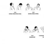
Visual gymnastics after a stroke
To restore vision, it is recommended to perform a set of special exercises. Gymnastics helps to maintain tone, strengthen muscles and brain function. All varieties of gymnastics are based on relaxing the eyes, reducing intraocular pressure, combating irritation and eye strain. Exercises can include any movement of the eyes, focusing the gaze at different distances, drawing objects and images in the air. Long-term therapy makes the picture clearer, eliminates distortion.
With partial vision loss after a stroke, exercise helps retrain the brain. Doctors include exercises in the physiotherapy complex. The simplest is the pencil exercise. You need to hold the pencil at a distance of 45 cm and follow it with your eyes, moving the pencil up, down and to the sides. You can't move your head. Also, the pencil is placed in front of the patient's face and moved to the sides.
After a stroke, it is recommended to work with puzzles and drawings. Patients can draw objects and figures, play games with words, and solve puzzles. Exercises like these help retrain the brain and help it identify objects through vision.
Even the simplest exercises help to strengthen the muscles of the eyeballs, train muscle memory and track objects. Muscle tone often disappears after a stroke. You can just keep your fingers upper eyelid and try to close your eyes. Such exercises can prevent visual fatigue and relieve stress, but exercise does not correct permanent structural damage in the brain.
Exercises can only be beneficial if they are systematically performed. It is necessary to repeat the full course daily. It is possible to achieve improvement only in cases where there is a slight loss of vision. For serious violations should be combined therapeutic gymnastics with medication. Only a comprehensive restoration will help restore vision.

Exercises to restore vision after a stroke
- Cover your eyes with your palms, inhale deeply and exhale several times. Gently press the palms on the upper and lower edges of the eye socket. Start with 3-4 reps, increasing the number of sets.
- Gently press your fingers on the recesses above the eyeballs. Make movements vibrating, abruptly release your fingers. Repeat 5 times.
- Gently press on the eyes, sharply releasing the fingers. Repeat 5 times.
- Close your eyes tightly and relax your eyes.
- Squeeze the bridge of the nose near the corners of the eyes. Hold for 5 seconds, then abruptly release your fingers. Repeat 5 times.
If the patient cannot do the exercises on his own, relatives or staff at the hospital can help him. Eye exercises are not effective in repairing damaged brain cells, but exercises help relieve eye strain, eliminate redness and burning.
It is recommended to combine exercises with drug therapy and training of other parts of the body that have been damaged (limbs, face, jaws). Only an integrated approach will allow you to fully rehabilitate after a stroke.
There are no diseases that would pass without consequences for the body. A stroke is one of those conditions that extremely negatively affect the quality of a person’s life, depriving him of his main functions: vision, speech, movement. A stroke causes an acute violation of blood circulation, including in the brain - an organ that is responsible for the functionality of the body. Vision problems occur if the lesion is localized in the area of \u200b\u200bthe vessels that supply blood to the visual center in the brain.
Visual impairment can be a complication of a stroke and an independent disease (stroke of the eye). Violation of blood circulation in the brain very often affects the visual centers. After a stroke, vision also deteriorates due to infections brought in intensive care.
There is a lot of information about diseases such as heart attack and stroke in medical reference books, they are regularly talked about on TV in programs, broadcast on radio stations. At the same time, information is presented in an accessible form, so that a person far from medicine can understand and assimilate it. But few people are aware that circulatory disorders can affect not only the heart muscle and brain, but also the retina. If the transportation of blood to the eyes stops due to blockage of the supplying artery, then an eye stroke occurs - a vascular pathology of the retina and optic nerve.
The conducted statistical studies forced doctors to draw disappointing conclusions. It turned out that an eye stroke can proceed unnoticed and not cause concern. A person may not even realize that he is in serious danger and not notice violations of his vision, the appearance of irreversible changes in the retina. Meanwhile, neurologists confirm that stroke of the visual organs is an age-related disease, and older people over 60 are at risk. It should be noted that not only the elderly are at risk of experiencing the consequences of a stroke. Sometimes the disease overtakes men and women in their prime, aged 30 to 50 years.
Causes of an eye stroke
There are several reasons that can cause transient ischemic attacks and vascular pathology of the retina.
- Hypertonic disease.
- Blood clotting disorders.
- Stressful situations and overwork.
- hereditary factor.
- High load on the organs of vision. For example, long work at the computer monitor.
- Diabetes mellitus type 2.
- Insufficiently balanced diet.
- Bad habits - smoking, frequent drinking.
What symptoms can develop with an eye stroke?
The centuries-old wisdom “Forewarned is forearmed” has not lost its relevance today. It is better to prevent the occurrence of an eye stroke than to endure the disease “on your feet” and eliminate its consequences all your life. The following are the symptoms - the first alarm signals that are given by the human body and indicate the development of pathology.
- Sudden narrowing and loss of visual field. The eyes of a person with a fixed position of the head, a fixed gaze, perceive a specific area of space. If an eye stroke occurs, then the human visual organs reduce the transmitted “picture”, and the fields of view narrow.
- The appearance of sparks, stars or flies. They flicker before your eyes, create visual interference, prevent you from taking a closer look at small objects, and do not allow you to calmly read a book or magazine.
- Can appear pain In eyes. Sometimes vision is completely lost.
Classification of eye strokes
The type of ischemic disorder that occurs in the retina largely depends on the treatment and rehabilitation of a person. To determine the type of stroke, electronic scanning of the vascular bed is prescribed. During the examination, the doctor who monitors the patient's condition receives a detailed idea of the state of the eyeballs, retina and nerve nodes, through which the eyes and the central nervous system are interconnected. Such a survey for early stage reveals places of vascular blockage, and it occurs due to thrombosis or spasm. Neurologists distinguish several probable cases that characterize the type of eye pathology in stroke.
- arterial occlusion and retinal detachment. This type of pathology is the most serious and severe. It is characterized by the fact that peripheral vision is partially or completely lost. This process occurs imperceptibly for a sick person and does not cause unpleasant symptoms. Sometimes retinal detachment is accompanied by spasm or narrowing carotid artery.
- Department of retinal veins. This type of pathology practically does not differ from the case described above. The only difference is the appearance of white spots that resemble glare of light. A stroke usually affects only one eye. With timely diagnosis, you can count on the success of treatment.
- Central artery occlusion. The disease develops suddenly, leads to unilateral loss of vision. A sick person is not able to distinguish colors. Treatment of pathology is complex and lengthy, but has a positive outcome if laser surgery methods are used.
Symptoms of deterioration of visual function should not be ignored. When they appear, you should consult a doctor and get his advice.
Among the causes of this pathology, the following can be distinguished:
- Stress at work, excessive workload.
- Diseases that lead to impaired blood clotting.
- Prolonged eye strain.
- Work at the computer, with books.
- Conditions leading to circulatory disorders.
- Diabetes mellitus, heart and vascular diseases, infections, allergies, eye injuries, glaucoma.
- Poor nutrition and bad habits.
- Long-term use of corticosteroids.
These conditions and diseases lead to disturbances in the body, as a result of which shifts occur in the coagulation system. As a result of these "breakdowns" in the body there is an increased thrombus formation. At some point, a blood clot breaks away from the vessel wall, is carried by blood flow to one or another organ, in this case, the eye.
Most often, in such conditions, patients need immediate treatment, but in rare cases, blood clots can resolve on their own.
Important! There are situations when you are at the computer for a long time, your work is directly related to eye strain. In the presence of concomitant diseases that can serve as aggravating factors, there is also a risk of ocular stroke. Do not neglect the advice of a specialist.
Eye stroke (apoplexy) is manifested primarily by visual impairment. The degree of its deterioration depends on the prevalence of the lesion. Along with this, characteristic "fireflies" appear before the eyes. Pain in the area of the diseased organ may be disturbing, but this is a non-permanent symptom.
When examining a diseased eye, small pinpoint hemorrhages can be seen, the eye sometimes becomes red. Some patients have hypertension (high blood pressure).
Eye stroke is characterized by signs:
- Acute or subacute, partial or complete loss of visual function.
- Characteristic white spots or glare before the eyes.
- Distortion of visual perception.
- Narrowing of the fields of central and peripheral vision.
- Color disturbances.
Treatment will largely depend on the type of hemorrhage, the nature and extent of the lesion, the causes that led to this outcome, and also on how timely medical care was provided.
The treatment of ocular stroke is mainly laser. It is produced by laser coagulation to destroy and remove the formed blood clot. As a result, blood circulation in the damaged area and the blood supply to the eye are normalized. It is also used to "strengthen" the retina in case of its detachment.
In rare cases, hyperbaric oxygen therapy is performed: the patient is placed in a sealed pressure chamber. Pressurized oxygen therapy is used.
The drugs are used under the supervision of a doctor and in a hospital setting. In this case, drugs are used:
- Preventing the formation of blood clots.
- Antispasmodics.
- Means that improve blood circulation.
- Angioprotectors.
- Antibiotics (in some cases, when an infection is attached or to prevent its development).
- Means that reduce blood pressure (in case of increased blood pressure).
- Drugs used to treat comorbid conditions that may exacerbate the course of the disease.
Important! When choosing drugs, it is not recommended to self-medicate, as this can harm your health and not give the desired effect. If pathological signs appear, you should consult a specialist. Do not forget: the earlier treatment is started, the better its result.
It should be regularly observed by a specialist in order to avoid complications!
With early detection of pathology, patients experience a fairly high percentage of vision recovery, however, some defects may remain in the form of flies before the eyes, white spots.
In some cases, if left untreated or with inadequate therapy, this condition can lead to retinal degeneration, which threatens with loss of vision.
The visual system plays an important role in human life. The eye is a paired sensory organ with a choroid responsible for its nutrition and metabolic processes. When one of the ophthalmic arteries is blocked, the blood supply to the organ is disrupted, which leads to pathological processes in the retina and optic nerve. This violation is conditionally called - stroke of the eye.
The disease is the most dangerous among all pathologies of the organ of vision. This is due to the latent nature of the course, since in almost 30% of cases there are no pronounced symptoms, therefore, more often minor visual impairments are mistaken for age-related changes and do not pay attention to them.
As a rule, an eye stroke occurs in people of the older age category (over 60 years), less often at a younger age (35-50 years).
Read about other types of stroke and their first signs here.

The first symptoms of an eye stroke can be determined by a visual examination of the eyeball: a hemorrhage appears or minor hemorrhages form. The first signs of the disease include:
- narrowing of the visual fields: in a healthy state, with the stillness of the head and the concentration of the gaze, a person can see a certain part of the space; with disturbed fields of visual function, the picture narrows and the person cannot cover the previously viewed area with vision;
- flies, sparks or stars very often appear before the eyes;
- discomfort (sometimes painful) in the eye area, in severe cases, the full ability to see with the affected eye is lost;
- hemorrhage in the eye, small hemorrhages;
- an eye stroke can develop with a sharp decrease in visual function, together with the appearance of white, blind spots.
Examination of the eye reveals hyperemia.
Visual impairment develops after precursors, which manifest themselves in the form of the following symptoms:
- fog, flickering or light glare appears before the eyes;
- in some cases, sensitivity is reduced in various parts of the skin.
Harbingers of this disease occur periodically, and with the necessary therapy they completely disappear. More often, such signs appear in people with a persistent increase in blood pressure, atherosclerosis and migraine.
Learn more about ocular migraine, its causes, symptoms and treatment here.
In the absence of timely medical care, serious consequences can occur that cannot be corrected:
- color vision disorders;
- narrowing of the visual fields;
- periodic appearance of flies and glare of light before the eyes;
- partial or complete loss of visual function.
Learn more about the effects of stroke here.
Human health and quality of life depend on the coordinated work of all organs and systems, in particular the visual one. If full visual perception is not possible, psychological changes occur, and in some cases constant help from outsiders is required. Therefore, at the first symptoms of visual impairment, it is necessary to contact a medical institution for diagnosis and adequate treatment.

The main signs of occlusion are a decrease in visual acuity and its distortion. But there are other manifestations of pathology that should disturb a person and cause an immediate visit to an ophthalmologist. Ignoring them is imprudent and dangerous. These include:
- organs of vision periodically hurt;
- from time to time there is double vision, bright flies, flashes and lightning;
- narrowing of the central and peripheral field of vision;
- color vision disorder.
With a severe degree of the disease on the whites of the eyes, pinpoint hemorrhages are noticeable - hemorrhages. The vascular network is dark red in color, clearly expressed, with extensive hemorrhages and weakened vessels, the entire protein may turn red. Sometimes there is an increase in intraocular and intracranial pressure.
The classification of the pathology is carried out depending on which of the vessels was damaged and how badly the retina was damaged. The most dangerous form of the disease is the combination of thrombus formation in the central artery with retinal detachment. The symptoms of the pathology are severe. Pain is usually absent. But at the same time, the following symptoms are noted:
- loss of peripheral vision;
- partial loss of the central;
- narrowing of the carotid artery, which is the most dangerous.
With the formation of a thrombus in the central retinal vein, accompanied by detachment, there is also a narrowing of the central and peripheral vision, the appearance of light spots resembling bright glare of light is noted. Before the eyes there is a feeling of a veil, objects are not clearly visible, pain rarely occurs. The pupils of the patient with this form of pathology are narrowed.
With centralized blockage of the artery, all of the above symptoms appear sharply and pronouncedly. Characteristic features this form of pathology:
- loss of central vision;
- distortion of the visual picture;
- severe pain;
- various oculomotor disorders - one eye squints or the eye does not open;
- pupil constriction.
This form is often accompanied by partial paralysis and impaired movement of the opposite arm and leg, in addition, other signs may be observed.
Regardless of the type of pathology, only surgery or laser treatment.

When blood flow to the eyeball is reduced due to blockage or rupture of one of the vessels, the situation is extremely dangerous. The organ of vision ceases to cope with its duties, which over time can lead to blindness.
An eye stroke is also dangerous because it is characterized by mild symptoms. Vision deteriorates imperceptibly, the injury causes only mild pain, and many people do not pay attention to the glare in front of their eyes.
As a result, the problem is often ignored - and completely in vain.
The human eye is a complex optical device that decodes information received in the form of light waves and transmits it to the optic nerve, after which the signal goes to the brain. This is a very important task, since a person receives about 90% of information about the world around him through his eyes.
Causes and risk factors
The main cause of the pathology is the blockage of the vessels of the eye. This condition leads to the formation of emboli or blood clots, which break off and penetrate into the bloodstream with blood flow. vascular system organ of vision, interfering with the normal blood supply. More often, blood clots occur in the coronary or carotid arteries, and under the influence of certain adverse factors, they break away from the walls and block the lumen of the central vessel of the visual system.
The main provoking factors are:
- arterial hypertension;
- frequent stressful situations;
- chronic fatigue syndrome;
- blood clotting disorders;
- constant eye strain;
- deterioration of blood circulation;
- diabetes ;
- frequent use spirits, smoking ;
- violation of the diet;
- long-term use of certain medications that adversely affect the eyes (corticosteroids, combined oral contraceptives, and others);
- damage to the arteries of the spinal column (more often occurs with a hernia of the spinal disc, trauma to the spinal column or osteochondrosis);
- jumps in intracranial pressure.
The relevance of considering this issue is that, according to statistics, about 30% of patients with an eye stroke do not notice or do not attach importance to the manifestations of the disease.
- All information on the site is for informational purposes and is NOT a guide to action!
- Only a DOCTOR can make an EXACT DIAGNOSIS!
- We kindly ask you DO NOT self-medicate, but book an appointment with a specialist!
- Health to you and your loved ones!
In most cases, people do not even realize that they are in serious danger and consider all the signs of visual impairment. age characteristics, because most often the disease occurs in people older than 50-60 years. In addition, neurologists note a more severe course of eye strokes in elderly patients.
But there are certain risk factors that contribute to the emergence and progression of vascular pathology of the retina and optic nerve in people of young and mature age:
- constant and prolonged overstrain of the organs of vision as a result of working with papers or behind a computer monitor;
- excessive fatigue, mental or physical fatigue;
- stress, nervous and psychological disorders;
- violation of the diet (excessive consumption of fried, spicy, fatty foods, pickled and salty foods);
- burdened heredity (predisposition to pathologies of vision, acute disorders of cerebral circulation, vasospasm);
- long-term use medicines contributing to the progression of vision pathology, vascular disorders (corticosteroids, oral contraceptives);
- bad habits (smoking, excessive alcohol consumption, drug addiction).
Ischemic cerebral disorders occur during vascular occlusion - blockage by blood clots, emboli, particles of atheromatous plaques, or as a result of prolonged spasm vessels of the eyeball and / or vessels of the brain and neck.
Also, the cause of vascular changes in the organ of vision are ruptures of the vessels of the retina or hemorrhagic strokes (bleeding into the substance of the brain).
| Diseases with vascular lesions, which are accompanied by their increased fragility, bleeding or thrombosis: |
|
| Diseases that contribute to degenerative changes in the vascular wall: |
|
Most common causes development of an eye stroke are considered:
- damage to the vascular wall in atherosclerosis, vasculitis, infectious-toxic changes in combination with arterial hypertension or frequent and prolonged spastic (vasomotor) paroxysms (with occlusive strokes or transient cerebrovascular accidents);
- combination hypertension or trauma to the eye and / or brain with congenital vascular anomalies (presence of aneurysms or malformations) with the hemorrhagic nature of the stroke (intracerebral or intraocular hemorrhages).
Occlusion of the central retinal artery causes the patient to develop such characteristic clinical signs:
- decreased visual acuity;
- the appearance of dark patches or livestock;
- pain in the eyeball;
- hemorrhages on the sclera;
- the appearance of flies or stars before the eyes;
- Strong headache;
- cataract;
- complete loss of vision due to atrophy of the optic nerve.
The mechanism of development of retinal occlusion is similar to circulatory disorders in any other organ. In the veins, arteries and their branches, areas of obstruction are formed due to blockage of the lumen by a blood or cholesterol clot. It breaks away from the inner surface of the carotid artery or heart valves and, together with the bloodstream, enters the retinal arteries, which leads to the development of pathology.

The causes of this condition may be spasms, thrombosis, embolism or collapse of the blood lines, which appear due to the following diseases:

Most often, retinal occlusion develops in older people (after 60-65 years) due to age-related changes in the cardiovascular system, which lead to the development arterial hypertension and atherosclerosis. At a young age, heart pathologies, eye injuries and other factors become the causes of the disease.
Fractures are a potential risk of retinal vascular occlusion tubular bones, increased blood viscosity, intravenous injections and massive bleeding.
In order to be able to restore vision in the future, in case of a stroke, you need to urgently consult a doctor and get qualified help. An ophthalmologist deals with the treatment of visual disorders after a stroke. All recovery steps must be approved by a doctor. It is very important to analyze the results and adjust the rehabilitation scheme in time.
- moisturizing the eyes with drops and gels;
- restoration of affected visual functions with the help of drugs;
- regular and correct exercise;
- diet with vitamins, especially vitamin A;
- taking food supplements.
After a stroke that caused loss of vision, you need to seriously deal with general rehabilitation and restoration of visual function. Without making an effort, the patient runs the risk of remaining blind for life, because medications are not able to restore vision completely. Rehab can include exercise, medication, and even surgery.
- Gymnastics. Special exercises help stop the progression of presbyopia at the initial stage of its development. Before starting therapy, the organs of vision should be prepared, it is recommended to hold the face over warm water and sprinkle it into the eyes. There are a large number of techniques, so the choice is up to the ophthalmologist.
- Medicines. Medications for visual impairment after a stroke are prescribed in the case when gymnastics is not effective enough. There are a large number of funds, so the choice should also be entrusted to the doctor. It will take into account the features of violations, concomitant pathologies and contraindications.
- Surgery. The operation is prescribed as a last resort. Typically, after a stroke, patients require lens replacement. In its place, a special lens is implanted, which performs all the functions of a natural lens and is combined with the structures of the eye. The operation to remove the lens is one hundred percent prevention of cataracts.
It must be remembered that the problem of vision after a stroke is very individual. The recovery program is developed for each individual patient, taking into account the degree of damage and consequences. However, absolutely everyone is recommended to moisturize the mucosa with the help of drops and gels.
Most often, the disease develops in people of the older age category (after 60 years). In such patients, neurologists note a more severe course of ocular stroke.
On the other hand, there are certain risk factors that contribute to the emergence and progression of pathology among young and mature people:
- constant and long work at the computer;
- stress, psychological disorders;
- excessive fatigue, physical and mental overwork;
- errors in nutrition (eating excessively salty and spicy foods, fried foods);
- burdened heredity;
- long-term use of corticosteroids and oral contraceptives;
- bad habits.
Often, partial or complete occlusion leads to clogging of blood vessels with blood plaques (emboli). In one hundred percent of cases, occlusion of the central retinal artery manifests itself as a consequence of acute hypertensive abnormalities. The underlying factors in the development of occlusion of the central artery or retinal veins are diseases such as arrhythmia, diabetes, antiphospholipid syndrome. Also provoking factors are hematomas and optic disc drusen, as well as the consequences of possible eye injuries.
Eye stroke
| There are three types of occlusive lesions of the eye: |
|
| Damage to the organ of vision can be observed with cardioembolic, atherothrombotic or hemodynamic strokes of the visual lobe of the brain, which can occur when circulatory disorders: |
|
| In the event of microocclusive and lacunar strokes or intracerebral hemorrhages in the area: |
|
A stroke does not always affect the entire left or right side brain, sometimes there is an ischemic stroke of individual parts of the brain. Read about
consequences of a cerebellar stroke
Read about restoring speech after a stroke at the link.
In the presence of characteristic symptoms during diagnostic measures the type of pathology is determined. For this purpose, dopplerography of the vessels of the eye is performed.
For some other reasons, the eyes of patients who have had a hemorrhage may not open, as well as how to deal with this condition, you will learn here.
Arterial occlusion and retinal detachment is a fairly serious condition, characterized by a painless course and a decrease in peripheral vision. Over time, in the absence of timely therapy, central vision is lost. In the majority of patients who have had a microstroke, during the diagnosis, a narrowing of the lumen of the carotid artery, CVS disease, and a persistent increase in blood pressure are detected.
With the progression of the disease, the patient ceases to distinguish colors and shades. timely laser treatment helps to prevent complications and eliminate visual impairments that have arisen.
In this case, there are signs of the disease in the form of the appearance of white spots before the eyes and deterioration of peripheral vision. Patients suffering from diabetes mellitus and arterial hypertension are at risk. The lack of timely medical care can provoke consequences such as blindness.

The development of the pathological process begins with a complete loss of peripheral vision, any pain missing. In rare cases, spastic pain may occur. Most patients do not distinguish the outlines of objects that are close to the affected eye and practically do not see bright light.
A new tool for the rehabilitation and prevention of stroke, which has a surprisingly high efficiency - Monastic Collection. The monastery fee really helps to fight the consequences of a stroke. Among other things, tea keeps blood pressure normal.
Occlusion or stroke of the eye is a condition of blockage of the vessels of the visual organ. As a result, the blood supply is disturbed, and an outpouring into the cavity of the eyeball occurs. This process develops due to exacerbations of pathologies of cardio-vascular system. In the early stages, the symptoms are imperceptible, but with development there is a chance of losing vision. Medical and laser therapy is used as a treatment.
80% of patients are people over the age of 60, but the disease may develop earlier. For childhood pathology is not typical.
Ocular stroke is an uncommon but very dangerous condition. The visual organs have a branched vascular scheme. Emboli and thrombi form in the bloodstream, they break thin walls and provoke hemorrhage. Such a process disrupts the nutrition of the eyes and brain, therefore, if the pathology is not noticed in time, the consequences can be adverse. The following conditions can provoke the formation of blood clots:
- chronic form of hypertension;
- problems with blood clotting;
- stress;
- eye strain;
- avitaminosis;
- alcoholism and smoking;
- long-term use of corticosteroid drugs;
- congenital pathologies retina.
Usually the tendency to such a pathology is inherited.
In the study of pathology, a hereditary character is traced. Patients with the second type are at risk diabetes. In this case, a microstroke of the eye is diagnosed, which is able to resolve itself. Patients with such a diagnosis should be registered with an ophthalmologist. With a microstroke against the background of a primary disease, the risk of vision loss increases by 2 times.
There are two forms of stroke:
- Ischemic;
- Hemorrhagic.
Ischemic stroke is caused by an acute decrease in blood flow (ischemia), usually due to a blood clot that clogs a vessel in the brain. This form of apoplexy is the most common type of stroke, accounting for 80 to 85% of all strokes.
Particularly serious consequences are given by an ischemic stroke in the brain stem, because there are vital centers that are responsible for controlling breathing and consciousness. An example of a brainstem infarction is thrombosis of the basilar artery: in severe cases, this causes complete paralysis of all limbs (tetraparesis) and coma or immediately leads to death.
Ischemia
Hemorrhagic stroke occurs when abnormalities in the coagulation system, high blood pressure, or pathological changes in the wall of blood vessels lead to bleeding in the brain. Between 15 and 20 percent of all apoplexies fall into this category.
Almost a million people in Russia suffer from apoplexy every year. 900,000 of them have their first stroke. Apoplexy occurs mainly in the elderly. As their proportion in the population is steadily increasing, the number of stroke patients is likely to increase as well.
Stroke of the brain affects mainly the elderly, but can also occur at a young age. Even newborn children in the mother's body can already have apoplexy. Possible reasons include coagulation disorders, cardiovascular pathologies. Sometimes an infectious disease causes a hemorrhage in the brain tissue in children.
In Russia, apoplexy is diagnosed every year in about 1000 children and adolescents. However, experts believe the actual number is much higher because diagnosing stroke in children is more difficult. The reason is that the maturation of the brain is not yet complete, so apoplexy in children is often detected only after a few months or years. For example, partial paralysis appears in infants only after six months.
Retinal vascular occlusion is divided into the following types:
- Occlusion of the central artery. With this type of pathology, a blockage of the main artery is formed, enriched with oxygen and delivering blood to the retina of the eye.
- Occlusion of the CAS branch. Occurs with a thrombus of small branches of the artery.
- Occlusion of the central retinal vein. This is a blockage of one of the veins.

Central vein occlusion is divided into two types:
- Blockage of the central retinal vein. In this disorder, blood flow disturbance occurs in the main vein.
- Blockade of the branch of the retinal vein. Observed with embolism on the branches of the vein.
Diagnostics
Eye stroke diagnostic methods
As a rule, the diagnosis of deviation is carried out in a hospital. As soon as a person has the first symptoms of pathology, he urgently needs to be examined by a neurologist. The doctor will evaluate the patient's reflexes, his speech, vision and orientation in space. If the suspicion of an ocular stroke is confirmed, then a consultation with an ophthalmologist is scheduled.
The following methods can be used in the diagnostic process:
- CT angiography (visualization of blood vessels).
These instrumental methods help to identify the location and degree of occlusion, and also help determine the treatment course. To detect malformations, vascular aneurysms and other pathologies, radiography is often prescribed, and ultrasound and ECG can be used to identify the causes of an eye stroke.
An eye stroke has visual signs, but they are not enough to confirm the diagnosis. The ophthalmologist studies a complete history, he needs information about the presence of chronic processes. The examination is complex, you need a consultation with a neurologist. Main diagnostic method- fluorescein angiography.

The procedure involves the study rear wall eye organ. With the help of a special apparatus, you can see changes in the vascular structure of the organ. To do this, the patient is injected intravenously with a substance that can change color when exposed to a beam. Thus, the doctor examines the problem area, determines the type and stage of the process.
Treatment of the disease
Within half an hour of the laser procedure, the patient's problem can be solved.
Stroke ophthalmic nerve requires urgent intervention. Treatment takes place in a specialized clinic under the supervision of a doctor. Most effective method considered laser coagulation. With the help of the device, you can eliminate traces of hemorrhage, and remove a blood clot without contact. The procedure is painless, lasts 20-30 minutes, the patient must ensure that the eye does not close. If the stage of the process does not require cardinal methods, drug therapy is used. After a stroke, a number of exercises are recommended.
Preparations
Medical therapy involves the use local funds in the form of drops to remove traces of inflammation, as well as drugs of general action. The scheme is determined individually depending on the patient's condition. During therapy, the emphasis is on the root cause of the process and drugs are introduced into the complex to eliminate the provoking factor. The therapeutic scheme works in several directions:
- normalization blood pressure;
- improvement of local blood circulation;
- spasm relief.
Folk recipes
In this case, the mummy can be used as a strengthening agent for the walls of blood vessels.
Initially, it is impossible to cure a stroke of the visual organ, it is worth using more specialized methods to eliminate the problem. However, non-traditional methods are used during the rehabilitation period. Folk recipes are used to restore and strengthen blood vessels. It is worth using such funds only with the permission of the doctor, if the stage allows. The following recipes have a beneficial effect:
- infusion of pine fingers (cones);
- decoction of birch leaves;
- mummy medicine;
- lemon balm based on pine needles;
- chestnut tincture.
Exercises
Gymnastics is very important to restore the functioning of the eyes. It is prescribed immediately after laser intervention, the duration of the course is determined individually. It is worth taking 10 minutes a day to practice. It is best to do gymnastics in the morning or when eye fatigue is felt. The complex includes several simple exercises:
- The injured eye closes, the other makes circular movements, then changes.
- Both eyes are tightly closed for 5 seconds, after which they open sharply.
- At the same time, sharp movements are made to the sides.
The effectiveness of therapy will increase if the patient begins to eat more fruits.
In order for the treatment to be more effective, it is necessary to supplement it proper nutrition. Fatty and spicy foods should be removed from the diet. Reduce the use of salt to a minimum. Eat plenty of vegetables and fruits every day. Additionally, the doctor may prescribe a vitamin course in tablets.
The goals of therapy for blockade of the central artery or vein retina are:
- restoration of venous circulation;
- resorption of hemorrhage;
- removal of puffiness;
- improvement of the trophism of the retina.
Urgent care
In case of occlusion, you should immediately contact a medical institution: further treatment may not bring results.

On the first day, the following measures should be taken:
- special massage of the eyeballs to move the embolus;
- paracentesis (incision) of the anterior cavity of the eye;
- special local remedies that improve blood circulation, stimulate cell nutrition and increase oxygen will help reduce intraocular pressure;
- to normalize the metabolism of the eye and microcirculation, vasodilating drops are used.
After the disease, the patient needs to undergo regular examinations by an ophthalmologist after 1, 3 months and six months.
A patient with CAS occlusion needs to be urgently hospitalized. Untimely seeking medical help can end in failure.
In the first hours, drug treatment is used:
- with spasms of arterioles, vasodilators are used - Nitroglycerin, Eufillin, Papaverine, oxygen inhalation, and oxygenation. To relieve smooth muscle tone, Atropine eye drops are also used;
- with thrombosis of the central artery, thrombolytics, anticoagulants, and dextrans are taken. To dissolve blood clots, antithrombotic drugs (Fibrinolysin) are prescribed;
- for any type of occlusion, retrobulbar and parabulbar injections are recommended with vasodilators that improve blood flow and reduce blood clotting;
- also use the intake of antioxidants, instillation of adrenoblockers;
- at weakening of a vascular tone analeptics (Caffeine) are shown;
- in the treatment of pathology, diuretics and anti-inflammatory drugs are also used, as well as medicines that stimulate blood circulation;
- it is mandatory to treat the corresponding disease that caused the blockage of the central artery or vein.
To eliminate occlusion and prevent complications, laser treatment is sometimes prescribed. In order to exclude the formation of a blood clot in the second eye, preventive therapy is used.
After a stroke, artificial tears are prescribed, Korneregel, Taufon, Normax, Taurine. Hydration is extremely important for patients who have been in a coma.
During the rehabilitation period, you need to review your diet. After a stroke, it is recommended to consume more carrots, yellow peppers, pumpkins, egg yolks and fish. Cataract prevention is provided by grapes, blueberries and onions.
After a stroke, it is helpful to massage with hot or cold compresses. This helps to relax the eyes and improve blood circulation. It is enough to moisten one towel in cold water and the other in warm water, and alternate them for 5-10 minutes.
The usual tossing of a ball or ball helps in restoring vision. You need to throw the object back and forth from the affected side. Focusing the eye on the object will help to establish the synchronism of movement and vision.
Computer programs will help to rehabilitate vision after a stroke. One such program shows a black square, at regular intervals a cluster of one hundred dots flashes from the side of the affected eye. Exercises on the computer take 15-20 minutes a day. The course of treatment is several months.
Comparative exercises allow you to check the extent of the violation of visual focus. They make it possible to determine the necessary degree of rehabilitation therapy. The patient should close their eyes and direct their gaze to the injured side of the body. After determining the correct (according to the patient) direction, the eyes are opened, and the doctor determines the proximity of the gaze from the desired direction.
Compensatory vision therapy allows you to stimulate the areas of the brain that provide vision. It includes scanning, visual field recognition, and prism exercises. The visual field can be adapted by moving images from invisible zones.
Prisms in ophthalmology are used to correct various visual disorders. The type and location of the prism will be determined by the symptoms. With doubling, the prism is placed on a glass lens, which allows you to align the direction of view. Spatial ignore requires the use of a prism on the left side of the visual field so that it reflects objects on different sides.
Restorative vision therapy aims to stimulate the nerve connections in the brain. There are different techniques for all types of visual impairments.
Usually, surgical treatment of the organs of vision does not help after a stroke, because the problem lies in the brain. Only in some cases, the operation corrects double vision by acting on the eye muscles.
The prognosis of the disease and the success of treatment depend primarily on the timeliness of the provision of medical care to the patient. The degree of spread of the lesion, the type of eye stroke, the reason for which it occurred also play a role. Therefore, treatment begins with the diagnosis of the underlying disease, then the affected vessel is identified and the type of occlusion is established.
For this, a visual examination of the eyeball and an examination of the fundus are carried out. If required, an electronic vascular scan is additionally performed, the patient is referred for a consultation with a neurologist.
AT modern medicine The method of laser coagulation is mainly used. A directed laser beam breaks a blood clot in the eye and then removes it. The integrity of the veins and arteries is not disturbed, blood supply and vision are restored. Also, with the help of such an operation, it is possible to fix the retina in case of its detachment and remove degeneration changes in the fundus area.
In some cases, it is more expedient to carry out hyperbaric oxygenation. The patient is placed in a special pressure chamber, after which oxygen is exposed at high pressure.

Non-surgical method, only with the use of medicines, it is impossible to cure a stroke of the organ of vision. But medications required in the postoperative period.
The following groups of drugs are used:
- Antispasmodics.
- Means that thin the blood and prevent the formation of blood clots.
- Drugs that stimulate blood circulation.
- Angioprotectors are medicines that strengthen and protect blood vessels from damage.
- With an increase in blood pressure - hypertensive drugs.
- Antibacterial drugs in case of infection.
- Medicines for the treatment of concomitant chronic diseases.
scheme drug therapy is only made by a doctor. He determines the combination of necessary drugs and their dosage. Self-medication in this case will not give positive result and only worsen the patient's condition. The sooner treatment is started, the better the prognosis. It is important to restore the normal blood supply to the organs of vision in the first hours after occlusion.
Our readers write
From the age of 45, pressure surges began, it became sharply ill, constant apathy and weakness. When I turned 63, I already understood that I didn’t have long to live, everything was very bad. An ambulance was called almost every week, all the time I thought that this time would be the last.
Everything changed when my daughter gave me one article to read on the Internet. You have no idea how grateful I am to her. This article literally pulled me out of the world. For the last 2 years, I have begun to move more, in spring and summer I go to the dacha every day, grow tomatoes and sell them on the market. Aunts are surprised how I manage to do everything, where so much strength and energy come from, they still won’t believe that I’m 66 years old.
Who wants to live a long and energetic life without strokes, heart attacks and pressure surges, take 5 minutes and read this article.
Possible complications of pathology
Even if there is no complete or partial loss of vision, without timely treatment, a person will suffer from loss of light perception, flies and glare before the eyes, narrowing of the peripheral or central fields of vision. In advanced cases, even surgery will not help restore visual function and fully recover.
Thus, an eye stroke is a serious pathological condition that almost never occurs in isolation. The main reason is circulatory disorders, provoked by diseases of the heart, blood vessels, metabolic disorders, congenital anomalies, etc. There are also factors that exacerbate the situation.

These are, first of all, bad habits and heavy loads on the eyes. Clearly, clogged arteries in the eyes can be prevented by being responsible for your health and getting regular check-ups with your ophthalmologist. At the first signs of pathological changes, the doctor will explain what to do, how to restore vision after an eye stroke and how to save it. Vitamins, special gymnastics, observing the rules of visual hygiene will help keep your eyes healthy until old age.
With proper treatment, you can get rid of the symptoms in 10 days, the full course, along with rehabilitation, lasts several months. However, if you ignore the disease, negative consequences are possible. It's about loss of vision. First, the patient develops a color dysfunction. On the advanced stage occlusion blindness can become irreversible.
When a rupture (hemorrhage) or blockage (occlusion) by a blood clot of an artery or vein stops the supply of blood to certain tissues of the eyeball or its outflow from the retina (retina). Conventionally, this process is called an eye stroke.
A similar situation can also be provoked by a spasm of the vessels of the brain, neck, and eyeball. This leads to various pathological processes, the result of which are:
- deterioration in peripheral vision;
- glare before the eyes;
- stroke of the optic nerve;
- strabismus;
- temporary or permanent color blindness;
- blindness.
In most cases, blockage or rupture of the vessel of the eye occurs after fifty years. However, the problem can also occur at a younger age. The provoking factors are:
- activities associated with eye strain - work with papers, prolonged sitting at a computer, in front of a TV;
- physical or mental strain, severe fatigue;
- constant stress, nervous tension, psychological illness, overwork;
- malnutrition - excessive consumption of spicy, fried, fatty foods, salty and pickled foods;
- hereditary predisposition to eye diseases, problems with blood vessels;
- taking drugs that adversely affect vision (oral contraceptives, corticosteroids);
- smoking, alcohol, drug addiction and other bad habits.
Pathological processes occurring in the body are capable of provoking blockage or rupture of blood vessels. The main causes are diseases associated with increased vascular fragility, bleeding or thrombosis. First of all, it is:
- Severe diseases of the cerebral vessels and myocardium - atherosclerosis, hypertension, arrhythmia, heart disease, endocarditis (inflammation of the inner lining of the heart muscle).
- Vascular diseases - congenital pathologies of the vessels of the eye or brain, stenosis, aneurysms, vasculitis, increased blood clotting.
- Diseases that provoked the destruction of the walls of blood vessels and problems with blood circulation. Among them are inflammatory or toxic diseases, brain tumors, metastases, pathological deposits, blood diseases, endocrine diseases (diabetes mellitus, problems with the adrenal glands, thyroid gland).
- Injury to the eye or brain.
- Ischemic stroke, in which there was a blockage of blood vessels in the brain.
- Hemorrhagic stroke - bleeding in the brain.
- Blockage or rupture of the vertebral arteries - observed with disc herniation, osteochondrosis, trauma.
- A sharp increase in intraocular or intracranial pressure.

A stroke is often triggered by a combination of several pathologies. For example, the cause may be changes in the vascular walls provoked by atherosclerosis, in combination with prolonged spastic (convulsive compression) of the arteries that occur with strokes. Another option is a combination of high blood pressure or eye injury with congenital vascular pathologies.
It is customary to distinguish three types of ischemic stroke that affects the organ of vision. This is:
- Blockage of the artery with subsequent detachment of the retina, which is responsible for the primary processing of the image and converting it into nerve impulses. A stroke leads to a decrease in peripheral vision.
- Separation from the retina of the veins - manifested by glare, spots before the eyes, unilateral damage to the organ of vision. The reason is a violation of the outflow of blood through the veins from the vessels of the retina. A stroke develops with problems with blood clotting, in diabetics, with atherosclerosis and other diseases that were triggered by changes in the vessels.
- Blockage of the central retinal artery, due to which a person ceases to distinguish colors, blind spots appear, complete blindness is possible. It develops against the background of a strong spastic narrowing of the carotid or vertebral arteries, through which blood flows to the brain. May be the result of severe heart disease, hypertension.
signs
Stroke of the organ of vision often occurs without pronounced signs, and therefore often goes unnoticed. In order to prevent pathology, it is necessary to pay attention to the symptoms of the disease, which are characteristic of all types of stroke:
- temporary or progressive decrease in visual acuity;
- gradual deterioration of peripheral vision;
- white spots, glare, other interference before the eyes;
- unexpected loss of parts of the field of view;
- problems with color perception;
- hemorrhage (bleeding in the eyeball).
With blockage of the artery of the retina, partial or complete loss of peripheral vision is possible, which over time can transform into complete blindness if retinal detachment occurs.
A stroke is accompanied by the appearance of blind spots, distorted perception of images.
The disease is rarely accompanied by pain, which is why patients do not pay attention to the gradual decrease in vision, and often realize when it cannot be restored.
A microstroke of the eye, known as an optic nerve stroke, is the result of a myocardial infarction or hemorrhagic stroke in the area of the brain that is responsible for the functioning of the eyes. This is a life-threatening situation that requires urgent medical intervention. The symptoms of a stroke are:
- sudden blindness or blurred vision in one eye, combined with loss of mobility of the muscles of the arm or leg with opposite side body (hemiparesis);
- the appearance of blind areas with preservation of color perception and visual acuity;
- sharp pain In eyes;
- constriction of the pupils;
- limitation of eye movement;
- nystagmus - involuntary oscillatory eye movements of high frequency;
- doubling of objects;
- strabismus.
Treatment
Finding symptoms that indicate a stroke of the organ of vision, you need to contact an ophthalmologist. To confirm or refute the diagnosis, after a visual examination of the eyeball, an electronic vascular scan (fluorescent hagiography) should be done, and a neurologist should be visited. Treatment depends on the extent and nature of the damage.
There are situations when the thrombus resolves on its own, which leads to the resumption of blood flow with a complete or partial restoration of vision. If this does not happen, treatment is prescribed to eliminate the cause of the disease and symptomatic therapy, whose task is to remove the manifestations of the disease. For this purpose, drugs of the following groups are prescribed:
- Thrombolytics are drugs that prevent the formation of blood clots.
- Antispasmodics - drugs that relieve spasms of blood vessels.
- Medications to improve circulation.
- Angioprotectors - cause vasodilation.
- Antibiotics - to prevent the development of a bacterial infection.
- Medications to lower blood pressure if hypertension is present.
- Medicines for the treatment of comorbidities that can cause eye complications.
If the use of medications does not help, more radical actions are needed. They provide:
- Laser coagulation of the retina. During the procedure, the thrombus is destroyed and removed, the detached retina is fused with choroid eyes. This leads to the normalization of blood circulation in the area damaged by a stroke, which helps prevent the development of dystrophies and further prevents retinal detachment.
- Hyperbaric oxygenation. A method that involves oxygen therapy under high pressure, which leads to an increase in the solubility of gases. The procedure is carried out in a hermetic pressure chamber. The method is effective if emboli are to blame for the blockage of blood vessels.
If the problem is not provoked by a serious pathology (for example, a cerebral stroke), in most cases, with timely diagnosis and correctly prescribed treatment, a complete restoration of all lost visual functions is observed. In a number of situations, the eyes are only partially restored: there are problems with glare, distorted perception of objects, and blind spots.
Visual impairment can become irreversible after a stroke, up to blindness, if timely assistance is not provided to the patient. This is the most important and dangerous complication diseases. No less formidable consequence is retinal detachment and macular defects, which also leads to loss of vision. The transition of the pathological process to the second eye, atrophy of the optic nerve occur less frequently.
Therapeutic measures
The treatment of a stroke always depends on several aspects. This includes the type of pathology, the features of its course, the causes, the patient's symptoms, and some other factors. Laser coagulation is considered the most effective and safest method. The use of a laser allows you to effectively get rid of a blood clot, which helps restore blood flow in the affected area and prevent dangerous complications. The same technique is used for retinal detachment.
Another type of therapy is hyperbaric oxygen therapy. Its essence lies in the use of oxygen under pressure. In this case, the patient is in a hermetic pressure chamber.
Drug therapy includes groups of drugs designed to eliminate a blood clot, normalize blood circulation, relieve spasm, and normalize blood pressure. Treatment includes:
- antihypertensive drugs. To normalize blood pressure, the patient is prescribed diuretics, beta-blockers, calcium antagonists, ACE inhibitors. The decision on the type of drug in a particular case is made by the attending physician, depending on the characteristics of the course of the disease;
- anticoagulants. The main goal of eye stroke therapy is to restore normal blood flow. Since the cause of circulatory disorders is most often blockage by a thrombus, to eliminate it, drugs are prescribed that help dissolve blood clots;
- antispasmodics. These drugs relieve spasm and pain that develops during an attack.
The tactics of treatment depends on the characteristics of the course of the pathology.
Depending on the associated pathologies, the patient may also be prescribed other medications, for example, non-steroidal anti-inflammatory drugs, corticosteroids, narcotic analgesics, and so on.
Only constant monitoring of vision will make it possible to identify its violations and promptly contact an ophthalmologist and neurologist. The method of treatment of the pathological process depends on the type of stroke, the severity of the symptoms that have arisen, the severity of the course, causes and concomitant diseases. Timely medical care is also of great importance in successful treatment.

Most effective treatment an eye stroke is considered laser correction.
The most commonly used and effective treatment for eye stroke is considered to be laser correction. During the procedure, laser radiation is applied to the clot that has formed and blocked the lumen of the vessel. As a result, the thrombus is destroyed, the blood supply to the organ of vision is restored.
In addition, laser treatment is performed with retinal detachment, as well as as a result of degenerative changes in the visual organ.
The use of laser therapy, together with the timely elimination of the main cause of the pathology in a short time, makes it possible to achieve restoration of vision without the development of negative consequences.
A less commonly used treatment is hyperbaric oxygen therapy (HBO), which is a unique technique for supersaturating the blood with oxygen. Therapy is carried out in specialized hyperbaric pressure chambers, where oxygen is used under high pressure.

To eliminate spasm, improve blood circulation, reduce blood pressure, drug therapy is prescribed. It is carried out under the supervision of an ophthalmologist at home or in an inpatient department.
Medical treatment consists of:
- drugs to prevent thrombosis;
- antispasmodics;
- vascular drugs to improve blood circulation;
- angioprotective agents;
- in cases of accession infectious agent antibacterial drugs are prescribed;
- medicines to normalize blood pressure and eye pressure.
To improve the effectiveness of the treatment, patients are advised to follow some rules:
- adjust the diet: give up fatty and fried foods, reduce salt intake, include a large amount of vegetables and fruits in the daily diet;
- perform exercises for the eyes;
- spend enough time outdoors, reduce television viewing;
- stick to good rest and sleep;
- avoid alcohol and smoking.
Complications and consequences
Certain symptoms are characteristic of a stroke, and a simple analysis of the clinical picture allows doctors to identify areas of the brain damaged by ischemia and hemorrhage. With a sudden onset of headache, nausea, weakness and deterioration of vision on the one hand, you should urgently contact a medical institution. A stroke provokes a sharp deterioration in the condition. This can happen in hours or even minutes, so help must be provided as quickly as possible.
Stroke symptoms:
- sudden headache, dizziness, nausea and vomiting;
- disorientation in space;
- disorder of consciousness (short-term or long-term);
- sometimes coma, loss of vision;
- muscle weakness on one side, paresis or paralysis.

It is possible to accurately diagnose, determine the affected lobes of the brain and analyze the extent of the process only after the examination. However, the doctor must be able to recognize a stroke and the symptoms of damage to a particular part of the brain, as this helps to navigate and provide first aid correctly.
It is believed that brain strokes are more common in men over 45 and 60 years old. The condition can cause irreparable harm to the body, but experts say that a third of stroke patients have the opportunity to restore vision and other impaired functions.
Large cerebral embolism in the vast majority of cases causes damage to the visual centers and permanent damage, while mild ischemia rarely causes long-term harm. Therefore, with a slight brain injury, it is possible to restore the work of the visual departments.
Most often, presbyopia (age-related farsightedness) develops with a stroke. It becomes difficult for the patient to work with small details at close range. If in healthy people age-related farsightedness is almost inevitable and does not require urgent treatment, presbyopia after a brain stroke can result in irreversible loss of vision. Loss of individual sections of the field of view is also noted.
To restore vision, it is recommended to perform a set of special exercises. Gymnastics helps to maintain tone, strengthen muscles and brain function. All varieties of gymnastics are based on relaxing the eyes, reducing intraocular pressure, combating irritation and eye strain. Exercises can include any movement of the eyes, focusing the gaze at different distances, drawing objects and images in the air. Long-term therapy makes the picture clearer, eliminates distortion.
With partial vision loss after a stroke, exercise helps retrain the brain. Doctors include exercises in the physiotherapy complex. The simplest is the pencil exercise. You need to hold the pencil at a distance of 45 cm and follow it with your eyes, moving the pencil down and to the sides. You can't move your head. Also, the pencil is placed in front of the patient's face and moved to the sides.
After a stroke, it is recommended to work with puzzles and drawings. Patients can draw objects and figures, play games with words, and solve puzzles. Exercises like these help retrain the brain and help it identify objects through vision.
Even the simplest exercises help to strengthen the muscles of the eyeballs, train muscle memory and track objects. Muscle tone often disappears after a stroke. You can simply hold your fingers on the upper eyelid and try to close your eyes. Such exercises can prevent visual fatigue and relieve stress, but exercise does not correct permanent structural damage in the brain.
Exercises can only be beneficial if they are systematically performed. It is necessary to repeat the full course daily. It is possible to achieve improvement only in cases where there is a slight loss of vision. In case of serious violations, therapeutic exercises should be combined with medication. Only a comprehensive restoration will help restore vision.

We have already partially touched possible consequences after an eye stroke. Now they can be listed:
- Loss of vision (complete or partial).
- Narrowing of fields of vision.
- Loss of color vision.
- Residual phenomena in the form of glare of light and flickering flies before the eyes.
To enjoy what is available to human visual perception, you should maintain your organ of vision at the proper level. For this, all necessary measures should be taken to avoid the disease.
Be healthy and save for yourself the breadth of attitude!
When blood flow to the eyeball is reduced due to blockage or rupture of one of the vessels, the situation is extremely dangerous. The organ of vision ceases to cope with its duties, which over time can lead to blindness. An eye stroke is also dangerous because it is characterized by mild symptoms.
What is an eye stroke
Causes of an eye stroke
signs
Drawing conclusions
Strokes are the cause of almost 70% of all deaths in the world. Seven out of ten people die due to blocked arteries in the brain. And the very first and main sign of blockage of blood vessels is a headache!
Blockage of blood vessels results in a disease under the well-known name "hypertension", here are just some of its symptoms:
- Headache
- Increased heart rate
- Black dots before the eyes (flies)
- Apathy, irritability, drowsiness
- blurred vision
- sweating
- Chronic fatigue
- swelling of the face
- Numbness and chills in fingers
- Pressure surges
Attention! If you notice at least 2 symptoms in yourself, this is a serious reason to think!
signs
With blockage of the artery followed by retinal detachment, visibility at close range worsens, the boundary of the visual angle gradually shifts inwards. Sometimes there is pain in the temples and front frontal lobes, sharp dizziness is possible, accompanied by blurred vision.
With blockage of the artery of the retina, partial or complete loss of peripheral vision is possible, which over time can transform into complete blindness if retinal detachment occurs. A stroke is accompanied by the appearance of blind spots, distorted perception of images. The disease is rarely accompanied by pain, which is why patients do not pay attention to the gradual decrease in vision, and often realize when it cannot be restored.
The separation of the veins from the retina is accompanied by a deterioration in peripheral vision, which eventually leads to blindness, and the signs of pathology progress rapidly. It is necessary to pay attention and urgently deal with treatment if objects suddenly become fuzzy, glare, clouding appear, blind spots are observed in the field of view.
The first warning signs of an eye stroke are:
- temporary and/or progressive loss of vision;
- gradual loss of peripheral vision;
- the appearance of white spots, "glare", other interference that block vision ("curtain effect");
- sudden loss of areas of the field of view;
- color disturbance.
It is important to remember that if any of these symptoms appear, you should immediately contact an ophthalmologist - timely diagnosis of pathological circulatory disorders of the eye and proper treatment prevent irreversible loss of vision and make it possible to fully or partially restore vision in case of an eye stroke.
Symptoms of an eye stroke with occlusion (blockage) of the central retinal artery:
- complete or partial loss of peripheral vision, which gradually develops into partial or complete loss of central vision (with retinal detachment);
- the appearance of blind spots, distorted outlines or distorted perception of the picture.
This pathology is considered one of the most dangerous and develops against the background of a significant spasm or narrowing (atherosclerotic) of the carotid arteries, pathology of the vertebral arteries, severe heart disease, and a malignant course of hypertension.
In most cases, the disease is not accompanied pain syndrome and patients do not pay attention to the manifestations of the disease, but prolonged occlusion of the central artery causes irreversible changes in the retina up to complete blindness.
This disease occurs when venous outflow from the vessels of the retina - blockage of the central vein of the retina. Most often, this pathology develops in violation of blood clotting, in patients with diabetes mellitus, atherosclerosis, or other vascular changes.
It notes:
- deterioration of vision, first peripheral with a gradual loss of central vision;
- blurred images of objects;
- the appearance of floating highlights, haze;
- loss of visual fields.
Symptoms of the disease appear in proportion to the degree of obstruction, they occur unexpectedly and gradually progress - from several hours to several days.
Centralized arterial occlusion occurs suddenly and is characterized by a unilateral loss of all visual functions (complete loss of vision).
With strokes of the optic nerve that develop during the formation of a focus of cerebral infarction or intracerebral hemorrhage in the region of the center of gaze in the precentral gyrus, the location of the oculomotor centers, optic chiasm (chiasm), visual lobes of the brain depends on:
- on the size and anatomical location of the stroke focus (cerebral hemispheres, trunk, cerebellum, parieto-occipital lobe);
- the degree of damage to the vascular basin;
- type of stroke (hemorrhagic or cerebral infarction);
- side of defeat.

This may cause symptoms:
- sudden blindness or a sharp decline vision in one eye with impaired movement (hemiparesis) and sensitivity of the arm and leg on the opposite side (cross syndrome);
- loss of visual fields (hemianopsia), often with preservation of color perception and visual acuity;
- acute pain, pupil constriction, limitation of eye mobility (ophthalmoplegia), nystagmus (swinging eye movements), doubling of objects, blindness in half of the eye on the side opposite to the focus;
- various oculomotor disorders, strabismus.
In the visual lobes of the brain, the formation of visual images occurs, therefore, when they are damaged, loss of the corresponding parts of the visual fields develops.
They are often accompanied by impaired skin sensitivity and paresthesias in various parts of the body.
The duration of the attack can last from several minutes to several hours, then vision is fully restored. This symptom often manifests itself against the background of hypertension, atherosclerosis, severe headache and nausea.
Why do eye changes occur?
In older people, hemorrhages in the eyes occur most often due to the natural weakening of blood vessels, slowing blood circulation and metabolic processes. But, unfortunately, young, quite healthy and active people are also subject to this phenomenon. The provoking factors are:
- prolonged and regular eye strain;
- stress and nervous overload at work or in the family;
- diseases that lead to circulatory disorders;
- conditions in which blood clotting changes;
- diabetes;
- allergies and infectious diseases;
- eye injury, glaucoma;
- pathologies of the heart and blood vessels, for example, ischemic stroke, atherosclerosis;
- malnutrition, alcohol and cigarette abuse;
- long-term use of hormonal drugs.
The main causes of hemorrhage in the organs of vision are diseases that provoke circulatory disorders, thickening of the blood, in which the vessels become brittle, weak, prone to thrombosis. These include:
- Pathologies of the heart and cerebral vessels: arterial hypertension, cerebral atherosclerosis, heart attacks, endocarditis of any form, birth defects hearts.
- Vascular diseases: vasculitis, aneurysms, hemorrhagic diathesis, stenosis, congenital pathologies of the vessels of the organs of vision or brain, infectious diseases and inflammatory processes affecting the elasticity of the vascular walls.
- Toxic diseases of the brain: meningitis, encephalitis, arachnoiditis.
- Atheromas, hemangiomas, brain metastases in malignant tumors.
- Endocrine system disorders: diabetes mellitus, hypothyroidism, adrenal dysfunction.
- Diseases of the hematopoietic system.
Medical practice shows that the most common cause of an eye stroke is a combination of arterial hypertension with trauma to the organs of vision or congenital anomalies of the cerebral vessels, atherosclerotic lesions of the vascular walls in combination with vasculitis or toxic infections of the brain. Sometimes a violation of the blood supply to the organs of vision occurs when the vertebral arteries rupture - a complication of osteochondrosis or a hernia of the vertebral discs.
The risk of developing a stroke of the organs of vision increases if several provoking factors and chronic diseases are combined at once, and this is exactly what happens most often. Often, occlusion occurs with a significant increase in intracranial pressure, if a person leans or stands up sharply. Often, pathology is noted in women who smoke, who regularly take oral contraceptives.
Any of these causes and factors, individually or together, lead to an increase in blood clotting, which provokes the formation of blood clots. If a blood clot breaks off, it is carried along with the bloodstream to an organ, in this case, to the eyes. The situation is aggravated if the walls of the vessels are thinned and fragile. Blockage of a vein or artery in the organs of vision is called an eye stroke.
In some cases, the thrombus does not come off, but resolves on its own. In this situation, the blood supply is restored, and vision is not impaired. But this happens very rarely. Therefore, if a person has crossed the 50-year mark, he has any chronic diseases or the organs of vision are constantly subjected to heavy loads, from time to time it is necessary to consult a specialist.
Clinical picture
The first "danger signals" indicating the possible development of the disease may be the following symptoms:

One of the main signs that a patient has had an ocular stroke is the simultaneous deterioration of vision (sharp) and the appearance of white spots in front of the eyes. With a visual examination of the visual organ, local redness, small hemorrhages are noticeable, the patient's blood pressure may jump.
Such a situation necessarily entails partial or complete violation the main function of the eye - vision is either greatly weakened, or the patient is completely blind.
Symptoms of an eye stroke due to blockage of the central retinal artery:

The course of the disease is not accompanied by pain, but if not diagnosed in time, it can lead to blindness.
With the separation of the central retinal vein, the following symptoms appear:
- blindness or lightning-fast unilateral loss of vision;
- violation of the movement of the visual organ with loss of sensitivity of the limbs on the opposite side (the so-called cross syndrome);
- loss of visual fields, subject to the preservation of visual function and color discrimination;
- sharp pain occurs in the eye, the pupil narrows;
- limited mobility or rocking movements of the organ of vision;
- doubling of objects;
- strabismus.
A typical harbinger of pathology is considered a symptom of flickering darkness, which manifests itself in the form of such visual impairments:

This symptom often accompanies severe hypertension, atherosclerosis, migraine.
Key Symptoms
The initial stages of the process are asymptomatic: there are no painful sensations, the eyes do not water. Minor disturbances of visual function do not disturb the patient. The pathology of the usual develops only on one side, while there is no difference between the vision of the left and right eyes. With the development, there are clearer symptoms of an eye stroke:
- Traces of petechial hemorrhages are formed on the surface of the white shell.
- Vision is impaired: clear contours of figures are lost, doubling occurs.
- The effect of "flies" appears after a sharp raise of the head.
- Opacification of the lens develops, a cataract is diagnosed.
To stop the process and restore vision, urgent diagnosis and treatment is necessary. First of all, the ophthalmologist determines the type of disease depending on the focus of the hemorrhage. With central occlusion, the signs develop rapidly: the transition of a microstroke to the acute phase occurs in 2-3 days.
When an artery is occluded, a patient without any symptoms can go blind.
An eye stroke is a vascular pathology of the retina, which causes a violation of the blood supply to the visual center. The resulting obstructions in the veins and arteries reduce and distort vision.
The main danger of this disease is that most people do not notice it, because it is painless, and, accordingly, they are in no hurry to treat it. However, the disease has consequences, up to the loss of peripheral vision.
The severity of the disease, and accordingly, the methods of its treatment depend on the degree of the problem and its location.
Among the causes of this pathology, the following can be distinguished:
- Stress at work, excessive workload.
- Diseases that lead to impaired blood clotting.
- Prolonged eye strain.
- Work at the computer, with books.
- Conditions leading to circulatory disorders.
- Diabetes mellitus, heart and vascular diseases, infections, allergies, eye injuries, glaucoma.
- Poor nutrition and bad habits.
- Long-term use of corticosteroids.
These conditions and diseases lead to disturbances in the body, as a result of which shifts occur in the coagulation system. As a result of these "breakdowns" in the body there is an increased thrombus formation. At some point, a blood clot breaks away from the vessel wall, is carried by blood flow to one or another organ, in this case, the eye. The blood supply is disturbed, resulting in ischemia, sometimes - rupture of the vessel. In people with age, the walls of blood vessels become thinner, degenerative changes occur in them. This aggravates the course of the disease.
Most often, in such conditions, patients need immediate treatment, but in rare cases, blood clots can resolve on their own.
Important! There are situations when you are at the computer for a long time, your work is directly related to eye strain. In the presence of concomitant diseases that can serve as aggravating factors, there is also a risk of ocular stroke. Do not neglect the advice of a specialist.
It is very important to seek medical help when the first signs appear, when the changes are still reversible.
The main manifestations of the disease
Eye stroke (apoplexy) is manifested primarily by visual impairment. The degree of its deterioration depends on the prevalence of the lesion. Along with this, characteristic "fireflies" appear before the eyes. Pain in the area of the diseased organ may be disturbing, but this is a non-permanent symptom.
When examining a diseased eye, small pinpoint hemorrhages can be seen, the eye sometimes becomes red. Some patients have hypertension (high blood pressure).
Eye stroke is characterized by signs:
- Acute or subacute, partial or complete loss of visual function.
- Characteristic white spots or glare before the eyes.
- Distortion of visual perception.
- Narrowing of the fields of central and peripheral vision.
- Color disturbances.
Types of disease
Blockage of an arterial vessel in combination with retinal detachment is the most dangerous and most severe type, often without pain. Patients notice a loss of peripheral visual fields, sometimes there is a partial loss of central vision. Sometimes accompanied by narrowing of the carotid artery.

An eye stroke can be painless, so the absence of pain does not mean that everything is fine!
Rehabilitation treatment partially helps, but changes in the form of white spots and narrowing of visual fields may remain. Pain symptoms fickle.
Detachment and occlusion of the central retinal vein is also accompanied by a narrowing of the visual fields on the periphery and the appearance of white spots (sometimes look like glare of light). The cause is a blood clot in a vein. The consequences are identical to those of arterial occlusion. Accompanied by edema of the optic nerve, retina.
Patients feel a deterioration in the quality of vision. First, the peripheral suffers, and then the central. Objects see fuzzy, blurry. Glare, spots, a feeling of fog, sometimes double objects can be observed before the eyes. There is also loss of visual fields. On examination, pupillary constriction. Sometimes these symptoms are accompanied by pain.
Through surgical treatment, in some cases, it is possible to partially restore a person's vision and improve the quality of life.
Central blockage of the artery is characterized by a sudden manifestation, the brightness of symptoms. Vision deteriorates sharply, the patient hardly distinguishes colors. Loss of central vision. The picture of perception is distorted. Often accompanied by severe pain.
In this case, the fastest onset has an important role in preserving vision. medical measures(laser treatment).
Therapy
Treatment will largely depend on the type of hemorrhage, the nature and extent of the lesion, the causes that led to this outcome, and also on how timely medical care was provided.
The treatment of ocular stroke is mainly laser. It is produced by laser coagulation to destroy and remove the formed blood clot. As a result, blood circulation in the damaged area and the blood supply to the eye are normalized. It is also used to "strengthen" the retina in case of its detachment. It is used to correct degenerative changes in the fundus area.
In rare cases, hyperbaric oxygen therapy is performed: the patient is placed in a sealed pressure chamber. Pressurized oxygen therapy is used.
The drugs are used under the supervision of a doctor and in a hospital setting. In this case, drugs are used:
- Preventing the formation of blood clots.
- Antispasmodics.
- Means that improve blood circulation.
- Angioprotectors.
- Antibiotics (in some cases, when an infection is attached or to prevent its development).
- Means that reduce blood pressure (in case of increased blood pressure).
- Drugs used to treat comorbid conditions that may exacerbate the course of the disease.
Important! When choosing drugs, it is not recommended to self-medicate, as this can harm your health and not give the desired effect. If pathological signs appear, you should consult a specialist. Do not forget: the earlier treatment is started, the better its result.

It should be regularly observed by a specialist in order to avoid complications!
With early detection of pathology, patients experience a fairly high percentage of vision recovery, however, some defects may remain in the form of flies before the eyes, white spots.
In some cases, if left untreated or with inadequate therapy, this condition can lead to retinal degeneration, which threatens with loss of vision.
What's next?
We have already partially touched on the possible consequences after an eye stroke. Now they can be listed:
- Loss of vision (complete or partial).
- Narrowing of fields of vision.
- Loss of color vision.
- Residual phenomena in the form of glare of light and flickering flies before the eyes.
To enjoy what is available to human visual perception, you should maintain your organ of vision at the proper level. For this, all necessary measures should be taken to avoid the disease.
Be healthy and save for yourself the breadth of attitude!
When blood flow to the eyeball is reduced due to blockage or rupture of one of the vessels, the situation is extremely dangerous. The organ of vision ceases to cope with its duties, which over time can lead to blindness. An eye stroke is also dangerous because it is characterized by mild symptoms. Vision deteriorates imperceptibly, the injury causes only mild pain, and many people do not pay attention to the glare in front of their eyes. As a result, the problem is often ignored - and completely in vain.
What is an eye stroke
The human eye is a complex optical device that decodes information received in the form of light waves and transmits it to the optic nerve, after which the signal goes to the brain. This is a very important task, since a person receives about 90% of information about the world around him through his eyes.
The flow of information is continuous, so the eye needs constant nutrition, which it receives through an extensive network of blood vessels. When a rupture (hemorrhage) or blockage (occlusion) by a blood clot of an artery or vein stops the supply of blood to certain tissues of the eyeball or its outflow from the retina (retina). Conventionally, this process is called an eye stroke. A similar situation can also be provoked by a spasm of the vessels of the brain, neck, and eyeball. This leads to various pathological processes, the result of which are:
- deterioration in peripheral vision;
- glare before the eyes;
- stroke of the optic nerve;
- strabismus;
- temporary or permanent color blindness;
- blindness.
Causes of an eye stroke
In most cases, blockage or rupture of the vessel of the eye occurs after fifty years. However, the problem can also occur at a younger age. The provoking factors are:
- activities associated with eye strain - work with papers, prolonged sitting at a computer, in front of a TV;
- physical or mental strain, severe fatigue;
- constant stress, nervous stress, psychological illness, overwork;
- malnutrition - excessive consumption of spicy, fried, fatty foods, salty and pickled foods;
- hereditary predisposition to eye diseases, problems with blood vessels;
- taking drugs that adversely affect vision (oral contraceptives, corticosteroids);
- smoking, alcohol, drug addiction and other bad habits.
Pathological processes occurring in the body are capable of provoking blockage or rupture of blood vessels. The main causes are diseases associated with increased vascular fragility, bleeding or thrombosis. First of all, it is:
- Severe diseases of the cerebral vessels and myocardium - atherosclerosis, hypertension, arrhythmia, heart disease, endocarditis (inflammation of the inner lining of the heart muscle).
- Vascular diseases - congenital pathologies of the vessels of the eye or brain, stenosis, aneurysms, vasculitis, increased blood clotting.
- Diseases that provoked the destruction of the walls of blood vessels and problems with blood circulation. Among them are inflammatory or toxic diseases, brain tumors, metastases, pathological deposits, blood diseases, endocrine diseases (diabetes mellitus, problems with the adrenal glands, thyroid gland).
- Injury to the eye or brain.
- Ischemic stroke, in which there was a blockage of blood vessels in the brain.
- Hemorrhagic stroke - bleeding in the brain.
- Blockage or rupture of the vertebral arteries - observed with disc herniation, osteochondrosis, trauma.
- A sharp increase in intraocular or intracranial pressure.
A stroke is often triggered by a combination of several pathologies. For example, the cause may be changes in the vascular walls provoked by atherosclerosis, in combination with prolonged spastic (convulsive compression) of the arteries that occur with strokes. Another option is a combination of high blood pressure or eye injury with congenital vascular pathologies.
It is customary to distinguish three types of ischemic stroke that affects the organ of vision. This is:
- Blockage of the artery with subsequent detachment of the retina, which is responsible for the primary processing of the image and converting it into nerve impulses. A stroke leads to a decrease in peripheral vision.
- Separation from the retina of the veins - manifested by glare, spots before the eyes, unilateral damage to the organ of vision. The reason is a violation of the outflow of blood through the veins from the vessels of the retina. A stroke develops with problems with blood clotting, in diabetics, with atherosclerosis and other diseases that were triggered by changes in the vessels.
- Blockage of the central retinal artery, due to which a person ceases to distinguish colors, blind spots appear, complete blindness is possible. It develops against the background of a strong spastic narrowing of the carotid or vertebral arteries, through which blood flows to the brain. May be the result of severe heart disease, hypertension.
signs
Stroke of the organ of vision often occurs without pronounced signs, and therefore often goes unnoticed. In order to prevent pathology, it is necessary to pay attention to the symptoms of the disease, which are characteristic of all types of stroke:
- temporary or progressive decrease in visual acuity;
- gradual deterioration of peripheral vision;
- white spots, glare, other interference before the eyes;
- unexpected loss of parts of the field of view;
- problems with color perception;
- hemorrhage (bleeding in the eyeball).
With blockage of the artery followed by retinal detachment, visibility at close range worsens, the boundary of the visual angle gradually shifts inwards. Sometimes there is pain in the temples and the anterior part of the frontal lobes, severe dizziness is possible, accompanied by blurred vision.
With blockage of the artery of the retina, partial or complete loss of peripheral vision is possible, which over time can transform into complete blindness if retinal detachment occurs. A stroke is accompanied by the appearance of blind spots, distorted perception of images. The disease is rarely accompanied by pain, which is why patients do not pay attention to the gradual decrease in vision, and often realize when it cannot be restored.
The separation of the veins from the retina is accompanied by a deterioration in peripheral vision, which eventually leads to blindness, and the signs of pathology progress rapidly. It is necessary to pay attention and urgently deal with treatment if objects suddenly become fuzzy, glare, clouding appear, blind spots are observed in the field of view.
A microstroke of the eye, known as an optic nerve stroke, is the result of a myocardial infarction or hemorrhagic stroke in the area of the brain that is responsible for the functioning of the eyes. This is a life-threatening situation that requires urgent medical attention. The symptoms of a stroke are:
- sudden blindness or blurred vision in one eye, combined with loss of mobility of the muscles of the arm or leg on the opposite side of the body (hemiparesis);
- the appearance of blind areas with preservation of color perception and visual acuity;
- sharp pain in the eyes;
- constriction of the pupils;
- limitation of eye movement;
- nystagmus - involuntary oscillatory eye movements of high frequency;
- doubling of objects;
- strabismus.
Finding symptoms that indicate a stroke of the organ of vision, you need to contact an ophthalmologist. To confirm or refute the diagnosis, after a visual examination of the eyeball, an electronic vascular scan (fluorescent hagiography) should be done, and a neurologist should be visited. Treatment depends on the extent and nature of the damage.
There are situations when the thrombus resolves on its own, which leads to the resumption of blood flow with a complete or partial restoration of vision. If this does not happen, treatment is prescribed to eliminate the cause of the disease and symptomatic therapy, the task of which is to remove the manifestations of the disease. For this purpose, drugs of the following groups are prescribed:
- Thrombolytics are drugs that prevent the formation of blood clots.
- Antispasmodics - drugs that relieve spasms of blood vessels.
- Medications to improve circulation.
- Angioprotectors - cause vasodilation.
- Antibiotics - to prevent the development of a bacterial infection.
- Medications to lower blood pressure if hypertension is present.
- Medicines for the treatment of comorbidities that can cause eye complications.
If the use of medications does not help, more radical actions are needed. They provide:
- Laser coagulation of the retina. During the procedure, the thrombus is destroyed and removed, the detached retina fuses with the choroid. This leads to the normalization of blood circulation in the area damaged by a stroke, which helps prevent the development of dystrophies and further prevents retinal detachment.
- Hyperbaric oxygenation. A method that involves high pressure oxygen therapy, which leads to an increase in the solubility of gases. The procedure is carried out in a hermetic pressure chamber. The method is effective if emboli are to blame for the blockage of blood vessels.
If the problem is not provoked by a serious pathology (for example, a cerebral stroke), in most cases, with timely diagnosis and correctly prescribed treatment, a complete restoration of all lost visual functions is observed. In a number of situations, the eyes are only partially restored: there are problems with glare, distorted perception of objects, and blind spots.
Occlusion or stroke of the eye is a condition of blockage of the vessels of the visual organ. As a result, the blood supply is disturbed, and an outpouring into the cavity of the eyeball occurs. This process develops due to exacerbations of pathologies of the cardiovascular system. In the early stages, the symptoms are imperceptible, but with development there is a chance of losing vision. Medical and laser therapy is used as a treatment.
80% of patients are people over the age of 60, but the disease may develop earlier. For childhood, pathology is not typical.
Etiology
Ocular stroke is an uncommon but very dangerous condition. The visual organs have a branched vascular scheme. Emboli and thrombi form in the bloodstream, they break thin walls and provoke hemorrhage. Such a process disrupts the nutrition of the eyes and brain, therefore, if the pathology is not noticed in time, the consequences can be adverse. The following conditions can provoke the formation of blood clots:
- chronic form of hypertension;
- problems with blood clotting;
- stress;
- eye strain;
- avitaminosis;
- alcoholism and smoking;
- long-term use of corticosteroid drugs;
- congenital pathologies of the retina.
Usually the tendency to such a pathology is inherited.
In the study of pathology, a hereditary character is traced. Patients with the second type of diabetes mellitus are at risk. In this case, a microstroke of the eye is diagnosed, which is able to resolve itself. Patients with such a diagnosis should be registered with an ophthalmologist. With a microstroke against the background of a primary disease, the risk of vision loss increases by 2 times.
Back to index
Key Symptoms
The initial stages of the process are asymptomatic: there are no painful sensations, the eyes do not water. Minor disturbances of visual function do not disturb the patient. The pathology of the usual develops only on one side, while there is no difference between the vision of the left and right eyes. With the development, there are clearer symptoms of an eye stroke:
- Traces of petechial hemorrhages are formed on the surface of the white shell.
- Vision is impaired: clear contours of figures are lost, doubling occurs.
- The effect of "flies" appears after a sharp raise of the head.
- Opacification of the lens develops, a cataract is diagnosed.
When an artery is occluded, a patient without any symptoms can go blind.
To stop the process and restore vision, urgent diagnosis and treatment is necessary. First of all, the ophthalmologist determines the type of disease depending on the focus of the hemorrhage. With central occlusion, the signs develop rapidly: the transition of a microstroke to the acute phase occurs in 2-3 days. With thrombosis of the veins, separation of the retina is possible. This type is characterized by complete loss of vision. The most dangerous form is arterial occlusion. In this case, the patient does not feel anything, but there is a possibility of loss of central vision.
Back to index
Diagnostics
An eye stroke has visual signs, but they are not enough to confirm the diagnosis. The ophthalmologist studies a complete history, he needs information about the presence of chronic processes. The examination is complex, you need a consultation with a neurologist. The main diagnostic method is fluorescein angiography. The procedure involves the study of the back wall of the eye organ. With the help of a special apparatus, you can see changes in the vascular structure of the organ. To do this, the patient is injected intravenously with a substance that can change color when exposed to a beam. Thus, the doctor examines the problem area, determines the type and stage of the process.
Back to index
Treatment of the disease
Within half an hour of the laser procedure, the patient's problem can be solved.
A stroke of the optic nerve requires urgent intervention. Treatment takes place in a specialized clinic under the supervision of a doctor. The most effective method is considered laser coagulation. With the help of the device, you can eliminate traces of hemorrhage, and remove a blood clot without contact. The procedure is painless, lasts 20-30 minutes, the patient must ensure that the eye does not close. If the stage of the process does not require cardinal methods, drug therapy is used. After a stroke, a number of exercises are recommended.
Back to index
Preparations
Drug therapy involves the use of local remedies in the form of drops to remove traces of inflammation, as well as general drugs. The scheme is determined individually depending on the patient's condition. During therapy, the emphasis is on the root cause of the process and drugs are introduced into the complex to eliminate the provoking factor. The therapeutic scheme works in several directions:
- normalization of blood pressure;
- improvement of local blood circulation;
- spasm relief.
Back to index
Folk recipes
In this case, the mummy can be used as a strengthening agent for the walls of blood vessels.
Initially, it is impossible to cure a stroke of the visual organ, it is worth using more specialized methods to eliminate the problem. However, non-traditional methods are used during the rehabilitation period. Folk recipes are used to restore and strengthen blood vessels. It is worth using such funds only with the permission of the doctor, if the stage allows. The following recipes have a beneficial effect:
- infusion of pine fingers (cones);
- decoction of birch leaves;
- mummy medicine;
- lemon balm based on pine needles;
- chestnut tincture.
Back to index
Exercises
Gymnastics is very important to restore the functioning of the eyes. It is prescribed immediately after laser intervention, the duration of the course is determined individually. It is worth taking 10 minutes a day to practice. It is best to do gymnastics in the morning or when eye fatigue is felt. The complex includes several simple exercises:
- The injured eye closes, the other makes circular movements, then changes.
- Both eyes are tightly closed for 5 seconds, after which they open sharply.
- At the same time, sharp movements are made to the sides.
The effectiveness of therapy will increase if the patient begins to eat more fruits.
For the treatment to be more effective, it is necessary to supplement it with proper nutrition. Fatty and spicy foods should be removed from the diet. Reduce the use of salt to a minimum. Eat plenty of vegetables and fruits every day. Additionally, the doctor may prescribe a vitamin course in tablets. After a stroke, it is important to reduce eye strain as much as possible.
Back to index
Effects
With proper treatment, you can get rid of the symptoms in 10 days, the full course, along with rehabilitation, lasts several months. However, if you ignore the disease, negative consequences are possible. It's about loss of vision. First, the patient develops a color dysfunction. At an advanced stage of occlusion, blindness can become irreversible. In addition, the eyes are in close proximity to the brain, so hemorrhages in this area can provoke stagnation in the central vessels.
Back to index
Prevention
First of all, you need to monitor the state of blood pressure. Jumps or increased rates of a regular nature put the patient at risk. In addition, the eyes should rest, it is worth taking breaks when reading or working on a computer. If you have problems with your eyesight, you should immediately consult a doctor, this will help protect yourself from a stroke.
An eye stroke or occlusion is not a diagnosis in ophthalmology, but only a conditional name for a pathological phenomenon in which there is a rupture or blockage of small vessels that are responsible for nourishing the retina of the eye. The organs of human vision have a branched vascular network with an intensive blood supply. If one of the vessels is damaged, occlusive pathologies of the retina and optic nerve develop, accompanied by hemorrhage. As a result, a person's vision is reduced or distorted.
The insidiousness is that the symptoms of an eye stroke may not appear for a long time, a person does not experience initial stages the development of pathology is no discomfort, vision remains normal until a blockage or rupture of the vessel occurs. Pathologies are usually affected by people of advanced age over 60 years old, but recently a microstroke of the eyeball can be diagnosed in young people aged 30–40 years. In the absence of timely treatment, the consequences of the pathological phenomenon can be the most serious, up to the complete loss of vision.
Note: Eye stroke does not occur in isolation. As a rule, this is a consequence of other pathological disorders in the human body. Therefore, it can be prevented if you monitor your health and do not start existing diseases.
Why do eye changes occur?
In older people, hemorrhages in the eyes occur most often due to the natural weakening of blood vessels, slowing blood circulation and metabolic processes. But, unfortunately, young, quite healthy and active people are also subject to this phenomenon. The provoking factors are:
- prolonged and regular eye strain;
- stress and nervous overload at work or in the family;
- diseases that lead to circulatory disorders;
- conditions in which blood clotting changes;
- diabetes;
- allergies and infectious diseases;
- eye injury, glaucoma;
- pathologies of the heart and blood vessels, for example, ischemic stroke, atherosclerosis;
- malnutrition, alcohol and cigarette abuse;
- long-term use of hormonal drugs.

Nervous shaking, tense emotional condition, excessive stress on the eyes when working at a computer - the main factors provoking an eye stroke
The main causes of hemorrhage in the organs of vision are diseases that provoke circulatory disorders, thickening of the blood, in which the vessels become brittle, weak, prone to thrombosis. These include:
- Pathologies of the heart and cerebral vessels: arterial hypertension, cerebral atherosclerosis, heart attacks, endocarditis of any form, congenital heart defects.
- Vascular diseases: vasculitis, aneurysms, hemorrhagic diathesis, stenosis, congenital pathologies of the vessels of the organs of vision or the brain, infectious diseases and inflammatory processes that affect the elasticity of the vascular walls.
- Toxic diseases of the brain: meningitis, encephalitis, arachnoiditis.
- Atheromas, hemangiomas, brain metastases in malignant tumors.
- Endocrine system disorders: diabetes mellitus, hypothyroidism, adrenal dysfunction.
- Diseases of the hematopoietic system.
Medical practice shows that the most common cause of an eye stroke is a combination of arterial hypertension with trauma to the organs of vision or congenital anomalies of the cerebral vessels, atherosclerotic lesions of the vascular walls in combination with vasculitis or toxic infections of the brain. Sometimes a violation of the blood supply to the organs of vision occurs when the vertebral arteries rupture - a complication of osteochondrosis or a hernia of the vertebral discs.
The risk of developing a stroke of the organs of vision increases if several provoking factors and chronic diseases are combined at once, and this is exactly what happens most often. Often, occlusion occurs with a significant increase in intracranial pressure, if a person leans or stands up sharply. Often, pathology is noted in women who smoke, who regularly take oral contraceptives.

Elderly people with chronic pathologies of the heart and blood vessels, everyone who falls into the risk group, it is recommended to visit an ophthalmologist at least once a year for a preventive examination.
Any of these causes and factors, individually or together, lead to an increase in blood clotting, which provokes the formation of blood clots. If a blood clot breaks off, it is carried along with the bloodstream to an organ, in this case, to the eyes. The situation is aggravated if the walls of the vessels are thinned and fragile. Blockage of a vein or artery in the organs of vision is called an eye stroke.
In some cases, the thrombus does not come off, but resolves on its own. In this situation, the blood supply is restored, and vision is not impaired. But this happens very rarely. Therefore, if a person has crossed the 50-year mark, he has any chronic diseases, or the organs of vision are constantly subjected to heavy loads, from time to time it is necessary to consult a specialist. It is recommended to visit an ophthalmologist at least once a year. The doctor examines the fundus, assesses the condition of the vessels and determines whether there is a need to start treatment.
Types of the disease and its manifestations
The main signs of occlusion are a decrease in visual acuity and its distortion. But there are other manifestations of pathology that should disturb a person and cause an immediate visit to an ophthalmologist. Ignoring them is imprudent and dangerous. These include:
- organs of vision periodically hurt;
- from time to time there is double vision, bright flies, flashes and lightning;
- narrowing of the central and peripheral field of vision;
- color vision disorder.
There are three main types of occlusion of the eye - occlusion of the vein, artery and centralized - any of them is dangerous for complete blindness in the absence of immediate medical attention
With a severe degree of the disease on the whites of the eyes, pinpoint hemorrhages are noticeable - hemorrhages. The vascular network is dark red in color, clearly expressed, with extensive hemorrhages and weakened vessels, the entire protein may turn red. Sometimes there is an increase in intraocular and intracranial pressure.
The classification of the pathology is carried out depending on which of the vessels was damaged and how badly the retina was damaged. The most dangerous form of the disease is the combination of thrombus formation in the central artery with retinal detachment. The symptoms of the pathology are severe. Pain is usually absent. But at the same time, the following symptoms are noted:
- loss of peripheral vision;
- partial loss of the central;
- narrowing of the carotid artery, which is the most dangerous.
Full restoration of vision after a stroke of the eye of this type is currently impossible, white spots and narrowing of the visual fields will still disturb throughout the rest of life.
With the formation of a thrombus in the central retinal vein, accompanied by detachment, there is also a narrowing of the central and peripheral vision, the appearance of light spots resembling bright glare of light is noted. Before the eyes there is a feeling of a veil, objects are not clearly visible, pain rarely occurs. The pupils of the patient with this form of pathology are narrowed.

Disturbances in the movements of the eyeball, strabismus, blindness of the eye are one of the signs of centralized blockage of the artery of the eye.
With centralized blockage of the artery, all of the above symptoms appear sharply and pronouncedly. Characteristic signs of this form of pathology:
- loss of central vision;
- distortion of the visual picture;
- severe pain;
- various oculomotor disorders - one eye squints or the eye does not open;
- pupil constriction.
This form is often accompanied by partial paralysis and impaired movement of the opposite arm and leg, in addition, other signs may be observed.
Regardless of the type of pathology, only surgery or laser treatment can help restore vision.
Treatment Methods
The prognosis of the disease and the success of treatment depend primarily on the timeliness of the provision of medical care to the patient. The degree of spread of the lesion, the type of eye stroke, the reason for which it occurred also play a role. Therefore, treatment begins with the diagnosis of the underlying disease, then the affected vessel is identified and the type of occlusion is established.

Timely laser therapy, medications, healthy lifestyle life allow you to fully restore vision after an eye stroke
For this, a visual examination of the eyeball and an examination of the fundus are carried out. If required, an electronic vascular scan is additionally performed, the patient is referred for a consultation with a neurologist.
In modern medicine, the method of laser coagulation is mainly used. A directed laser beam breaks a blood clot in the eye and then removes it. The integrity of the veins and arteries is not disturbed, blood supply and vision are restored. Also, with the help of such an operation, it is possible to fix the retina in case of its detachment and remove degeneration changes in the fundus area.
In some cases, it is more expedient to carry out hyperbaric oxygenation. The patient is placed in a special pressure chamber, after which oxygen is exposed at high pressure.
Non-surgical method, only with the use of medicines, it is impossible to cure a stroke of the organ of vision. But medications are needed in the postoperative period.
The following groups of drugs are used:
- Antispasmodics.
- Means that thin the blood and prevent the formation of blood clots.
- Drugs that stimulate blood circulation.
- Angioprotectors are medicines that strengthen and protect blood vessels from damage.
- With an increase in blood pressure - hypertensive drugs.
- Antibacterial drugs in case of infection.
- Medicines for the treatment of concomitant chronic diseases.
After an eye stroke, only an integrated approach, including the use of eye drops and vitamins, as well as special exercises, will help restore and maintain vision.
The scheme of drug therapy is only a doctor. He determines the combination of necessary drugs and their dosage. Self-medication in this case will not give a positive result and will only worsen the patient's condition. The sooner treatment is started, the better the prognosis. It is important to restore the normal blood supply to the organs of vision in the first hours after occlusion.
If therapy is carried out on time and correctly, vision can be fully restored. Perhaps there will be small defects in the form of flies and specks, but the quality of life will not be impaired. If the symptoms are ignored, treatment will not begin or will be carried out incorrectly, degenerative changes in the retina will progress, which will eventually lead to loss of vision.
Consequences and complications
Even if there is no complete or partial loss of vision, without timely treatment, a person will suffer from loss of light perception, flies and glare before the eyes, narrowing of the peripheral or central fields of vision. In advanced cases, even surgery will not help restore visual function and fully recover.
Thus, an eye stroke is a serious pathological condition that almost never occurs in isolation. The main reason is circulatory disorders, provoked by diseases of the heart, blood vessels, metabolic disorders, congenital anomalies, etc. There are also factors that exacerbate the situation. These are, first of all, bad habits and heavy loads on the eyes. Clearly, clogged arteries in the eyes can be prevented by being responsible for your health and getting regular check-ups with your ophthalmologist. At the first signs of pathological changes, the doctor will explain what to do, how to restore vision after an eye stroke and how to save it. Vitamins, special gymnastics, observing the rules of visual hygiene will help keep your eyes healthy until old age.
Eye stroke is one of the manifestations of retinal vascular catastrophes.
According to international classification diseases, the pathology is called retinal vascular occlusion.
Condition associated with rupture, blockage, compression, or extreme spasm blood vessel. As a result, the nutrition of the eye is significantly limited, and symptoms of visual impairment appear. With untimely treatment, we are talking about irreversible loss of vision.
Occlusion of the vessels of the eye is rarely found as an independent nosology. The condition is often a manifestation or complication of another, general somatic, pathology. Diseases and conditions that provoke occlusion of retinal vessels:
- atherosclerosis;
- increased blood pressure;
- violation of carbohydrate metabolism (diabetes mellitus);
- autoimmune inflammatory diseases vessels (giant cell arteritis);
- increased blood viscosity (polycythemia, multiple myeloma);
- thrombophilia - congenital or acquired;
- endocarditis;
- viral and bacterial vascular diseases;
- congenital vascular anomalies;
- pathology of the heart valves (rheumatism, heart defects);
- heart rhythm disturbances;
- myocardial infarction, acute coronary syndrome;
- extensive and chronic blood loss;
- acute disorders of cerebral circulation (strokes, transient ischemic attacks);
- hyperthyroidism;
- long-term use of diuretics, oral contraceptives that increase blood clotting agents;
- invasive diagnostic methods associated with the introduction of contrast agents (angiography).
Local provoking conditions:
- calcifications of the optic disc (drusen);
- edema of the optic disc;
- eye tumors;
- retrobulbar hematomas.
In 20-30% of cases, it is not possible to establish the cause of an eye stroke.
At-risk groups
 Most of the patients who underwent an eye stroke suffered from atherosclerosis and its manifestations (cardiac ischemia, angina pectoris, hypertension), diabetes mellitus. The next position as a direct etiological factor is occupied by rheumatic pathologies, then - temporal arteritis, vascular disease.
Additional risk groups:
- people whose working activity associated with working at a computer (eye strain);
- people exposed to frequent and prolonged stressful situations (doctors, lawyers, employees of military units and emergency response workers);
- age over 40 years;
- smokers;
- people who abuse alcohol;
- frequently ill people (decreased immunity);
- burdened heredity for atherosclerosis, hypertension, diabetes, heart and vascular diseases.
Retinal vein thrombosis is more common in people aged 40 and older. Retinal artery occlusion is more common in men aged 60 to 65 years.
Varieties of eye stroke
An eye stroke can be localized in the venous or arterial bed of the retinal circulatory system. If the catastrophe occurred in a venous vessel, they speak of retinal vein thrombosis. Blockage can occur in the central retinal vein (CRV) or in its branches (the superior temporal branch is affected in 82% of cases). A large role in retinal vein occlusion is assigned to a violation of the internal vascular epithelium, predisposing to the formation of blood clots.
Obstruction of the outflow leads to stagnation of blood and an increase in intravenous pressure. In this case, the liquid begins to seep through the walls of the vessel. They may also break. Blood fills the eye space, swelling increases, intraocular pressure rises even more. This is fraught with compression of small capillaries, which further limits the outflow of fluid and exacerbates retinal hypoxia. Such a cycle of mechanisms of the pathological process is called a "vicious circle".
The closure of the lumen of an arterial vessel is called arterial occlusion. Distinguish occlusion of the central retinal artery (CAS), its branches and cilioretinal artery. Often blockage occurs in the CAS, the branches are affected with a lower frequency. The cause of blockage of the artery in most cases is a cholesterol, fibrin thrombus or calcification embolism.
The embolus closes the access of nutrition to the retina - ischemia develops. Clinical symptoms of visual impairment develop rapidly. If medical care is provided within the first 40 minutes of the onset of symptoms, vision can still be fully restored. The delay in therapy threatens with irreversible consequences.
Symptoms
 The main symptom of retinal artery occlusion is visual field loss. The sign is disturbing either in one or both eyes. The symptom develops suddenly. Sometimes the first signs of the disease are the appearance of glare, sparks, flickering, unsharp pain in the orbit. Rarely worried about quickly passing blindness.
As with arterial occlusion, with vein thrombosis, symptoms develop suddenly, against the background of good health. Before the main symptom, patients are often disturbed by a fog before their eyes, a dark spot, image distortion, dull pains in the depth of the orbit.
Eye stroke symptoms are painless, may be mistaken for minor temporary aggravation due to fatigue or stress. This is an erroneous opinion. If any of the above symptoms appear, call immediately medical care.
Treatment
An eye stroke is an absolute indication for hospitalization by ambulance and treatment under round-the-clock supervision.
There are no significant differences in the treatment of arterial and venous stroke. The goal of therapy for these conditions is general, it consists in cleansing the lumen of the vessel, strengthening the vascular wall, restoring vision, eliminating intraocular hemorrhage (if any), and preventing relapse. For these purposes, several groups of drugs are used:
- Thrombolytics. Thrombus agents. For the best effect, the drugs are administered under the eye, under the conjunctiva or directly into the cavity of the eyeball.
- Preparations for expanding the lumen of the vessel. They can also reduce the "sticking" of red blood cells and platelets to the vessel wall, preventing blockage.
- Means for protecting and restoring the inner lining of blood vessels. They are used in the treatment and as a prevention of a repeated episode of pathology.
- Blood thinners. They help in the resorption of a blood clot and prevent recurrence.
- With retinal edema, treatment is often added glucocorticosteroids.
- Drugs to reduce intraocular pressure. If the pressure remains high, or it needs to be reduced as soon as possible, resort to a puncture of the cornea.
With high blood density, hemodilution is performed - a method of thinning the blood by intravenous administration solutions similar in composition to blood plasma. To surgical treatment they resort to the ineffectiveness of conservative therapy and in difficult cases (compression of the optic disc, retinal detachment, compression of the vessel from the outside).
 With arterial occlusion in the first 8 hours, you need to massage the eyeball. This helps to move the clot to the periphery. The head is low. The eyelids are closed. You have to press your fingers eyeball over upper eyelid for 3-5 seconds, at first weakly, then stronger. Pressure on the eye increases intraocular pressure in small arterioles, which slightly increases blood flow to the eye.
When the pressure on the eyeball stops, the pressure drops sharply. Changes in pressure set the clot in motion, it moves, freeing up blood flow. This is not a curative measure, but a way to extend the time before starting therapy so as not to aggravate retinal hypoxia.
With any type of stroke, treatment of the underlying disease that caused the embolism is necessary.
Complications and consequences
Visual impairment can become irreversible after a stroke, up to blindness, if timely assistance is not provided to the patient. This is the main and most dangerous complication of the disease. No less formidable consequence is retinal detachment and macular defects, which also leads to loss of vision. The transition of the pathological process to the second eye, atrophy of the optic nerve occur less frequently.
When the retina lacks nutrition, the body begins to come up with workarounds. This is how the formation of new blood vessels occurs - neovascularization. This is not a healing process - the new vessels are too brittle and tortuous, and the risk of their rupture is high. Cases of neovascularization of the optic disc and neovascular glaucoma are known.
Forecast
With timely assistance, the prognosis is favorable. Ischemic strokes are more likely to develop complications. Patients who have undergone arterial occlusion have a 2-fold increased risk of cardiovascular complications.
Prevention
Prevention is reduced to the treatment and prevention of influencing diseases. Regular visits to the therapist, checking vision, fundus should be made at least 1 time per year.
Precautionary measures:
- avoid eye strain, wear safety glasses when working with a computer and telephone;
- avoid smoking, drinking alcohol;
- dose physical, emotional stress;
- observe sleep and wakefulness;
- do not take any drugs without first consulting a doctor;
- know and treat chronic and hereditary pathologies;
- take courses of vitamin complexes to increase immunity, especially in the spring and autumn.
Observance of precautionary measures and knowledge of the main symptoms and methods for recognizing an eye stroke will help to avoid such a formidable condition. If this happens, respond in time and correctly and maintain your health.