Retinal detachment - what is dangerous detachment, causes. What should not be done in case of retinal detachment? What is the disease called when the retina peels off
Retinal detachment is one of the most severe diseases of vision, which is characterized by separation of the retina from choroid(or choroid). For a healthy eye, it is characteristic that the retina tightly attaches to the choroid, and receives the main nutrition from it.
In modern ophthalmology, retinal detachment is an intractable disease, as it is a serious disease in terms of surgical intervention, and an unpredictable outcome of the operation (possible pathological conditions). Already over the past 10 years, the development of retinal detachment has increased, and is 1 case of the disease per 10,000 people. This is one of the main causes of blindness and disability (about 70% of all patients are people of working age).
This disease is not treated with injections, drops, tablets. The only thing that can help is an emergency operation, thanks to which it will be possible to restore and save vision, save eyes.
Causes of retinal detachment

To understand the causes, consider the mechanism of the appearance of the disease. The development of the disease can provoke physical overstrain, a sharp pressure on the surface of the retina, as a result of which small defects appear, with the appearance of which fluid from vitreous body moves into the space below the retina. The resulting fluid begins to separate the retina. The more liquid seeps out, the larger the area of detachment will be.
Basically, such a disease is observed only in one of the eyes, but at the same time, it negatively affects the other eye. Therefore, both eyes are examined with equal care.
- The disease can occur through trauma to the eye (penetrating injury). In addition to the retina, other membranes of the eye can also be injured.
- Another reason for the appearance of the disease is diseases of the organs of vision (uveitis, tumors of the vascular membranes, retinitis, age-related macular degradation, diabetic retinopia and other diseases).
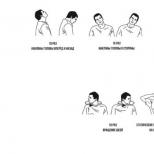
- The occurrence of peripheral vitreochorioretinal dystrophies, which cause visual impairment and can occur even in healthy people. In such cases, the disease can be detected using a three-mirror Goldman lens.
Risk factors that provoke the disease:
- eye injury;
- retinal detachment in the fellow eye;
- Availability this disease close relatives;
- peripheral vitreochorioretinal retinal dystrophy;
- activities associated with constant physical stress and heavy lifting;
- Availability various pathologies retina.
Who is at risk?
- people diagnosed with diabetes;
- people suffering from high myopia, astigmatism (since these conditions provoke thinning of the retina);
- athletes involved in injury dangerous sports (weightlifting, boxing, wrestling).
Symptoms
- The appearance of "flashes", "sparks", floating points, "lightning", floating points resembling "soot flakes".
- The appearance of "curtains", "shrouds" before the eyes. Washing with tea or other special preparations does not give a result. At this point, it is important to remember how the disease began to manifest itself, from which side it began (where the “curtain” appeared).
- Local loss of visual fields, narrowing.
- The objects that you are looking at begin to distort, the size and shape change, and subsequently the object vision decreases.
- With the rapid development of the disease, a veil may appear before the eyes.

When a retinal vessel ruptures, black dots, flies appear before the eyes of a person, in addition, pain and discomfort may manifest themselves.
When cobwebs, floating spots appear before the eyes, this may indicate that a retinal detachment has occurred, which caused hemorrhage in the middle of the vitreous body.
In some cases, patients notice that after a night's rest, their vision returned to normal. This is due to the fact that the fluid that has accumulated overnight under the retina has resolved and the retina has returned to its original place. But after a few hours, the disease will again manifest itself.
A detachment in the lower parts of the eye is very dangerous, since we can not notice it for a long time.
Diagnosis of the disease. Treatment Methods
If you notice at least the slightest signs of the disease, be sure to contact an ophthalmologist, so you prevent the development of the disease and save your eyesight.
If the symptoms began to manifest through a traumatic brain injury, in addition to the ophthalmologist, a consultation with a neuropathologist will also be necessary.
Basically, the disease is localized on the periphery of the retina (there is the lowest blood supply). That is why direct and indirect ophthalmoscopy of the fundus is performed.
To better examine the pupil, it is dilated with the help of special drops (instilled into the eyes).
With this inspection, you can determine:
- localization of detachment;
- how many breaks are there, and what are they;
- is there retinal dystrophy and where is it localized;
- if the detachment has a connection with the vitreous body.

To clarify or confirm the diagnosis, additional methods must also be used:
- check visual acuity (vision in the affected eye disappears abruptly, especially in cases where it is the central localization of the detachment);
- determination of intraocular pressure (usually this indicator does not change, but this does not apply to cases when an injury or stroke occurs, then the pressure becomes higher);
- ophthalmic perimeters (determination of visual fields). If a disease occurs, the fields will be narrowed;
- conducting ultrasound research(applied if there are contraindications to other methods, or you need to more accurately establish the diagnosis);
- laser tomography (the method is used when it is necessary to study the state of the optic nerve).

Treatment
The only treatment is surgical intervention. The choice of method will depend on the localization of the defect, its complexity, and size.
There are several types of operations:
- sclerosis (damage is sealed with diathermy (laser or current)). The tissue that is around the retina is scarred and does not allow fluid to penetrate under the retina;
- pneumatic retinopexy (the defect is sclerosed by freezing or using a laser). Further, air is introduced into the eye into the vitreous cavity, which puts the retina in place;
- vitrectomy (two small holes are made in the sclera in order to have the correct illumination of the surgical field, and insert a cutting emitter and thin tweezers inside). The removed vitreous body is replaced by gas. After the expiration of time, the gas is absorbed and filled with its own moisture;
- local filling (a small silicone filling is sewn onto the sclera, so that the sclera is pulled inward and together with the choroid, and approaches the retina);
- ballooning of the retina (the essence of the method: a catheter with a balloon is temporarily sutured into the sclera in the area of the projection of the detachment). The balloon is inflated and the same effect is obtained as when filling the sclera.
Possible consequences of retinal detachment
The most terrible and often occurring consequence of this disease is blindness. That is why it is necessary to resort to the help of ophthalmologists as early as possible. Only surgical intervention will stop the development of the disease, restore vision and avoid its loss.
The disease also threatens to drop a certain area out of sight, and a veil may appear in front of the eye.
In addition, retinal detachment is dangerous because it can cause loss of visual acuity, distortion of images of objects. A dangerous decrease in vision occurs during the course of the disease (the macula appears).
Preventive actions
People who have diabetes mellitus, have received eye or head injuries, have retinal dystrophy, myopia, must undergo examinations by ophthalmologists in order to detect the problem in a timely manner. A separate risk group includes pregnant women (in some cases, retinal detachment may occur after childbirth).
In addition, people who are at risk should observe the correct mode of sleep and wakefulness, not lift heavy, and avoid heavy physical exertion.
The retina provides primary information processing. It is on it that an image is formed after the rays are refracted in the optical media of the eye. At the same time, the retina is also the most fine structure eyeball. Even a small injury can lead to its detachment. Consider the features of this pathological process.
The retina, or inner membrane, lines eyeball from within. Light rays, refracted after passing through the vitreous body, cornea and lens, fall on the retina and form an image. These light impulses are converted into nerve impulses and transmitted to the brain. So in general view can describe the process of seeing. The retina is also the thinnest structure of the eye. Various factors can lead to a violation of its integrity. Any damage to the retina is accompanied by a violation of visual functions.
Retinal detachment (retinal detachment) is the separation of the retina from the choroid. Light-sensitive receptors, which are located in the inner shell, do not receive nutrients due to disruption of connections with blood vessels. As a result, these receptors cannot perform their functions, that is, ensure the normal functioning of vision. Gradually, the places where the retina has detached are filled with fluid. Ultimately, this pathological process can lead to complete and irreversible blindness. Why does it start? We list the causes and symptoms of the disease.
Retinal detachment: causes
The main, or primary, cause of retinal detachment is its rupture. It subsequently leads to the accumulation of fluid, due to which detachment occurs. Rupture can be caused various diseases- systemic and ophthalmic. They can be considered secondary causes, or disposing factors. These include:
- Myopia. In patients with myopia, the eyeball is larger than in people without visual impairment. Because of this, the light rays after refraction are not at the center of the retina, but in front of it. The reasons for the abnormal growth of the organs of vision can be different. The main one is a genetic predisposition to this refractive error. As the eyeball increases in size, the retina expands. Unlike the cornea, vitreous, and other structures, the retina does not grow. With a high degree of myopia, it is in constant tension. It gradually thins out. As already noted, this is the thinnest shell of the eye. Any minor injury, physical activity, stress can lead to its rupture and subsequent detachment.
- Diabetes. This disease is one of the main culprits of retinal detachment. Diabetics are often diagnosed with diabetic retinopathy. It develops as a result of a violation of the integrity blood vessels. An increase in blood glucose levels affects the state of the entire vascular system. It becomes more vulnerable, and the vessels are fragile. When they break, blood enters the retina. In other words, there is a hemorrhage in the inner shell of the eye. The accumulation of blood is accompanied by the formation of scars. They become the cause of delamination.
- Retinal dystrophy is the process of dying of the tissues of the inner shell. There is various reasons and varieties of this degenerative disease. Almost all of them eventually lead to retinal detachment.
Detachment can also occur due to other factors. Start this pathological process can:
- mechanical injury of the eyeball;
- eye surgery;
- diseases that affect circulatory system;
- inflammatory, infectious, viral ophthalmic diseases;
- excessive physical exercise;
- pregnancy and childbirth;
- stress.
Now let's find out how this disease manifests itself.

Retinal detachment: symptoms
If you consult a doctor when the first symptoms of detachment appear, the risk of a quick recovery and a favorable outcome of treatment increases significantly. The disease is accompanied by the following symptoms:
- the appearance of a veil before the eyes;
- shadows in the field of vision, especially when moving the head;
- floating black dots / spots, which indicate hemorrhage into the vitreous body;
- flashes, lightning, sparks in the eyes;
- curvature of shapes, outlines of objects.
Also, in the presence of this pathology, people complain of constant fatigue. For some, vision improves in the morning and gets worse in the evening. This is due to the fact that at night the liquid accumulating in the areas of exfoliation is partially absorbed.
Symptoms of retinal detachment can be severe or moderate. It depends on the type of pathological process and its localization. So, with detachment in the upper part of the eye, vision deteriorates quickly, due to the fact that the fluid begins to descend, provoking the separation of the lower parts of the inner membrane from the choroid. With the development of the pathological process from below, signs of detachment appear gradually. The disease is asymptomatic for a long time. This form of pathology is more dangerous.
Types of retinal detachment: symptoms
Depending on the causes, provoking factors, the degree of development, the location of detachment or ruptures, the duration of the course of the disease, several types of retinal detachment are distinguished. According to the type of occurrence, there are the following varieties of this pathology:
- primary;
- secondary;
- traumatic.
Primary, or dystrophic, is a consequence of a violation of the integrity of the inner membrane, as a result of which fluid from the vitreous body penetrates into it. In this case, degenerative processes in the retina become a provoking factor in the development of the disease. Another name for this type of detachment is rhegmatogenous detachment. Translated from Greek, the word "regma" means break. In most cases, it is he who causes the separation of the layers of the inner shell. More often it happens in older people. This is due to the fact that with age, the vitreous body loses its elasticity.
Traumatic detachment is a consequence of mechanical damage to the eye or surgical operation on the eyeball. The pathological process can occur immediately at the time of tissue damage and even several months or a year after injury. Therefore, the symptoms of detachment may not appear immediately, which is why the disease is diagnosed untimely.
Secondary detachment develops against the background of various systemic ailments or ophthalmic pathologies. Along with signs of retinal detachment, symptoms of the underlying disease are observed. The secondary form is further divided into two types:
- traction;
- serous.
Traction retinal detachment is caused by abnormal adhesive processes her with a vitreous body. In other words, detachment occurs in places of tension of the retina in the area of the vitreous substance, which is provoked by the growth of blood vessels. Serous detachment is the result of penetration into the inner part of the eye of fluid from the vessels of the retina. This usually happens when arterial hypertension, vasculitis, swelling of the optic disc and other severe pathological processes. Detachment is accompanied by symptoms characteristic of the diseases that cause it.
According to the severity of the pathology, 4 types of it are distinguished:
- Local. Detachment is characterized by damage to ¼ of the entire retina.
- Common is a pathological process covering 1/2 of the inner shell.
- Subtotal leads to the spread of the disease by ¾.
- Complete detachment, or total, captures the entire plane of the considered eye structure.
There are other classifications as well. So, if the fluid under the retina has collected in a bubble, a vesicular detachment is diagnosed. The formation of folds indicates a flat delamination. According to the mobility of the pathological process, there are:
- mobile detachment, localized in the lower layers;
- rigid retinal detachment, in which adherence to the lower or upper layers is not observed.
Determining the type and form of the disease is the main task of diagnosing retinal detachment. The method of treatment depends on its results. Find out what procedures are prescribed for suspected this pathology.
Diagnosis of retinal detachment
Diagnostics takes place in several stages, including three types of research methods:
- standard;
- special;
- laboratory.
Standard procedures include:
- visual acuity check - visometry;
- measurement of intraocular pressure - tonometry;
- examination of the anterior part of the eye - biomicroscopy;
- examination of the fundus - ophthalmoscopy;
- study of visual fields and detection by cattle - perimetry.
Special methods can also be prescribed, for example, an ultrasound of the eyes, which allows you to examine them in detail, evaluating the functional features of the retina. In some cases, electrophysiological procedures (EPS) are required - electrooculography (EOG), electroretinography (ERG) and electroencephalography with the study of the potentials of the visual cortex.
Laboratory methods include blood tests - general, syphilis, hepatitis, biochemistry, as well as urinalysis (general and sugar).
The main of these studies is ophthalmoscopy of the fundus. Using this diagnostic method, the doctor can examine the retina in great detail, assess the degree of the disease, the spread of detachment. Under an ophthalmoscope, retinal detachments appear grayish-white against a reddish-pink background. If the pathological process has been developing for months, scars and folds are observed. Areas of discontinuity appear in red. However, they have non-standard outlines. After the ophthalmologist determines the cause and type of pathology, a treatment method is selected. Consider the main ones and move on to the prevention of retinal diseases.

Retinal Detachment: Treatment
Surgery is indicated for retinal detachment. However, surgery is not always required. There are several ways to treat this pathology, which can be combined into three groups:
- extrascleral;
- endovitreal;
- laser.
Extrascleral include extrascleral ballooning and extrascleral filling. The essence of the first operation is as follows. A balloon with a catheter is inserted behind the eyeball. With its help, pressure is created on the sclera. After that, laser fixation of the retina is carried out, that is, coagulation. After 5-7 days the balloon is removed. Contraindications to the procedure:
- extensive gaps with their location in the back of the eye;
- folds that occupy almost the entire fundus of the eye;
- hemorrhages in the vitreous body.
Extrascleral filling is also performed on the surface of the sclera. The purpose of the operation is to bring the detached part of the retina closer to pigment epithelium. The surgeon makes incisions on the conjunctiva and on certain parts of the sclera, in places of detachment, applies seals from a silicone sponge. They are biocompatible. If necessary, the doctor removes the accumulated fluid from the eye. Vision after such an operation returns after a few months. Sometimes there are complications - a cataract, a change in refraction towards myopia, an increase in intraocular pressure.
Endovitreal methods for the treatment of retinal detachment
These techniques involve treating the eye from the inside. One of the commonly prescribed procedures of this type is vitrectomy, during which part or all of the vitreous body is removed. This gives the doctor access to back wall eyes. After that, he performs laser coagulation - soldering the exfoliated retina. The removed substance is replaced by a viscous transparent material - saline solution, gas, polymers. There are a number of contraindications to vitrectomy:
- pathology of the optic nerve;
- clouding of the cornea;
- severe retinal disease.
Vitrectomy is one of the most effective ways treatment of detachment of the inner membrane of the eye. However, after such an operation, there are complications:
- hemorrhages in the retina;
- increased intraocular pressure;
- change in the curvature of the cornea;
- repeated detachment;
- development inflammatory processes;
- lens damage.

Treatment of retinal detachment: laser-type operations
Laser coagulation can be a stage of one of the procedures and a separate method for the treatment of retinal detachment. This procedure is called peripheral restrictive laser photocoagulation. It can also be considered as a prevention of retinal detachment. During the procedure, the doctor acts on the thinned areas of the inner shell with a laser, soldering it to the underlying tissues. This achieves:
- increase in blood flow;
- normalization of blood supply;
- nutrition of injured parts of the retina;
- blocking the flow of fluid under the inner shell.
If the patient has myopia, then two weeks after such an operation, laser correction can be performed. Peripheral restrictive laser coagulation is not prescribed for:
- gross changes in the fundus of the eye;
- the presence of films on the retina;
- clouding of optical media.

Detachment prevention
So, the causes of exfoliation are primary and secondary. In the presence of disposing factors, a person must take all measures to prevent the occurrence of a pathological process. It is necessary to limit physical activity, visit an ophthalmologist more often, wear corrective means for existing myopia. No specific prevention of retinal detachment has been developed. At the first sign, you should consult a doctor. The first symptom that usually appears is floating spots in the visual field. They are noticeable when you look at light objects. This symptom cannot be ignored. It may also indicate other severe pathologies, including systemic ones.
Now you know what retinal detachment is, causes, symptoms, treatment methods and preventive measures for this disease. A few words about its consequences. The most dangerous of them is blindness. There may be other complications:
- eye hypotension;
- cataract;
- subatrophy of the eyeball;
- chronic iridocyclitis;
- loss of visual functions.
Vision - the most important body feelings, without it it is impossible to imagine the normal existence of man. The eyes are complex and are the main instrument of visual function. Pro retinal detachment almost everyone has heard it today. What can be done to avoid vision loss?
What is "retinal detachment"?
The tissue lines the inside of the eyeball. Her function is to perceive a light beam and convert it into nerve impulses that follow further along optic nerves into the human brain. This structure of the visual organ is responsible for the coordinated and high-quality work of the vision analyzer.
The retina is fed through the thinnest vascular network, with which it is securely connected. medical name retina - retina.
The retina consists of unevenly distributed nerve cells of two different types are cones and rods. Cells- cones concentrated in the center of the retina. They are responsible for visual acuity and help color perception of the surrounding world. sticks located on the periphery of the organ and allow people to be able to navigate in space, perceive light and see the outlines of objects in the dark.

Detachment from the network of vessels leads to disruption of the entire visual apparatus. This means that a person loses the ability to transform and transmit light signals. That is, blindness sets in.
Retinal detachment corresponds to class H00-H59 code according to ICD-10.
Causes
The pathological process of retinal detachment is caused by several reasons:


- Exudative . When fluid accumulates behind the retina, it can also detach, while the retinal tissue itself remains completely unharmed.

- Secondary . Functional diseases of the eye structures on an inflammatory and infectious background can also cause retinal detachment.
- traumatic . The occurrence of this pathology provokes a serious injury, while detachment can occur immediately at the time of receipt of the traumatic factor, or after it. There are frequent cases of detachment after surgery.

Another peeling process eye retina accepted to classify according to the extent of its distribution:
- Local concentrated within one quadrant:

- Common localized on the area of two quadrants:

- Subtotal covers the territory of three quadrants
- Total detachment - distributed throughout the length:

Symptoms
Elderly age and the presence of high degrees in a person, people subject to eye operations, and hypertonic disease- these are the main risk groups for detachment of the retina. The emerging symptoms in the early stages will allow you to urgently respond to the pathology and prevent vision loss in a timely manner.
Signs of retinal detachment:
- Light shadows, flashes, glare, lightning and spots occur at the very initial stage of the peeling process.
- Trembling and blurring of the boundaries of objects, curvature of outlines, vibration of objects when they are considered.
- Flashing flies before the eyes, which is a hindrance to concentration. The symptom does not disappear even after good rest and sleep.
- A black line or spot forms in the eyes, this is the visible area of the detachment. This symptom tends to grow to a complete narrowing of the field of view.
- When reading, individual letters or sections of the text being read fall out of the visual field.
- Partial or complete loss of peripheral vision.
- A progressive decline in visual acuity, blindness can occur in just a few hours.
Important point! If a veil has appeared in the eyes, then it is necessary to concentrate and understand from which side it spread. It is important for a doctor to know about the source of the pathological process - it went from the center, or started from the edges of the retina.
Treatment
The insidious disease does not cause pain, so people usually do not rush to the doctor, attributing the resulting symptoms to severe fatigue and life circumstances. Meanwhile, the disease does not sleep, it kills the retina without a chance for recovery.

What to do?- If you feel the very first signs of detachment, a visit to the ophthalmologist should be immediate , only this will help to keep the ability to see.

Today, the disease is treated exclusively operational way There are no effective conservative treatments for retinal detachment. The sooner the diagnosis begins and the operation is performed, the higher the chance of the patient returning to a full life.
Diagnostics
medical examination detachment of the retinal epithelium includes :

Operation
The statistics are relentless - 70% of cases of blindness occur due to detachment and lack of timely medical care.
Distinguish two types surgical intervention in the retina:
- extrascleral a method that is carried out by a laser through the surface of the eye sclera. The laser leads to a sharp increase in temperature, which causes coagulation (coagulation) of the tissue, due to which the retina strengthens and fuses with the vascular network, that is, its further damage stops.

- Endovitreal a method in which the object of access to the retina is the vitreous body.

The principle is the same in both cases. blocking the site of the pathology of the retina and bringing it closer to the supplying vascular network. Restoration of vision after surgery for retinal detachment is possible only if the full trophism of the organ is resumed.
Recipe for success surgical treatment- This is a timely appeal to a specialist. Delay is unacceptable when retinal detachment has occurred, an operation whose cost varies within from 50,000 to 100,000 rubles required for the preservation and restoration of the function of vision.
The final price of the procedure will depend on the nature of the occurrence and the extent of the damage to the visual organ as a whole.
Video about the operation:
AT clinical practice, among patients with retinal detachment (OS), there is a group of patients with old rigid flat retinal detachments extending to the two lower quadrants of the eyeball.
Lower flat retinal detachments with multiple small perforated breaks in myopia associated with PCRD tend to slowly progress and develop local preretinal proliferative vitreoretinopathy (PVR), retinal rigidity, and a tendency to self-limitation. At the same time, the functions of the organ of vision in patients are preserved until the process spreads to the central zone. The absence of complaints makes early detection of retinal detachment difficult.
As a rule, these are young patients with a preserved high central and peripheral field of vision, with myopia of more than 3 diopters, who turned to an ophthalmologist in connection with a planned diagnostic examination or about laser correction myopia. Retinal detachment was diagnosed after examination of the fundus with a three-mirror Goldman lens. All patients had multiple perforated breaks against the background of lattice retinal dystrophy in the meridians from 4 to 5 hours, varying degrees of PVR (stages B–C 1–3), spread of retinal detachment from the extreme periphery to the central zone. The solution to the question of the tactics, nature, volume of surgical intervention for lower flat retinal detachments is ambiguous.
Target
Analysis surgical treatment lower flat retinal detachments with multiple perforated breaks, correction of indications for minimal extrascleral surgery and vitreoretinal interventions.
Materials and methods
The material was 30 operations performed for the above retinal detachment in the Department of Federal State Institution "MNTK "MG" named after. acad. S. N. Fedorova Rosmedtekhnologii. The age of patients is from 12 to 35 years. The statute of limitations for retinal detachment was over six months.
Etiology in all patients with myopia varying degrees-peripheral lattice dystrophy of the retina.
1st group of patients: 10 people were operated on by extrascleral
local filling, in 2 cases with circling. The stages of PVR in patients were A, B, C 1–3. The macular area remained fixed.
2nd group of patients: 20 people were operated on endovitreal. Stage
PVR-C 1–3; in 2 cases, the OS extended above the inferior vascular arcade with the involvement of the macular zone. In 3 cases, subretinal cords passed through the central zone without retinal contraction. In all cases, a rigid widespread flat retinal detachment with preretinal membranes was observed. Desolation of the extreme peripheral vessels was noted. All patients underwent surgical intervention using the Millennium ophthalmosurgical system - subtotal posterior closed vitrectomy, removal of preretinal membranes (mobilization of the retina), tamponade with a perfluoroorganic compound (perfluorodikalin) - adaptation of the retina, endolaser coagulation (fixation of the retina). After 7–14 days, the perfluoroorganic compound (PFOS) was replaced with silicone oil.
In 5 cases, in order to adapt the retina, a laxative, extended up to 180°, local (in 2-3 meridians) retinotomy was performed on the middle periphery of the retina in the lower quadrants.
Results and discussion
Given the good visualization of the retina, flat retinal detachment with a height of not more than 3.5 mm with a demarcation line without capturing the central zone, patients of the 1st group managed to block the breaks in all cases, the subretinal fluid resolved in the period from 10 days to 3 months. In 2 patients, despite the complete blockade of the breaks, retinal detachment from the depression shaft, flat, up to 1–1.5 mm, remained. The second stage was an endovitreal operation with local retinotomy, and complete adaptation of the retina was achieved. In all patients of the 1st group, the macular area was fixed before surgery. The functional results after the operation are good, the values of the index of visual acuity with correction are from 0.7 to 1.0, the visual fields are full. In the postoperative period, all patients (stage PVR-C 1–3) underwent transpupillary laser coagulation.
In the second group, in 15 cases, after complete removal preretinal membranes, the retina was mobilized, fixed with short-term PFOS tamponade (7–14 days), followed by replacement with silicone oil. In 2 cases, after the simultaneous replacement of PFOS with silicone, a relapse of retinal detachment occurred with increased progression of PVR, which led to repeated endovitreal intervention with retinotomy, endolaser coagulation (ELC) of the retina and subsequent tamponade of PFOS and silicone oil.
In 3 cases during the operation, despite the complete mobilization of the retina, it was not possible to achieve anatomical fit on the vitreopress tamponade. A lower quadrant laxative mid-peripheral retinotomy followed by adaptation, retinal ELK, and PFOS tamponade with silicone oil was performed. In all cases, the silicone was removed after 1 month.
Functional results (15 cases) with a fixed macular area are good, visual acuity was fully restored after 1–3 months. after operation. In 5 cases, there was a detachment of the macular zone, subretinal cords in the central zone, the values of the visual acuity index decreased by 0.1–0.2 from the initial ones. Visual fields were restored in all patients.
findings
1. Despite the optimal conditions for minimal extrascleral surgery - good visualization of the retina, transparency of the optical media, low height of the lower retinal detachments, the possibility of complete blocking of all breaks - adaptation and mobilization of the retina can be difficult, which requires repeated surgical endovitreal intervention. The main indicators for extraocular surgery are PVR stages A, B, C 1–3, the absence of detachment in the macular zone.
2. Taking into account the rigidity of old lower retinal detachments, stages PVR-C 1–3, in some cases it is necessary to perform local laxative retinotomy in order to fully adapt and fix the retina, PFOS tamponade for 7–10 days, followed by replacement with silicone.
3. It is advisable to combine extrabulbar and endovitreal surgical intervention at the stage of PVR-C 1-3 with the capture of the central zone.
One of the most important components of the organs of vision is the retina - the thin back shell of the eye, which is responsible for the passage of light impulses. The retina is responsible for the formation of a holistic clear image, so any damage to it negatively affects visual functions. Retinal detachment is a pathological process characterized by separation of the retina from the choroid. This disease is accompanied by a rapid loss of vision.
What is retinal detachment? This concept is a common pathological condition in which the retina is separated from the eye wall. Due to the influence of a number of factors, a retinal rupture occurs, through which fluid flows under the retina, provoking detachment.
The following diseases can lead to retinal detachment:
- hyperglycemia;
- inflammatory processes;
- myopia;
- damage to the organs of vision;
- hypertension;
- kidney dysfunction;
- infectious diseases.
provoke development pathological condition also factors such as:
- ophthalmic surgery;
- excessive visual load;
- genetic predisposition;
- abnormal structure of the eyeball;
- pregnancy;
- physical or emotional stress;
- body intoxication.
The risk of retinal detachment increases with age. Most often, pathology is a consequence degenerative changes in the structure of the eye and is diagnosed in elderly people. This disease often leads to disability.
Signs of retinal detachment

At the initial stage of the development of the disease, the following signs are observed:
- flashes of light before the eyes;
- curvature of straight lines;
- a veil before the eyes;
- dark spots in the field of view.
As the pathological process progresses, the quality of vision deteriorates significantly, the following symptoms of retinal detachment appear:
- flashing "flies" before the eyes;
- narrowing of the field of view;
- blurred vision;
- the formation of blind spots;
- loss of central vision;
- pain, discomfort at break.
At the initial stages of retinal detachment, the patient may notice an improvement in visual functions in the morning. However, during the day, the symptoms increase, and by the evening a person can hardly distinguish objects. A black veil forms in front of the eyes, due to which visibility disappears. Such a pathological process is characterized by rapid progression, a person loses sight in a matter of days.
The rate of loss of visual functions is markedly reduced if the lower part of the fundus is affected.
Classification
Taking into account the mechanism of origin, there is the following classification of the pathological process:
- Rhegmatogenous. The most common type of disease that occurs as a result of a rupture of the retina, through which the interstitial space is filled with fluid from the vitreous body. Rhegmatogenous detachment most often occurs after 40 years and is diagnosed mainly in men. Most common cause this form of the disease is the separation and liquefaction of the vitreous body.
- Exudative. Occurs as a result of other pathological processes, accompanied by the accumulation of fluid in the subretinal space. No rupture of the shell is observed. Serous retinal detachment can occur due to various inflammatory, infectious or idiopathic diseases.
- Traumatic. It is the result of an injury to the organs of vision, it can occur as a complication after eye surgery. The first signs of pathology may appear both immediately and many years after the injury.
- Traction. Occurs as a result of tension (traction) of the membrane from the side of the vitreous body, caused by the ingrowth of newly formed vessels or fibrinous cords. Most often, a traction variety develops against the background of an eye injury or.
Depending on the degree of mobility, retinal detachment is mobile or rigid. In the first case, the retina is completely adjacent to the fundus, and in the second, no adhesion areas were found.
Diagnostic methods

To detect the pathological process and select the most appropriate method of therapy in adults, the ophthalmologist prescribes the following diagnostic measures:
- ophthalmoscopy;
- computer perimetry;
- visometry;
- tonometry;
- electrophysiological study;
- biomicroscopy;
Additionally, other laboratory and instrumental diagnostic methods, consultations of specialists in related fields may be prescribed. The most important in making the correct diagnosis is the ophthalmoscopic picture, for which various modern equipment can be used.
Only a combination of several diagnostic methods allows you to study all the details of the pathological condition and choose the right treatment tactics.
Retinal detachment and pregnancy
Pregnancy is one of the predisposing factors for the development of retinal detachment. The risk of a pathological process increases if a woman has high blood pressure or severe toxicity. If before conception there was a problem of myopia, then during pregnancy it is necessary to undergo monthly observation by an ophthalmologist, since the walls of the retina become thinner, and the risk of ruptures increases significantly.
First visit to an ophthalmologist future mom should take place in the first trimester of pregnancy. If subtotal retinal detachment occurred during the process of bearing a baby, then immediate surgical intervention is necessary. This disease is an indication for caesarean section, as during childbirth, a strong load is placed on the eyes
Possible Complications
If the treatment of retinal detachment was not started in a timely manner, then separation of all quadrants occurs, which ultimately leads to complete loss of vision. How often do people go blind with retinal detachment? In the absence of timely therapy, this occurs in more than 50% of cases. In addition, such a pathological process can lead to the development of diplopia,. Often occurs with retinal detachment.
Types of surgical treatment

Retinal detachment is a serious ophthalmic disease that leads to a sudden loss of vision. Surgery should be performed as early as possible to avoid severe complications. The following methods can be used to treat this pathological process.
Laser coagulation
This is the least traumatic method of surgical treatment of retinal detachment, which is performed on initial stages development of pathology. With the help of a laser beam, tissues are soldered, as a result of which the area of the gap is limited, microcirculation improves, and material metabolism is restored.
Laser surgery is performed on an outpatient basis under local anesthesia, is easily tolerated and passes without complications.
Extrascleral filling
The essence of the operation is to connect the exfoliated part of the membrane with the surface epithelium using a special silicone seal, which blocks the gaps and ensures the absorption of fluid by the epithelium and capillaries. The operation is carried out in stationary conditions, recovery period takes a long time, complications may occur. Sometimes a second operation is required.
Extrascleral ballooning
A special balloon catheter is sutured into the area of the shell rupture, which puts pressure on the sclera, fixing its inner shell. The catheter is removed within a week after the operation. Extrascleral ballooning is not performed in the presence of total detachment, hemorrhages in the vitreous body, as well as in the presence of a large rupture at the back.
Vitrectomy
One of better ways elimination of retinal detachment, the essence of which is to remove part or all of the vitreous body with its further replacement with a special transparent material. Vitrectomy provides the most effective results, but is time-consuming and often accompanied by complications. Replacement of the lens after retinal detachment is not carried out with severe or serious pathologies of the eyeball.
Postoperative regime
Recovery of vision after retinal detachment surgery depends on many factors and averages 2-4 months. To avoid postoperative complications and in order to restore vision as soon as possible during rehabilitation period the following recommendations must be followed:
- avoid excessive physical and visual stress;
- avoid stressful situations, experiences;
- avoid getting water into the operated eye;
- observe bed rest;
- wear an eye patch;
- stop drinking alcohol and smoking;
- limit salt intake;
- wear sunglasses;
- bury special eye drops prescribed by the doctor.
A few days after the operation, you need to visit a doctor and, if necessary, correct vision. In some cases, skilled nursing care is required.
Treatment with folk remedies

Retinal detachment is a dangerous pathological process that leads to rapid loss of vision. The only true method of treating this disease in ophthalmology is surgery. Medical therapy and folk remedies can be prescribed only as an auxiliary method of treatment for the speedy recovery after surgery. To do this, it is recommended to fill your diet with antioxidants, vitamins and omega-3 fatty acids.
To increase immunity and normalize metabolic processes, it is recommended to take coniferous decoction or infusion of white mistletoe.
Forecast and prevention
With timely treatment, retinal detachment is favorable, vision is restored by more than 85%, repeated retinal detachment after surgery occurs extremely rarely. On the late stages the prognosis for vision is unfavorable. It is possible to prevent the development of the pathological process if the following preventive measures are observed:
- avoid excessive visual stress;
- control the level of blood pressure and sugar;
- avoid heavy lifting;
- observe eye hygiene;
- treat existing diseases in a timely manner;
- undergo annual preventive medical examinations.
In order to avoid rupture of the retina, strong physical and emotional stress should be avoided, extreme sports should be abandoned. When the first signs of pathology appear, it is necessary to immediately make an appointment with an ophthalmologist.
Video