Deciphering the ecg, the norm of indicators
Deciphering the ECG is the business of a knowledgeable doctor. With this method of functional diagnostics, the following is evaluated:
- heart rhythm - the state of the generators of electrical impulses and the state of the heart system that conducts these impulses
- condition of the heart muscle itself (myocardium), the presence or absence of its inflammation, damage, thickening, oxygen starvation, electrolyte imbalance
However, modern patients often have access to their medical documents, in particular, to electrocardiography films on which medical reports are written. With their diversity, these records can bring even the most balanced, but ignorant person. Indeed, often the patient does not know for certain how dangerous for life and health what is written on the back of the ECG film by the hand of a functional diagnostician, and there are still a few days before an appointment with a therapist or cardiologist.
To reduce the intensity of passions, we immediately warn readers that with no serious diagnosis (myocardial infarction, acute arrhythmias), the functional diagnostician of the patient will not let the patient out of the office, but at least send him for a consultation with a specialist colleague right there. About the rest of the "secrets of the Open" in this article. In all unclear cases of pathological changes on the ECG, ECG control, daily monitoring (Holter), ECHO cardioscopy (ultrasound of the heart) and stress tests (treadmill, bicycle ergometry) are prescribed.
Numbers and Latin letters in ECG decoding
PQ- (0.12-0.2 s) - time of atrioventricular conduction. Most often, it lengthens against the background of AV blockade. Shortened in CLC and WPW syndromes.
P - (0.1s) height 0.25-2.5 mm describes atrial contractions. Can talk about their hypertrophy. 
QRS - (0.06-0.1s) - ventricular complex
QT - (no more than 0.45 s) lengthens with oxygen starvation (myocardial ischemia, infarction) and the threat of rhythm disturbances.
RR - the distance between the apexes of the ventricular complexes reflects the regularity of heart contractions and makes it possible to calculate the heart rate.
The decoding of the ECG in children is shown in Fig. 3
Options for describing the heart rate
Sinus rhythm
This is the most common inscription found on the ECG. And, if nothing else is added and the frequency (HR) is indicated from 60 to 90 beats per minute (for example, heart rate 68`) - this is the most successful option, indicating that the heart works like a clock. This is the rhythm set by the sinus node (the main pacemaker that generates electrical impulses that cause the heart to contract). At the same time, sinus rhythm implies well-being, both in the state of this node, and the health of the conduction system of the heart. The absence of other records denies pathological changes in the heart muscle and means that the ECG is normal. In addition to sinus rhythm, it can be atrial, atrioventricular or ventricular, indicating that the rhythm is set by the cells in these parts of the heart and is considered pathological.
sinus arrhythmia
This is a variant of the norm in young people and children. This is a rhythm in which impulses exit the sinus node, but the intervals between heartbeats are different. This may be due to physiological changes (respiratory arrhythmia, when heart contractions slow down on exhalation). Approximately 30% of sinus arrhythmias require observation by a cardiologist, as they are threatened by the development of more serious rhythm disturbances. These are arrhythmias after rheumatic fever. Against the background of myocarditis or after it, against the background of infectious diseases, heart defects and in people with a history of arrhythmias.
Sinus bradycardia
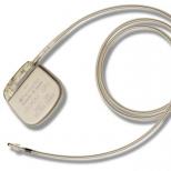
These are rhythmic contractions of the heart with a frequency of less than 50 per minute. In healthy people, bradycardia occurs, for example, during sleep. Also, bradycardia is often seen in professional athletes. Pathological bradycardia may indicate sick sinus syndrome. At the same time, bradycardia is more pronounced (heart rate from 45 to 35 beats per minute on average) and is observed at any time of the day. When bradycardia causes pauses in heart contractions of up to 3 seconds during the day and about 5 seconds at night, leads to impaired oxygen supply to tissues and manifests itself, for example, by fainting, an operation is indicated to install a heart pacemaker, which replaces the sinus node, imposing a normal rhythm of contractions on the heart.
Sinus tachycardia
Heart rate more than 90 per minute - is divided into physiological and pathological. In healthy people, sinus tachycardia is accompanied by physical and emotional stress, drinking coffee, sometimes strong tea or alcohol (especially energy drinks). It is short-lived and after an episode of tachycardia, the heart rate returns to normal in a short period of time after the cessation of the load. With pathological tachycardia, palpitations disturb the patient at rest. Its causes are temperature rises, infections, blood loss, dehydration, anemia,. Treat the underlying disease. Sinus tachycardia is stopped only with a heart attack or acute coronary syndrome.
Extrasystole
These are rhythm disturbances, in which foci outside the sinus rhythm give extraordinary heart contractions, after which there is a pause doubled in length, called a compensatory one. In general, heartbeats are perceived by the patient as uneven, rapid or slow, sometimes chaotic. Most of all, failures in the heart rhythm are disturbing. They can occur in the form of jolts, tingling, feelings of fear and emptiness in the abdomen.
Not all extrasystoles are dangerous to health. Most of them do not lead to significant circulatory disorders and do not threaten either life or health. They can be functional (against the background of panic attacks, cardioneurosis, hormonal disruptions), organic (with IHD, heart defects, myocardial dystrophy or cardiopathy, myocarditis). They can also lead to intoxication and heart surgery. Depending on the place of occurrence, extrasystoles are divided into atrial, ventricular and antrioventricular (arising in a node on the border between the atria and ventricles).
- Single extrasystoles most often rare (less than 5 per hour). They are usually functional and do not interfere with the normal blood supply.
- Paired extrasystoles two each accompany a number of normal contractions. Such a rhythm disturbance often indicates pathology and requires additional examination (Holter monitoring).
- Allorhythmias are more complex types of extrasystoles. If every second contraction is an extrasystole, it is bigymenia, if every third is trigynemia, and every fourth is quadrihymenia.
It is customary to divide ventricular extrasystoles into five classes (according to Laun). They are evaluated during daily ECG monitoring, since the indicators of a conventional ECG in a few minutes may not show anything.
- Class 1 - single rare extrasystoles with a frequency of up to 60 per hour, emanating from one focus (monotopic)
- 2 - frequent monotopic more than 5 per minute
- 3 - frequent polymorphic (of different shapes) polytopic (from different foci)
- 4a - paired, 4b - group (trigymenia), episodes of paroxysmal tachycardia
- 5 - early extrasystoles
The higher the class, the more serious the violations, although today even grades 3 and 4 do not always require medical treatment. In general, if there are less than 200 ventricular extrasystoles per day, they should be classified as functional and not worry about them. With more frequent, ECHO of the COP is indicated, sometimes - MRI of the heart. They do not treat extrasystole, but the disease that leads to it.
Paroxysmal tachycardia
In general, paroxysm is an attack. Paroxysmal acceleration of the rhythm can last from several minutes to several days. In this case, the intervals between heartbeats will be the same, and the rhythm will increase over 100 per minute (on average from 120 to 250). There are supraventricular and ventricular forms of tachycardia. The basis of this pathology is the abnormal circulation of an electrical impulse in the conduction system of the heart. Such a pathology is subject to treatment. From home remedies to eliminate an attack:
- breath holding
- increased forced cough
- face immersion in cold water
WPW syndrome
Wolff-Parkinson-White syndrome is a type of paroxysmal supraventricular tachycardia. Named after the names of the authors who described it. At the heart of the appearance of tachycardia is the presence between the atria and ventricles of an additional nerve bundle, through which a faster impulse passes than from the main pacemaker.
As a result, an extraordinary contraction of the heart muscle occurs. The syndrome requires conservative or surgical treatment (with ineffectiveness or intolerance of antiarrhythmic tablets, with episodes of atrial fibrillation, with concomitant heart defects).
CLC - Syndrome (Clerk-Levy-Christesco)
It is similar in mechanism to WPW and is characterized by an earlier excitation of the ventricles compared to the norm due to an additional bundle along which the nerve impulse travels. The congenital syndrome is manifested by attacks of rapid heartbeats.
Atrial fibrillation
It can be in the form of an attack or a permanent form. It manifests itself in the form of flutter or atrial fibrillation.
Atrial fibrillation

Atrial fibrillation
When the heart flickers, it contracts completely irregularly (intervals between contractions of very different durations). This is due to the fact that the rhythm is not set by the sinus node, but by other atrial cells.
It turns out a frequency of 350 to 700 beats per minute. There is simply no full-fledged atrial contraction; the contracting muscle fibers do not provide effective filling of the ventricles with blood.
As a result, the release of blood by the heart worsens and organs and tissues suffer from oxygen starvation. Another name for atrial fibrillation is atrial fibrillation. Not all atrial contractions reach the ventricles of the heart, so the heart rate (and pulse) will either be below normal (bradysystole with a frequency of less than 60), or normal (normosystole from 60 to 90), or above normal (tachysystole more than 90 beats per minute). ).
An attack of atrial fibrillation is difficult to miss.
- It usually starts with a strong heartbeat.
- It develops as a series of absolutely non-rhythmic heartbeats with a high or normal frequency.
- The condition is accompanied by weakness, sweating, dizziness.
- The fear of death is very pronounced.
- There may be shortness of breath, general arousal.
- Sometimes observed.
- The attack ends with the normalization of the rhythm and the urge to urinate, in which a large amount of urine leaves.
To stop the attack, they use reflex methods, drugs in the form of tablets or injections, or resort to cardioversion (stimulation of the heart with an electric defibrillator). If an attack of atrial fibrillation is not eliminated within two days, the risks of thrombotic complications (pulmonary embolism, stroke) increase.
With a constant form of heartbeat flicker (when the rhythm is not restored either against the background of drugs or against the background of electrical stimulation of the heart), they become a more familiar companion of patients and are felt only with tachysystole (rapid irregular heartbeats). The main task when detecting signs of tachysystole of a permanent form of atrial fibrillation on the ECG is to slow down the rhythm to normosystole without trying to make it rhythmic.
Examples of recordings on ECG films:
- atrial fibrillation, tachysystolic variant, heart rate 160 in '.
- Atrial fibrillation, normosystolic variant, heart rate 64 in '.
Atrial fibrillation can develop in the program of coronary heart disease, against the background of thyrotoxicosis, organic heart defects, with diabetes mellitus, sick sinus syndrome, with intoxication (most often with alcohol).
atrial flutter
These are frequent (more than 200 per minute) regular atrial contractions and the same regular, but more rare ventricular contractions. In general, flutter is more common in the acute form and is better tolerated than flicker, since circulatory disorders are less pronounced. Trembling develops when:
- organic heart disease (cardiomyopathies, heart failure)
- after heart surgery
- on the background of obstructive pulmonary disease
- it almost never occurs in healthy people.
Clinically, flutter is manifested by rapid rhythmic heartbeat and pulse, swelling of the jugular veins, shortness of breath, sweating and weakness.
Conduction disorders
Normally, having formed in the sinus node, electrical excitation goes through the conduction system, experiencing a physiological delay of a fraction of a second in the atrioventricular node. On its way, the impulse stimulates the atria and ventricles, which pump blood, to contract. If in some part of the conduction system the impulse lingers longer than the prescribed time, then the excitation to the underlying sections will come later, which means that the normal pumping work of the heart muscle will be disrupted. Conduction disorders are called blockades. They can occur as functional disorders, but are more often the result of drug or alcohol intoxication and organic heart disease. Depending on the level at which they arise, there are several types of them.
Sinoatrial blockade
When the exit of the impulse from the sinus node is difficult. In fact, this leads to a syndrome of weakness of the sinus node, a decrease in contractions to severe bradycardia, impaired blood supply to the periphery, shortness of breath, weakness, dizziness and loss of consciousness. The second degree of this blockade is called the Samoilov-Wenckebach syndrome.
Atrioventricular block (AV block)
This is a delay in excitation in the atrioventricular node of more than the prescribed 0.09 seconds. There are three degrees of this type of blockade. The higher the degree, the less often the ventricles contract, the more severe the circulatory disorders.
- At the first delay allows each atrial contraction to maintain an adequate number of ventricular contractions.
- The second degree leaves part of the atrial contractions without ventricular contractions. It is described in terms of PQ prolongation and ventricular beat prolapse as Mobitz 1, 2, or 3.
- The third degree is also called a complete transverse block. The atria and ventricles begin to contract without interrelation.
In this case, the ventricles do not stop, because they obey the pacemakers from the underlying parts of the heart. If the first degree of blockade may not manifest itself in any way and be detected only with an ECG, then the second is already characterized by sensations of periodic cardiac arrest, weakness, fatigue. With complete blockades, cerebral symptoms (dizziness, flies in the eyes) are added to the manifestations. Morgagni-Adams-Stokes attacks may develop (when the ventricles escape from all pacemakers) with loss of consciousness and even convulsions.
Conduction disturbance within the ventricles
In the ventricles to the muscle cells, the electrical signal propagates through such elements of the conduction system as the trunk of the bundle of His, its legs (left and right) and the branches of the legs. Blockades can occur at any of these levels, which is also reflected in the ECG. In this case, instead of being covered by excitation at the same time, one of the ventricles is delayed, since the signal to it goes around the blocked area.
In addition to the place of origin, a complete or incomplete blockade is distinguished, as well as permanent and non-permanent. The causes of intraventricular blockades are similar to other conduction disorders (IHD, myo- and endocarditis, cardiomyopathies, heart defects, arterial hypertension, fibrosis, heart tumors). Also, the intake of antiarthmic drugs, an increase in potassium in the blood plasma, acidosis, and oxygen starvation also affect.
- The most common is the blockade of the anteroposterior branch of the left leg of the bundle of His (BPVLNPG).
- In second place is the blockade of the right leg (RBNB). This blockade is usually not accompanied by heart disease.
- Blockade of the left leg of the bundle of His more characteristic of myocardial damage. At the same time, complete blockade (PBBBB) is worse than incomplete blockade (NBLBBB). It sometimes has to be distinguished from the WPW syndrome.
- Blockade of the posterior inferior branch of the left leg of the bundle of His may be in persons with a narrow and elongated or deformed chest. Of the pathological conditions, it is more characteristic of right ventricular overload (with pulmonary embolism or heart defects).
The clinic of blockades at the levels of the bundle of His is not expressed. The picture of the main cardiac pathology comes first.
- Bailey's syndrome - two-beam blockade (of the right leg and posterior branch of the left leg of the bundle of His).
Myocardial hypertrophy
With chronic overloads (pressure, volume), the heart muscle in some areas begins to thicken, and the heart chambers stretch. On the ECG, such changes are usually described as hypertrophy.
- (LVH) - typical for arterial hypertension, cardiomyopathy, a number of heart defects. But even in normal athletes, obese patients and people engaged in heavy physical labor, there may be signs of LVH.
- Right ventricular hypertrophy- an undoubted sign of increased pressure in the pulmonary circulation system. Chronic cor pulmonale, obstructive pulmonary disease, cardiac defects (pulmonary stenosis, Fallot's tetralogy, ventricular septal defect) lead to HPZh.
- Left atrial hypertrophy (HLH)) - with mitral and aortic stenosis or insufficiency, hypertension, cardiomyopathy, after.
- Right atrial hypertrophy (RAH)- with cor pulmonale, tricuspid valve defects, chest deformities, pulmonary pathologies and pulmonary embolism.
- Indirect signs of ventricular hypertrophy is the deviation of the electrical axis of the heart (EOC) to the right or left. The left type of EOS is its deviation to the left, that is, LVH, the right type is LVH.
- Systolic overload- this is also evidence of hypertrophy of the heart. Less commonly, this is evidence of ischemia (in the presence of angina pain).
Changes in myocardial contractility and nutrition
Syndrome of early repolarization of the ventricles
Most often, it is a variant of the norm, especially for athletes and people with congenitally high body weight. Sometimes associated with myocardial hypertrophy. Refers to the peculiarities of the passage of electrolytes (potassium) through the membranes of cardiocytes and the characteristics of the proteins from which the membranes are built. It is considered a risk factor for sudden cardiac arrest, but it does not give a clinic and most often remains without consequences.
Moderate or severe diffuse changes in the myocardium
This is evidence of myocardial malnutrition as a result of dystrophy, inflammation () or. Also, reversible diffuse changes accompany disturbances in the water and electrolyte balance (with vomiting or diarrhea), taking medications (diuretics), and heavy physical exertion.
Nonspecific ST changes
This is a sign of deterioration in myocardial nutrition without pronounced oxygen starvation, for example, in violation of the balance of electrolytes or against the background of dyshormonal conditions.
Acute ischemia, ischemic changes, T wave changes, ST depression, low T
This describes the reversible changes associated with oxygen starvation of the myocardium (ischemia). It can be either stable angina or unstable, acute coronary syndrome. In addition to the presence of the changes themselves, their location is also described (for example, subendocardial ischemia). A distinctive feature of such changes is their reversibility. In any case, such changes require comparison of this ECG with old films, and if a heart attack is suspected, rapid troponin tests for myocardial damage or coronary angiography should be performed. Depending on the variant of coronary heart disease, anti-ischemic treatment is selected.
Developed heart attack
It is usually described as:
- by stages: acute (up to 3 days), acute (up to 3 weeks), subacute (up to 3 months), cicatricial (lifelong after a heart attack)
- by volume: transmural (large-focal), subendocardial (small-focal)
- according to the location of the infarct: there are anterior and anterior-septal, basal, lateral, lower (posterior diaphragmatic), circular apical, posterior basal and right ventricular.
In any case, a heart attack is a reason for immediate hospitalization.
All the variety of syndromes and specific ECG changes, the difference in indicators for adults and children, the abundance of reasons leading to the same type of ECG changes do not allow a non-specialist to interpret even a ready-made conclusion of a functional diagnostician. It is much more reasonable, having an ECG result in hand, to visit a cardiologist in a timely manner and receive competent recommendations for further diagnosis or treatment of your problem, significantly reducing the risks of emergency cardiac conditions.