What not to do before ecg
For a thorough and in-depth study of the work of the heart, doctors have been using one of the most reliable diagnostic methods for many years - electrocardiography (or ECG for short). Thanks to this diagnosis and the correct interpretation of the cardiogram, much can be said about the nature and cause of deviations in the work of the heart.
In a new series of articles, we will tell you about the specifics of the ECG procedure, as well as how to properly prepare for it and be able to independently decipher the results obtained by comparing your own indicators with the norm.
EKG how to prepare
Contrary to the opinion of cardiologists, it is generally accepted that the ECG does not require special preparation. The study of the work of the heart muscle involves the avoidance of stress, fatigue, and requires complete rest. On the day of the procedure, you need to sleep well, and ignore the morning exercises. If the procedure is scheduled for the morning, then you should avoid a heavy breakfast, but it is better to completely abandon it. With the upcoming daily procedure, you should limit yourself to a light snack. in 2 hours before the session.
Don't forget to cut down on the amount of fluids that affect muscle function. Avoid coffee, tea, and other energy drinks. They will help stimulate cardiac activity, and the results will be distorted.
It is advisable to take a shower. It is not necessary to apply care products to the body, because the components of creams and lotions will contribute to the formation of a greasy film on the surface, which will adversely affect the contact of the electrodes with the skin.
Try to relax as much as possible just before the ECG. Sit with your eyes closed, restore your breathing - this will ensure an even pulse and objective readings of the device.
Is an EKG harmful?
The logical question of whether ECG is harmful can be answered based on the advantages of this diagnostic method:
- reliability of information
- security and comfort of the session
- efficiency (10min)
- no health or pregnancy restrictions
As you already understood, it is impossible to harm the health of the ECG, since this method is based on the removal of heart rate indicators and does not produce any radiation or effects on the body. Moreover, for people whose work is associated with constant physical exertion, electrocardiography is done almost daily, which once again confirms its absolute harmlessness.
The order of the procedure
How is an ECG done? , if the appointed time has come?
You will be asked to take off your outer clothing so that nothing obstructs access to the chest, and free your shins. The places where the electrodes will be attached will be treated with alcohol, on which a special gel will be applied.
The next step is to attach the cuffs and suction cups. They are fixed on the arms, in the ankles and chest. Ten electrodes will track the rhythm of the heart, and give an encrypted result.

The interpretation of the results will be effective if the patient has followed all the instructions for preparing for the ECG
The heart plays the role of an electrical generator. The tissues of the body have a high degree of electrical conductivity, which allows you to mark the electrical impulses of the heart by applying electrodes to parts of the body. The indications of biopotentials are processed by the electrocardiograph, and output data in the form of a summary picture - the propagation of excitation signals through the muscle in a graphic image. More specifically, the difference in electrical voltage.
The spread of the impulse throughout the heart is facilitated by depolarization of myocardial cells, during which part of the cells acquires a positive charge, the other part - a negative one. This creates a potential difference. In the case of complete depolarization (contraction) or repolarization (relaxation) of the cell, no voltage difference was noted. The device records EMF - the electromotive force of the heart.
After an ECG is performed, the doctor gets an idea about the work of the organ and the existing deviations.
An electrocardiogram can reveal:
- arrhythmia
- ischemia
- myocardial infarction
vse-o-gipertonii.ru
The principle of operation of the electrocardiograph
The device for recording an ECG consists of electrodes that are attached to the patient's body, a galvanometer, an amplifier, a recorder and a switch for leads. The impulses that are formed in the heart muscle must first be amplified, then they are perceived by the galvanometer. It converts electrical waves into mechanical vibrations.
The registrar records with the help of recorders on thermal paper a typical graphic curve, which is called an electrocardiogram.

With the help of an ECG study, one can judge the state of the heart muscle by the following indicators:
- impulse conduction;
- rhythmic heartbeats;
- an increase in one or more parts of the heart;
- myocardial blood supply;
- areas of necrosis (infarction) their size, depth and duration of occurrence.
How to properly prepare for an ECG, what not to do
Electrocardiography does not require long-term preparation, which is one of the advantages of this method. It is removed according to emergency indications in any condition of the patient. But if a planned study is scheduled, then before conducting it, it is recommended:
- Do not eat or drink caffeinated drinks for at least 3 hours before the procedure.
- Before the study, you need to have a good rest.
- Eliminate physical and emotional stress.
- Take a shower, after which do not use the cream.

Clothing is selected so that it is easy to attach the electrodes to the skin of the ankles, wrists and chest.
On the day of the study, it is strictly forbidden to take alcoholic beverages, smoking, you need to give up sports and a hearty breakfast. As a drink, ordinary drinking water, weak tea or fruit juice is best.
How an EKG is done
In order to take an electrocardiogram, the patient is placed on the couch, the medical worker places electrodes on the shins, wrists and chest. If there is difficulty in breathing in a horizontal position, then the procedure is performed while sitting.
Rules for the procedure
For good contact between the skin and the electrode, the attachment point is degreased with ethyl alcohol and a special conductive gel is applied. After that, readings are taken using an ECG diagnostic device.
The whole procedure takes about 10-15 minutes.
In order to get a reliable result, you need to be in a calm, relaxed state, do not hold your breath. Muscle tremors from excitement or cold can distort the data.
Common leads are 3 standard, 3 reinforced and 6 chest. Each lead will record at least 4 cardiac cycles. After that, the device is turned off, the electrodes are removed and the signed tape is issued to the doctor of functional diagnostics, which he must decipher.
For more information on ECG registration, see this video:
Are there any features during pregnancy
In the body of a pregnant woman, the load on the heart muscle changes, as  it must provide blood supply to the fetus in the uterus. An electrocardiogram may show abnormalities that are not indicative of heart disease.
Therefore, starting from 3-4 months, when deciphering the testimony, an amendment is made for the presence of a gestation process.
In preparing and conducting the procedure itself, standard research methods are used.
How is an EKG done for women?
For women, the rules for installing electrodes are the same as for men. They should be located in the region of the heart, directly on the skin, therefore, before the ECG, you must completely remove all clothing from the chest, including the bra. Keep in mind that pantyhose or stockings will prevent you from attaching the sensors to your lower leg.
Deciphering ECG indicators
On the tape, the curve obtained after taking the cardiogram has 5 teeth. They occur when the atria and ventricles contract in succession. The following designations have been adopted:
- The P wave is an indicator of the work of the right (first half) and left atrium.
- P Q - the interval of passage of the impulse to the ventricle along the bundle of Giss.
- QRST - the complex occurs when the ventricles contract, while the highest R wave reflects the excitation of the ventricular myocardium, and Q and S are the partitions between them, T - occurs during the recovery period of the myocardium after systole.
 Prongs and intervals
 Normal in adults
A doctor can fully evaluate the electrocardiogram, since diagnosis requires knowing the symptoms of the disease and data from other research methods (blood tests, ultrasound, echocardiography). The general characteristics that a specialist evaluates in a healthy person are as follows:
- Rhythm of contractions from 60 to 80 per minute.
- The size of the intervals should not exceed normal values, or be shorter than the average values.
- Electrical axis - normally R exceeds S in all leads except aVR, V1 - V2, sometimes V3.
- The ventricular complex is not more than 120 ms.
- T is positive and longer than the QRS complex.
 ECG (normal)
 During pregnancy
As the uterus grows, it raises the dome of the diaphragmatic septum and after 24-24 weeks, the apex of the heart shifts to the left. This is reflected on the electrocardiogram by an increase in the amplitude of R in the first, and S and Q in the third lead, the ventricular complex decreases along with the ST segment. Changes in conduction in the heart muscle are also associated with the influence of hormones produced by the placenta.
Characteristic signs:
- Displacement of the axis of the heart to the left.
- T biphasic and negative in the right chest leads.
- The ventricular complex is wider than normal.
- Accelerated rhythm, single extraordinary contractions.
 Respiratory arrhythmia in pregnant women
 Deviations that the device is able to detect
With the help of removing and decoding the electrocardiogram, signs of such diseases can be detected:
- angina pectoris and heart attack;
- type of arrhythmia, location of the pacemaker;
- blockade due to reduced conductivity;
- myocardial hypertrophy and its localization;
- signs of myocarditis and pericarditis;
- thromboembolism of the pulmonary artery;
- symptoms of pulmonary hypertension;
- violations of the electrolyte composition of the blood.
 3rd degree AV block
 Disadvantages of conducting ECG examinations
Despite the high diagnostic value, a conventional ECG cannot fix changes in the work of the heart outside the time of its removal. Therefore, along with the traditional method, the patient may be assigned additional monitoring during the day according to Holter, tests with physical activity.
Using this method, it is impossible to recognize heart murmurs, therefore, if valvular or septal defects are suspected, echocardiography, phonocardiography, or ultrasound of the heart should be performed.
If it is planned to install a stent or shunt for myocardial ischemia, then coronary angiography is required to determine the localization of the narrowing of the coronary arteries. Tumor processes are diagnosed by x-ray or MRI examination.
Actual questions of patients
The ECG method is traditional and has been used in medical practice for a long time. But patients often have concerns about its appointment. The most common questions:

Thus, an ECG is a time-tested, affordable type of diagnostics that is used both for a preventive examination during a medical examination and for making a diagnosis in the presence of complaints of a violation of the heart. Such a study is safe and informative.
cardiobook.ru

Surely there are only a small number of people who have never had a cardiogram taken in their lives, which means that most of us know about it firsthand.
But it turns out that such a familiar and seemingly simple study has a number of their pitfalls.
So let's try to "overcome" them together.
In order for the cardiogram to turn out to be ideal, attention should be paid to preparing for it, which means that you need to sleep well the day before, and in the morning do not give the body unnecessary physical activity in the form of jogging or swimming for certain distances. After all, the goal of doctors is to fix the work of your heart in the usual, and not in extreme conditions.
At breakfast, it is advisable to refuse coffee or strong brewed tea, because it is known that caffeine can accelerate the heart rate, and the doctor, misled in this way, will search in vain for the cause of tachycardia. But you should not abuse other drinks, such as water or juice, because the overload of the body with liquid can not be reflected in the cardiogram in the best way.
The best option would be one in which breakfast or any other meal will take place no earlier than two hours before an EKG and it is even better to conduct the study on an empty stomach, because eating can also cause a rapid heartbeat.
On the day of the ECG, you should take a shower, but after this hygiene procedure, you should not use creams and lotions, as they disrupt the contact of the electrodes with the skin.
If the cardiogram is for a man whose chest is not deprived of abundant vegetation, then you should not shave it off, but still it would not be superfluous to take a razor with you, so to speak, just in case of an emergency. But lovely ladies should not wear pantyhose under trousers before conducting the study, since they will still have to be removed, it is also better to prefer a blouse with a fastener on the chest as a “top”.
Do not forget that the medical staff does not always have the necessary amount of time to comply with all the necessary formalities, and therefore you should be careful in monitoring the "cleanliness of the process" so that, if necessary, you can give the doctor an explanation about certain deviations on the film.
Do not do it go for an EKG earlier than two hours after undergoing any physiotherapy procedures.
Be sure to rest for ten to fifteen minutes before the study, you are unlikely to be able to do this in the office, but do not rush to enter there, it is better to sit in the corridor for a while.
In the place where the electrodes are applied, the skin is preliminarily degreased with alcohol, and then a special gel is applied to it.
As soon as you receive the results of the study, you should not immediately worry if there are any deviations, since often the worries are simply unjustified.
For example, sinus tachycardia
or palpitations, when the frequency of beats exceeds ninety beats per minute, may be caused by anxiety, smoking, exercise, dehydration, drinking coffee or tea, the presence of pain, etc.
In the event that at rest your pulse is kept within the normal range, then there is no reason to worry.
At sinus bradycardia
there is a slowdown in heart rate (HR).
This can happen as a result of not getting enough sleep, or taking sleeping pills at night, or inclement weather outside.
Most experts are inclined to believe that bradycardia can be discussed when the heart rate is less than fifty beats per minute.
sinus arrhythmia
Also called respiratory.
In this case, irregular heartbeats are observed, in which the time between individual heartbeats fluctuates by a fraction of a second.
In principle, this is the norm, and in this case we can talk about the features of the autonomic nervous system and respiration.
If we talk about axis deviation heart left or right, then this is not at all special, since such a deviation should be interpreted only in conjunction with other indicators of the cardiogram. Since this can also be a variant of the norm.
represents an individual feature of the electrical potentials of your heart muscle and is considered the norm.
The atrial rhythm normally originates in the sinus node, but in some cases it can also occur in the immediate vicinity of it.
In most cases, such a deviation does not in any way affect the operation of your “motor”.
The rhythm cradle can periodically migrate, making its way from the atria to the sinus node and vice versa.
With incomplete blockade of the right (left) leg of the bundle of His there is a violation of intraventricular conduction, but at the moment such a pathology is not considered as such, but belongs to the category of the norm.
www.blog-bliss-s.ru
What are the problems with taking an electrocardiogram (ECG)? But it turns out that in such a simple matter there are also pitfalls.
When should an EKG be done?
- An ECG is prescribed by a doctor if the patient has complaints of pain in the heart of a different nature, as well as to check the condition of a patient with diseases of the cardiovascular system.
- An ECG is performed when the patient complains of shortness of breath, the occurrence of arrhythmia, before surgery of any nature. In addition, an ECG is recommended if there are diseases of other internal organs.
- An ECG is also performed when there is an occupational risk.
 Before the study, it is better to sleep well, and in the morning not to give the body unnecessary physical exertion. It is important to fix the work of your heart in normal, not extreme conditions. Also in the morning it is better not to drink a lot, overloading the heart with fluid can affect the cardiogram. And even more so to give up coffee and strong tea - caffeine speeds up the heart rate. It is better to have a snack no later than 2 hours before the study, if possible, take an ECG on an empty stomach.
On the day of the study, you need to take a shower, after which oily and fatty creams and lotions should not be used - they worsen the contact of the electrodes with the skin. Prefer clothes that are comfortable, so to speak, for quick access to the body.
The study is carried out lying on the back after a 10-15 minute rest with calm breathing. At a minimum, it is important to have at least 10 minutes to relax in front of the office, and not lie down on the couch, having just "run" to the 4th floor. Before the study, the patient undresses to the waist, the shins should also be freed from clothing, since the electrodes should be in direct contact with the skin.
The skin in the place where the electrodes are applied is degreased (rubbed with alcohol) and moistened with special gels. 10 electrodes are attached to the human body, which are attached to the upper and lower extremities, as well as to the chest with suction cups and cuffs.
With the help of an ECG, you can evaluate:
- Source of heart rate;
- Regularity of heartbeats;
- heart rate;
- Changes in cardiac conduction;
- Changes in the terminal part of the gastric complex, which make it possible to determine ischemic changes in the heart.
For competent interpretation of the ECG it is necessary to know the nature of each of its teeth, the intervals between the teeth and packages of teeth of one cardiac cycle, as well as the nature of the relationship of the curves in various leads. Therefore, the interpretation of the ECG should be carried out only by a specialist with experience in such work.
Often, the conclusion describes a deviation from the norm that makes us worry. Considering that most often unrest is unnecessary, I will decipher the most frequent “non-terrible” deviations from the ideal.
- Sinus tachycardia- increased heart rate above 90 beats per minute. Can be caused by anxiety, smoking, exercise, dehydration, coffee, tea, pain, and more. If you normally have a normal resting heart rate, it's okay.
- Sinus bradycardia- slowing the heart rate to less than 60 beats per minute. Perhaps you just didn’t get enough sleep, drank sleeping pills at night, and gray rainy weather outside. By the way, recently more and more experts believe that bradycardia begins when the pulse slows down to 50 beats per minute.
- Sinus (respiratory) arrhythmia- slightly uneven heartbeats, when the time between individual heartbeats fluctuates by a fraction of a second. The variant of the norm is most often associated with the work of the autonomic nervous system, the influence of breathing.
- Deviation of the electrical axis of the heart to the left / right- in itself does not carry a semantic load and is interpreted only "in total" with other ECG indicators. It is often a variant of the norm.
- Syndrome of early repolarization of the ventricles- this is an individual feature of the electrical potentials of your heart, a variant of the norm.
- atrial rhythm- Normally, the rhythm originates in the sinus node, but sometimes - somewhere in the immediate vicinity of it. Most often, this does not affect the work of the heart in any way, the cradle of the rhythm can periodically migrate from the atria to the sinus node and back.
- Incomplete blockade of the right / left leg of the bundle of His- a violation of intraventricular conduction, which in modern medicine is considered as a variant of the norm.
An ECG is a fairly simple but informative study, which is included in the minimum examination of patients. But sometimes a simple ECG is not enough to make a diagnosis. In this case, the doctor may consider it appropriate to conduct functional tests or daily monitoring. Functional tests with physical activity or with the use of special medications make it possible to detect disorders that, for various reasons, could not be registered with a conventional electrocardiographic study (latent insufficiency of blood supply to the heart muscle, non-permanent rhythm disturbances).
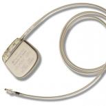
ECG taken during the day, is called "24 hour monitoring" or "Holter monitoring". To do this, a miniature ECG machine and electrodes are attached to the patient's body. During the day, he leads his usual way of life, but makes entries in his diary, where he notes all the events that occurred during the day. Daily monitoring allows not only to identify transient heart rhythm disturbances and ischemic changes, but also to associate their appearance with any events (physical stress, psychological experiences), with the time of day.
In some cases, in addition to the ECG, the doctor may prescribe an ultrasound to diagnose heart disease - echocardiography. This method makes it possible to “see” the heart, assess the thickness of its walls and their ability to contract, the condition of the valves and blood flow.
Khannanov Z.A., therapist, doctor of the highest category.
What is sinus rhythm of the heart Aortic aneurysm diagnosis