Cardiogram of the heart
The cardiogram of the heart is the result of a study that occurs in the process of cardiac activity of the electric field. The study is carried out using a special device - an electrocardiograph.
Content
It gives the result in the form of a graph printed on special paper. The method is so common today that it can be carried out not only in a specialized office, but also with the help of a portable device at the patient's home and in an ambulance.
With the help of this study, you can get a complete picture of the processes occurring in the tissue of the heart, the presence or absence of pathological processes in it. Currently, there is a whole direction, the main task of which is to study additional diagnostic possibilities using the method. You can make a cardiogram of the heart in any clinic. What is a method?
The essence of the ECG method
If you look closely at the graph, you can trace the sequence of identical depressions and teeth. In this way, the activity of the heart muscle is recorded at various periods of its work. It is on their characteristics that the decoding of the cardiogram is based.

The process of performing electrocardiography (ECG)
It seems to an ordinary person that the contraction of the heart occurs at once, but in fact, this is not so. Heart contraction is a process that is cyclically repeated throughout human life.
The heart consists of four sections, which are commonly called chambers: two atria and two ventricles. During contraction, first the impulse is formed in the atria (atrial systole), then in the ventricles (ventricular systole), followed by diastole - a period of relaxation.
In each of these stages, the cells of the heart muscle generate bioelectrical impulses that are recorded by the electrocardiograph. A normal cardiogram of the heart looks approximately the same in all cases. The slightest changes in cardiac activity are expressed in changes in the shape and size of the teeth of the cardiogram of the heart.
What is the study for?
ECG is an absolutely safe and painless way to study cardiac activity.

ECG device (electrocardiograph)
To conduct it, the patient must be laid on a couch, special electrodes should be placed in the necessary places, which will record the impulses. They are generated in the process of work by the heart muscle.
The tissues of the human body are, to one degree or another, conductors of electric current, so it can be recorded in different parts of the body. The study is conducted in twelve standard leads. How to decipher the cardiogram of the heart?
Deciphering the cardiogram
Deciphering a cardiogram is a technique that every graduate of a medical university knows the basics of. The cardiogram of a healthy person contains information about the heart rate. The teeth of such a cardiogram are even, the same in size and repeating with the same interval.

Teeth, segments and intervals of the ECG (decoding of the cardiogram)
Deciphering the cardiogram gives an idea, first of all, of the presence or absence (increased heart rate) or bradycardia (decrease in the number of heartbeats per unit of time). If the size and shape of the teeth themselves begin to change, we are talking about more serious violations.
If serious changes occur in the composition of tissues that occur during the development of such conditions as, their ability to conduct electrical impulses also changes. This is what a cardiogram looks like.

Electrocardiograms of the heart on thermal tape (sample)
Such changes will immediately be reflected in the result of the ECG. The cardiogram for myocardial infarction will differ significantly from the result of a study of a healthy person. Moreover, these changes will become noticeable almost from the very beginning of the development of the process.
In some cases, a more detailed study of the activity of the heart muscle is required. In certain pathologies, changes can be noticed only during long-term follow-up.
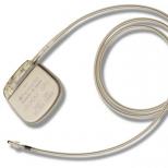
For a long-term study of the activity of the heart muscle, a method of daily monitoring of the heart according to Holter was developed. In this case, the examination is carried out at home, a mini-device for recording heart impulses is fixed on the patient's body.

Method of daily monitoring of the heart by Holter
This device "remembers" the data of the cardiogram of the heart - the decoding of the data and the graphic image of the study will later be obtained by the specialist conducting the study.
How to decipher a cardiogram? In order to independently decipher the cardiogram, you need to know what each tooth means, what shape it should be, what size.
Each wave on the graph has its own designation. These are Latin letters P,Q.R,S,T,U. The shape and size of the P wave characterizes atrial contraction, the Q, R, S complex characterizes the work of the ventricles, and the T wave shows what happens during diastole. The teeth pointing up are positive and those pointing down are negative. Both the height and the shape of the teeth are evaluated.
In some cases, the result obtained with electrocardiography is insufficient. In such a situation, various additional studies are assigned, they allow you to get a more complete picture of the processes occurring in the human heart.
These studies are especially important in pediatrics, for the early detection of various malformations, and in the study of blood vessels for the presence of a blood clot.