How to decipher the cardiogram of the heart yourself
ECG interpretation of an electrocardiogram is considered a complex process that only a diagnostician or cardiologist can do. They carry out decoding, revealing various defects and disorders of the human heart muscle. This diagnostic method is widely used today in all medical institutions. The procedure can be done both in the clinic and in the ambulance.
Electrocardiography is a science in which the rules of the procedure are studied, how to decipher the results obtained and explains the unclear points and situations. With the development of the Internet, ECG decoding can even be done independently, using special knowledge.
The electrocardiogram is deciphered by a special diagnostician who uses the established procedure that determines normal indicators and their deviations.
Heart rate and heart rate are assessed. In the normal state, the rhythm should be sinus, and the frequency should be from 60 to 80 beats per minute.
Intervals are calculated that characterize the duration of the moment of contraction. This is where special formulas come into play.
The normal interval (QT) is 390 - 450 ms. If the interval is violated, if it lengthens, the diagnostician may suspect that the patient has atherosclerosis, rheumatism or myocarditis, as well as coronary artery disease. Also, the interval may be reduced, and this indicates the presence of hypercalcemia disease. These parameters are calculated by a specialized automatic program that provides a reliable result.
The location of the EOS is calculated from the isoline along the height of the teeth. If the indicators are significantly higher than each other, a deviation of the axis is noticed, defects in the vital activity of the right or left ventricle are suspected.
An indicator showing the activity of the ventricles, the QRS complex, is formed during the passage of electrical impulses to the heart. It is considered normal when there is no defective Q wave and the distance does not exceed 120 ms. When the specified interval is shifted, it is customary to speak of a conduction defect, or it is also called the blockade of the legs of the His bundle. With incomplete blockade, RV or LV hypertrophy can be suspected, depending on the location of the line on the ECG. The interpretation describes ST particles, which are reflectors of the recovery time of the initial position of the muscle relative to its complete depolarization. Under normal conditions, the segments should fall on the isoline, and the T wave, which characterizes the work of both ventricles, should be asymmetric and directed upwards. It must be longer than the QRS complex.
Correctly deciphering ECG indicators can only be done by doctors who are specially involved in this, but often an ambulance paramedic with extensive experience can easily recognize common heart defects. And this is extremely important in emergency situations.
When describing and decoding the diagnostic procedure, various characteristics of the work of the heart muscle are described, which are indicated by numbers and Latin letters:
- PQ is an indicator of atrioventricular conduction time. In a healthy person it is 0.12 - 0.2 s.
- R - description of the work of the atria. It may well tell about atrial hypertrophy. In a healthy person, the norm is 0.1 s.
- QRS - ventricular complex. In the normal state, the indicators are 0.06 - 0.1 s.
- QT is an indicator that can indicate cardiac ischemia, oxygen starvation, heart attack, and rhythm disorders. The normal indicator should be no more than 0.45 s.
- RR is the gap between the upper points of the ventricles. Shows the constancy of heart contractions and allows you to count their frequency.
Cardiogram of the heart: decoding and main diagnosed diseases
Deciphering a cardiogram is a long process that depends on many indicators. Before deciphering the cardiogram, it is necessary to understand all the deviations of the work of the heart muscle.
Atrial fibrillation is characterized by irregular contractions of the muscle, which can be quite different. This violation is dictated by the fact that the beat is set not by the sinus node, as it should happen in a healthy person, but by other cells. The heart rate in this case ranges from 350 to 700. In this condition, the ventricles do not fully fill with incoming blood, which causes oxygen starvation, from which all organs in the human body suffer.
 An analogue of this condition is atrial fibrillation. The pulse in this state will be either below normal (less than 60 beats per minute), or close to normal (from 60 to 90 beats per minute), or above the specified norm.
On the electrocardiogram, you can see frequent and constant contractions of the atria and less often of the ventricles (usually 200 per minute). This is atrial flutter, which often occurs already in the exacerbation phase. But at the same time, it is easier for the patient to tolerate than flicker. Circulatory defects in this case are less pronounced. Trembling can develop as a result of surgery, with various diseases, such as heart failure or cardiomyopathy. At the time of examination of a person, flutter can be detected due to rapid rhythmic heartbeats and pulse, swollen veins in the neck, increased sweating, general impotence and shortness of breath.
Conduction disorder - this type of heart disorder is called blockade. The occurrence is often associated with functional disorders, but it can also be the result of intoxications of a different nature (against the background of alcohol or taking medications), as well as various diseases.
There are several types of disorders that the cardiogram of the heart shows. Deciphering these violations is possible according to the results of the procedure.
Sinoatrial - with this type of blockade, there is difficulty in the exit of the impulse from the sinus node. As a result, there is a syndrome of weakness of the sinus node, a decrease in the number of contractions, defects in the circulatory system, and as a result, shortness of breath, general weakness of the body.
Atrioventricular (AV blockade) - characterized by a delay in excitation in the atrioventricular node longer than the set time (0.09 seconds). There are several degrees of this type of blocking.
The number of contractions depends on the magnitude of the degree, which means that the defect in the blood flow is more difficult:
- I degree - any compression of the atria is accompanied by an adequate amount of compression of the ventricles;
- II degree - a certain amount of atrial compression remains without ventricular compression;
- III degree (absolute transverse blockade) - the atria and ventricles are compressed independently of each other, which is well shown by the decoding of the cardiogram.
Conduction defect through the ventricles. An electromagnetic impulse from the ventricles to the muscles of the heart propagates through the trunks of the bundle of His, its legs and branches of the legs. Blocking can occur at every level, and this will immediately affect the electrocardiogram of the heart. In this situation, it is observed how the excitation of one of the ventricles is delayed, because the electrical impulse goes around the blockage. Doctors divide the blockage into complete and incomplete, as well as permanent or non-permanent blockade.
Myocardial hypertrophy is well shown by the cardiogram of the heart. Decoding on an electrocardiogram - this condition shows a thickening of individual sections of the heart muscle and stretching of the chambers of the heart. This happens with regular chronic overload of the body.
- Syndrome of early repolarization of the ventricles. Often, it is the norm for professional athletes and people with congenital large body weight. It does not give a clinical picture and often passes without any changes, so the interpretation of the ECG becomes more complicated.
- Various diffuse disorders in the myocardium. They indicate a myocardial malnutrition, as a result of dystrophy, inflammation or cardiosclerosis. Disorders are quite susceptible to treatment, often associated with a disorder of the body's water and electrolyte balance, taking medications, and heavy physical activity.
- Non-individual ST changes. A clear symptom of myocardial supply disorder, without bright oxygen starvation. Occurs during hormonal imbalances and electrolyte imbalances.
- T wave distortion, ST depression, low T. Cat's back on the ECG shows the state of ischemia (oxygen starvation of the myocardium).
In addition to the disorder itself, they also describe their position in the heart muscle. The main feature of such disorders is their reversibility. The indicators, as a rule, are given for comparison with old studies in order to understand the patient's condition, since it is almost impossible to read the ECG on your own in this case. If a heart attack is suspected, additional studies are carried out.
There are three criteria by which a heart attack is characterized:
- Stage: acute, acute, subacute and cicatricial. Duration from 3 days to a life-long condition.
- Volume: large-focal and small-focal.
- Location.
Whatever the heart attack, it is always a reason to place a person under strict medical supervision, without any delay.
ECG results and options for describing the heart rhythm
 The results of the ECG provide an opportunity to look at the state of the work of the human heart. There are different ways to decipher the rhythm.
sinus is the most common signature on an electrocardiogram. If, apart from heart rate, no other indicators are indicated, this is the most successful forecast, which means that the heart is working well. This type of rhythm suggests a healthy state of the sinus node, as well as the conduction system. The presence of other records proves the existing defects and deviations from the norm. There is also atrial, ventricular or atrioventricular rhythm, which indicate which cells in specific parts of the heart set the rhythm.
sinus arrhythmia is often normal in young adults and children. This rhythm is characterized by the exit from the sinus node. However, the intervals between contractions of the heart are different. This is often associated with physiological disorders. Sinus arrhythmia should be carefully monitored by a cardiologist to avoid the development of serious diseases. This is especially true for people with a predisposition to heart disease, as well as if the arrhythmia is caused by infectious diseases and heart defects.
Sinus bradycardia- characterized by rhythmic contraction of the heart muscle with a frequency of about 50 beats. In a healthy person, this condition can often be observed in a state of sleep. Such a rhythm can manifest itself in people professionally involved in sports. They have ECG teeth that are different from the teeth of an ordinary person.
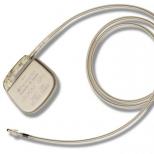
Constant bradycardia may characterize the weakness of the sinus node, manifested in such cases by more rare contractions at any time of the day and in any condition. If a person has pauses during contractions, then a surgical intervention is prescribed to install a stimulator.
Extrasystole. This is a rhythm defect that is characterized by extraordinary contractions outside the sinus node, followed by ECG results showing an extended pause, called a compensatory pause. The patient feels the heartbeat as uneven, chaotic, too fast or too slow. Sometimes patients are disturbed by pauses in the heart rhythm. Often there is a feeling of tingling or unpleasant jolts behind the sternum, as well as a feeling of fear and emptiness in the stomach. Often such conditions do not lead to complications and do not pose a threat to a person.
 Sinus tachycardia- with this disorder, the frequency exceeds the normal 90 beats. There is a division into physiological and pathological. Under the physiological understand the onset of such a state in a healthy person under certain physical or emotional stress.
It can be observed after taking alcoholic beverages, coffee, energy drinks. In this case, the condition is temporary and passes quite quickly. The pathological type of this condition is characterized by periodic heartbeats that disturb a person at rest.
The causes of the pathological appearance can be elevated body temperature, various infectious diseases, blood loss, long periods without water, anemia, etc. Doctors are treating the underlying disease, and tachycardia is stopped only in case of a heart attack in a patient or an acute coronary syndrome.
Paroxysmal tachycardia- in this condition, a person has a rapid heartbeat, expressed in an attack lasting from several minutes to several days. The pulse may increase to 250 beats per minute. There are ventricular and supraventricular forms of such tachycardia. The main reason for this state is the defect in the passage of the electric pulse in the conducting system. This pathology is quite susceptible to treatment.
You can stop the attack at home with the help of:
- Holding the breath.
- Forced cough.
- Immersion in cold water of the face.
WPW syndrome It is a subspecies of supraventricular tachycardia. The main provocateur of an attack is an additional nerve bundle, which is located between the atria and ventricles. To eliminate this defect, surgical intervention or medication is required.
CLC- very similar to the previous type of pathology. The presence of an additional nerve bundle here contributes to the early excitation of the ventricles. The syndrome, as a rule, is congenital and manifests itself in a person with attacks of an accelerated rhythm, which is very well shown by ECG teeth.
Atrial fibrillation May be episodic or permanent. A person feels pronounced atrial flutter.
ECG of a healthy person and signs of changes
The ECG of a healthy person includes many indicators by which a person's health is judged. The ECG of the heart plays a very important role in the process of detecting abnormalities in the work of the heart, the worst of which is myocardial infarction. Exclusively with the help of electrocardiogram data, it is possible to diagnose necrotic infarct zones. Electrocardiography also determines the depth of damage to the heart muscle.
ECG norms of a healthy person: men and women
ECG norms for children
 The ECG of the heart is of great importance in the diagnosis of pathologies. The most dangerous heart disease is myocardial infarction. Only an electrocardiogram will be able to recognize necrotic infarction zones.
ECG signs of myocardial infarction include:
- the zone of necrosis is accompanied by changes in the Q-R-S complex, resulting in a deep Q wave;
- the damage zone is characterized by a displacement (elevation) of the S-T segment, smoothing the R wave;
- the ischemic zone changes the amplitude and makes the T wave negative.
Electrocardiography also determines the depth of damage to the heart muscle.
How to decipher the cardiogram of the heart yourself
Not everyone knows how to decipher the cardiogram of the heart. However, having a good understanding of the indicators, you can independently decipher the ECG and detect changes in the normal functioning of the heart.
First of all, it is worth determining the indicators of the heart rate. Normally, the heart rhythm should be sinus, the rest indicate the possible development of arrhythmia. Changes in sinus rhythm, or heart rate, suggest the development of tachycardia (speeding up) or bradycardia (slowing down).
Abnormal data of teeth and intervals are also important, since you can read the cardiogram of the heart yourself by their indicators:
- Prolongation of the QT interval indicates the development of coronary heart disease, rheumatic disease, sclerotic disorders. Shortening of the interval indicates hypercalcemia.
- An altered Q wave is a signal of myocardial dysfunction.
- The sharpening and increased height of the R wave indicates hypertrophy of the right ventricle.
- A split and dilated P wave indicates left atrial hypertrophy.
- An increase in the PQ interval and a violation of the conduction of impulses occurs with atrioventricular blockade.
- The degree of deviation from the isoline in the R-ST segment diagnoses myocardial ischemia.
- Elevation of the ST segment above the isoline is a threat of acute infarction; a decrease in the segment registers ischemia.
The cardio line consists of divisions (scales) that determine:
- heart rate (HR);
- QT interval;
- millivolts;
- isoelectric lines;
- duration of intervals and segments.
This simple and easy-to-use device is useful for everyone to independently decipher the ECG.


