How many salivary glands does a person have? Where is the salivary gland located in humans? Topography of the parotid salivary gland
Digestion begins long before food enters the esophagus. The process starts in the oral cavity: food is exposed to saliva produced by the glands. Other functions of the salivary glands are no less important.
What is saliva?
The defining function of the salivary glands is the secretion of saliva, a viscous liquid that has complex composition: water, acid salts, trace elements, enzymes, vitamins, proteins.
Enzymes contained in the liquid break down food, and fat digestion begins. The particles are enveloped and glued together to facilitate movement through the esophagus.
The composition varies depending on the time of day, foods and drinks consumed, diseases, age, and environmental conditions. An indicator of oral health is the PH level. Normal values: 6.5 - 7.5.
The structure of the human salivary glands
The structure of organs is determined by their type. They come in big and small; Based on the type of secretion, mucous, protein, and mixed are distinguished. Dislocation of small ones - mucous membrane of the lips, tongue, cheeks, palate. Large salivary glands - paired - are of three types:
- Parotid - large, weighing 20-30 grams, located under auricle, on the side of the lower jaw. Covered with a sheath of connective tissue, divided into lobules. The main function is the production of liquid saliva (one third of the total volume) with a high concentration of sodium and potassium chlorides.
- Submandibular (15 grams) with the upper edge adjacent to the lower jaw. An excretory duct departs from them, opening near the frenulum of the tongue. The secretion of low acidity is isolated.
- The sublinguals weigh 5 grams and are located at the bottom of the oral cavity under the mucous membrane. The secretion produced is proteinaceous, rich in mucin, and highly alkaline.

The brain gives the command to produce saliva.
The brain gives the command to produce saliva. The centers located in the posterior region begin to work in certain situations - when thinking about food, chewing, appetizing smells, during stress. A large amount of secretion is produced when chewing: the muscles put pressure on the glands, forcing them to work more intensely.
New research notes interesting fact: parotid major salivary glands are enlarged in those who like to talk on the phone; The amount of saliva produced is also above average.
Functions
The major and minor salivary glands perform the same functions.
- endocrine – production of biologically active substances similar to hormones;
- exocrine - secretion of mucus and proteins;
- excretory – removal of metabolic products;
- filtration – filtration nutrients from blood.

The functions of the salivary glands make them an essential element of the digestive system.
The functions of the salivary glands make them an essential element of the digestive system. The oral cavity is wetted and food becomes available for chewing. Constant hydration is a condition for normal articulation and enhanced taste of foods. Thanks to its chemical composition, saliva protects tooth enamel from damage, preventing the appearance of caries.
With reduced secretion production, xerostomia is diagnosed - dry mouth syndrome. Soft fabrics become irritated and vulnerable to infections. Dryness causes an unpleasant odor, changes in taste, difficulty swallowing.
Sources:
- Kurepina M.M., Ozhigova A.P., Nikitina A.A. Human anatomy. Moscow, 2010.
- Fedyukovich N. Anatomy and physiology of man. Tutorial. Rostov-on-Don, 2003.
Saliva plays a very important role in the human body. With its help, chewed food is glued together, swallowed, as well as the perception of taste and the protection of tooth enamel from damage. And special glands secrete saliva, which will be discussed further.
Types of organs that produce saliva
The excretory ducts of the salivary glands flow into the oral cavity, divided into large (they have the structure of an organ) and small, which are located in different areas of the mucosa.
The minor ones include: labial, buccal, molar, lingual and palatal. The large ones are called the two parotid, submandibular and sublingual. The largest are a pair of parotid glands.
Physiology
The salivary glands, in the process of salivation, excrete a secretion through a system of ducts into the oral cavity, which contains enzymes involved in digestion: amylase, proteinase, lipase, etc. The secretions of all the organs that produce them mix in the human mouth and form saliva, which forms a food bolus and provides the beginning digestion process.
Parotid salivary glands
These two glands are considered the most important. They lie around the jaw branch and take part in the initial phase of digestion, secreting the required amount of secretion. They are of the serous type and produce ptyalin. Their secretions enter the oral cavity through the ducts of the parotid salivary glands.
These organs are located behind the rami of the lower jaw and in front of the mastoid process, which extends from the temple bone. They are closely related to the functioning of the branching facial nerve, therefore, if their functioning is disrupted, serious dysfunction in the movement of the facial muscles may occur.
Almost a fifth of the total volume of saliva enters the oral cavity through the excretory ducts. The weight of each of them ranges from 20-30 g.
Submandibular gland
The submandibular salivary glands produce a mixture that includes mucus and Despite the fact that they are smaller than the parotid glands, the proportion of salivary fluid they produce is 70%. It enters the oral cavity from these secretory organs through the submandibular canal, which is the duct for these salivary glands.
Description of the sublingual gland
Sublingual or sublingual are the large glands located under the tongue. They are mainly involved in the secretion of mucus. Unlike other large glands, the duct system of the sublingual salivary gland is simpler. It is not so diverse and branched. It does not include intercalary ducts and jet flow outlets.
From the sublingual glands, 8 to 20 salivary ducts open into the oral cavity. Up to 5% of all saliva passes through them.

The structure of the parotid glands
The parotid glands are a complex alveolar gland. Each of them has a lobular structure and is covered with fascia, which closes them into a separate capsular formation.
The excretory duct of the parotid salivary gland opens into the oral cavity in the form of a small hole located in front of the second large molar on the upper jaw. Its length is 6 cm and on its way to the oral cavity it passes through the surface of the masseter muscle, the fatty tissue of the cheek and the buccal muscle. Sometimes this duct can bifurcate.
The structure of the submandibular gland
In its anatomy, it acts as a complex alveolar-tubular gland, the second largest among the large organs that secrete saliva. It, like the parotid, has a lobular structure and is located in the submandibular fossa, extending beyond the posterior border of the mylohyoid muscle. The base of the duct of the salivary gland, located under the jaw, is located near the posterior edge of this muscle and, bending around its surface, opens on the sublingual papilla.

The structure of the sublingual gland
The structure of this gland is the same as that of the submandibular gland. It is located just under the oral mucosa on top of the mylohyoid muscle. There it forms a sublingual fold, located between the surface of the lower jaw and the tongue. The number of ducts of this gland can vary from 18 to 20. They open into the oral cavity along the sublingual fold. The main duct of the salivary gland passes near the submandibular ducts and opens with a common opening or next to it.
Functions
The main purpose of the glands described is to produce a special secretion. The ducts of the salivary glands are designed to remove it from the oral cavity. Thus, the functioning of the salivary ducts ensures the following:
- saliva moistens the oral cavity;
- food liquefies;
- articulation is ensured;
- taste sensations are enhanced;
- teeth are protected from damage (thermal or mechanical);
- the oral cavity is cleansed.

Possible diseases
There are many diseases that can disrupt the functioning of the salivary glands and the ducts that extend from them. Among them, the most dangerous are:
- Dilation of the ducts. It leads to disruption of the removal of secretions into the oral cavity and causes the formation of stones and purulent inflammation in the ducts of the salivary glands.
- Abscesses. This disease affects and therefore requires urgent hospitalization followed by surgery.
- Formation of intraglandular stones. As the disease develops, the system of salivary gland ducts becomes filled with stones, which impede the passage of secretions.
- Sialadenitis. With the onset of the disease, there is a decrease in the activity of secretion by the gland, leading to inflammatory processes spreading in the gland itself and its ducts.
- The formation of polyps that block the path of secretion. As a result of constant stagnation of fluid, infection and inflammation begin to develop.
- Sialolithiasis. The course of the disease involves filling the gland ducts with stones, leading to the same consequences as polyps.
- Mucocele. There is stagnation of saliva accumulated in the ducts due to polyps or stones.
- Papillary stenosis. Due to the disease, the ducts of the salivary glands narrow in places where the secretion enters the oral cavity, which leads to its stagnation and development inflammatory process.

Treatment methods
In the vast majority of cases, diseases affecting the salivary glands and their ducts are treated by surgical intervention. The reason is that patients rarely seek help in the early stages of the disease, and since delays in treatment lead to complications of the disease, only a surgeon can get rid of them.

Surgical treatment provides for the following activities:
- Lithotripsy. During this procedure, the doctor, using a special apparatus, crushes or ducts and then removes them.
- Marsupialization of the ducts. Treatment is carried out by opening the salivary duct, from which stones or polyps are removed. Since more gentle methods now exist, marsupialization is used very rarely and only in cases where large stones or formations are found on the floor of the mouth. After removing the pathological formation, duct plastic surgery is performed.
- Therapeutic sialoendoscopy. It is an option for endoscopic surgery and makes it possible to remove small stones that have formed, as well as get rid of strictures (narrowing of the lumen) of the ducts. The procedure is performed under local anesthesia by inserting a tube (or several) into the duct.
- Extracorporeal lithotripsy. It is planned to influence the stones formed in the duct from the outside using a special emitter. During this treatment, stones are destroyed, regardless of their size. After crushing, the stones are removed and the ducts are washed with a special solution to prevent the development of inflammation.
- Endoscopic laser lithotripsy. This method is based on direct impact on stones in the duct. Crushing is carried out using a laser emitter. At the end of the procedure, the stones are removed.
- Removal of polyps endoscopically. The procedure is performed using a laser, which cuts off polyps. It is very popular because the laser, after cutting off the polyp, cauterizes and disinfects the area where the growth was located. In addition, there is no bleeding of the salivary gland ducts, which prevents the development of purulent complications.
- Endoscopic dilatation. It is used when it is necessary to cut adhesions in the gland or duct that form on scar tissue during the process. The procedure allows you to restore the outflow of secretions without damaging the walls of the ducts.
Endoscopic methods for treating diseases affecting the salivary glands and ducts are very popular, as they are highly effective and do not require further hospitalization. In addition, they prevent the development of various complications, which allows patients to recover quickly.

Since the salivary ducts play a very important role in the process of salivation, any disruption in their functioning leads to serious consequences. Therefore, at the first feeling of discomfort in the area of the salivary system, you must consult a doctor who can make the correct diagnosis and prescribe effective method treatment.
Knowing the structural features of the salivary glands, you can better understand how the salivary glands function. human body and why this paired secretory organ is needed.
Saliva also helps to moisturize the oral cavity and has protective bactericidal properties, helping to cope with various harmful microorganisms.
Structural features
At the first stage of food entering the human body, it is influenced by the secretion produced by the large and small (small) salivary glands.
Thanks to the enzymes and mucus in saliva, the breakdown of food begins at the initial stage of digestion, and a food bolus is formed.
Small salivary glands are located throughout the oral mucosa, including its submucosa. Based on their location, they are divided into lingual, buccal, molar, and palatal. They are also located on the mucous membrane of the tonsils and nasopharynx. The size can vary from 1 to 5 mm in diameter.
The parotid, submandibular (submandibular) and sublingual are the three pairs of major major salivary glands. The most voluminous of them are the parotid ones, which are involved in the production the largest number saliva. Depending on their location, each of them has its own special name.
For inflammation of the submandibular gland, see.
Salivary glands: anatomy and features of the parotid salivary gland
 It predominantly produces saliva enriched with protein; it is the largest glandular organ among the three large ones and has a lobular structure.
Covered on the outside with a dense connective shell in the form of a capsule. It is located under the auricle, going deep under the zygomatic arch (parotid-masticatory zone). Its weight is 20-30 grams.
Consists of a superficial and deep lobe. The main excretory duct is on average about 2-4 cm long, usually does not exceed 5-7 cm, with a diameter of 2-3 mm, enters the oral cavity at the site of the second molar on the upper jaw, and can have a straight, arched or bifurcated shape (rarely). In older people it is wider.
Depending on the blood flow, the color of the gland can be either pink or yellowish-gray. The structure of a healthy organ is of uniform, moderate density with a bumpy surface. It is practically not palpable upon palpation.
The ratio of saliva production is about ¼ of the total volume. In one hour, on average, about 5 ml of saliva is produced.
Substances contained in the secretion contribute to the breakdown of starch during chewing.
Anatomy of the human salivary glands: features of the submandibular gland
 It is medium in size (from 8 to 10 grams), paired alveolar, and has a lobular structure.
It is located under the lower jaw, in part of the angle of the lower jaw, located near the parotid gland, and in back contact with the sublingual gland.
Also covered with a capsule, dense on the outside and thin on the inside. The space between the capsule and the gland is filled with fatty tissue.
It is moderately dense in consistency, pink or gray in color with a yellowish tint. With age, it decreases in size, its structure and color change.
The excretory duct, called the Wharton duct, has a length of about 5-7 cm, the lumen diameter is from 2 to 4 mm. The duct emerges into the oral mucosa on the side of the frenulum of the tongue, forming an elevation in the form of a sublingual papilla.
The submandibular gland produces saliva mixed type(serous-mucous), rich in protein. About 12 ml of saliva is produced per hour; normally, the numbers can vary from 1 to 22 ml.
The mental, facial and lingual arteries are involved in supplying blood to the submandibular gland.
When performing surgery on the duct, the location of the lingual and facial nerves must be taken into account.
Anatomical features of the sublingual gland
The smallest glands are the sublingual glands, weighing from 3 to 5 grams, located respectively under the tongue, namely under the mucous membrane of the oral cavity above the maxillofacial muscle, forming a fold.
This paired organ consists of lobules, is characterized by a tubular-alveolar structure, and has a gray-pink color. Covered with a thin capsule.
The length of the excretory duct (Bartholin's duct) is 1-2 cm, the width of the lumen (diameter) is 1-2 mm. At the exit into the oral mucosa, the lumen of the Bartholin duct in most people connects to the terminal part of the duct. It is not uncommon for the duct opening to emerge independently, but this phenomenon is not observed often. Also, many small ducts branch off from them, having access to the sublingual fold.
Blood supply occurs due to the mental and sublingual arteries, blood outflow occurs due to the sublingual vein.
In the saliva secreted from the sublingual gland, the mucous component predominates. The secretion produced makes up 5% of the total amount of saliva produced by all large glands.
In an adult, according to average indicators over 24 hours, all salivary glands secrete from one to one and a half liters of saliva, depending on various factors, including the degree of stimulation of secretion production with the help of one or another food.
Salivary glands in children
 The formation of the parotid and submandibular glands occurs in the sixth week of intrauterine development of the fetus; in the seventh week, the formation of the sublingual salivary gland occurs. They develop from the epithelium of the oral cavity.
Infants have weak development of the salivary glands; active growth occurs from the fourth month of life until the child reaches two years of age, during which time the weight of these paired organs increases significantly. Later, it grows in length, and branching of existing ducts is noted. The area of the first molar is the site of exit of the parotid duct. This is slightly lower compared to adults.
Among the main characteristics of a child’s salivary glands, the following are worth highlighting:
- low level of secretion production;
- achieving sealing of the oral cavity during sucking;
- low concentration of amylase in saliva;
- neutral or slightly acidic reaction.
Babies begin to produce saliva from the first hours of life. A secretion is produced in an amount of 0.6 to 6 ml per hour; during active sucking, about 24 ml of saliva per hour can be released. In a newborn, the secreted secretion contains certain substances that promote the breakdown of components such as glycogen and starch.
Uncontrolled drooling in children of the first year of life is caused by the immaturity of the processes of swallowing saliva and salivation.
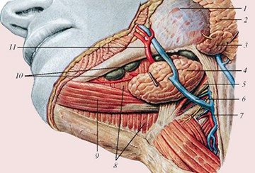
Salivary glands location according to photo


It is known to be a formation resulting from blood clots or other causes of blockage of the glandular ducts. For example, too thick gland secretion can settle on flow vessels and cause blockage.
Read about inflammation of the sublingual salivary gland here.
Diseases and pathologies associated with the structure and functioning of the glands
The most common occurrence is sialadenitis, an inflammatory process characterized by acute or chronic form with all the attendant symptoms and manifestations.Various diseases affecting the nervous, endocrine, digestive system, can affect the normal functioning of the glands and lead to disruptions in their functioning.
Also a common occurrence is the development benign tumor, which, depending on the size, structure and location, is removed on an outpatient basis or in a hospital.
In addition, diseases such as atrophy or hypertrophy, abscess, fistula, sialolithiasis, mucocele, impaired secretion, etc. may occur.
An excellent preventive aid that helps avoid the development of many diseases is adherence to basic rules of personal hygiene aimed at systematic oral care.
What is a salivary gland? The salivary gland (glandulae salivariae) is an external secretion gland that produces a special substance called saliva. These glands are located throughout the oral cavity, as well as in the maxillofacial area. The ducts of the salivary glands open in different places oral cavity.
In the definition of the term “salivary gland” there is a mention that it is an organ of external secretion - this means that the products synthesized in it enter the cavity associated with the external environment (in this case it is the oral cavity)
Types and functions
There are several classifications.
The sizes of glandulae salivariae are:
- large;
- small.
According to the nature of the secreted secret:
- serous - saliva is enriched with a large amount of protein;
- mucous - the secretion contains predominantly a mucous component;
- mixed - they can secrete serous and mucous secretions.
The main function of glandulae salivariae is the production of saliva.
Saliva is a transparent, slightly viscous, slightly alkaline substance. More than 99.5% of its composition is water. The remaining 0.5% are salts, enzymes (lipase, maltase, peptidase, etc.), mucin (mucus), lysozyme (antibacterial substance).

All functions of saliva are divided into 2 types - digestive and non-digestive. Digestive ones include:
- enzymatic (breakdown of certain substances, for example, complex carbohydrates starts in the mouth);
- formation of a food bolus;
- thermoregulatory (cooling or heating food to body temperature).
Non-digestive functions:
- moisturizing;
- bactericidal;
- participation in the mineralization of teeth, maintaining a certain composition of tooth enamel.
Note. The study of the function of glandulae salivariae was carried out by Academician Pavlov during experiments on dogs at the end of the 19th century.

Minor salivary glands
They make up the bulk of all glandulae salivariae. They are located throughout the oral cavity.

Depending on the location, small glands are called:
- buccal;
- palatal;
- lingual;
- gingival;
- molar (located at the base of the teeth);
- labial
In terms of secretion, most of them are mixed, but there are serous and mucous ones.
The main function is to maintain normal level saliva in the mouth. This prevents the mucous membrane from drying out between meals.
Major salivary glands
The number of large salivary glands in humans is six. Among them are:
- 2 parotid;
- 2 submandibular;
- 2 sublingual.

Note. The glands are formed in the 2nd month of embryonic development from the epithelium of the oral mucosa and initially have the appearance of small cords. Subsequently, their size increases, and future ducts appear. At the 3rd month, a channel appears inside these outflow tracts, connecting them to the oral cavity.
During the day, large glandulae salivariae synthesize a small amount of saliva, but when food arrives, its amount increases sharply.
Parotid gland
It is the largest of all salivary glands. It is serous in appearance of secretion. Weight about 20 grams. The volume of secretion released per day is about 300-500 ml.

This salivary gland is located behind the ear, mainly in the retromaxillary fossa, limited in front by the angle of the lower jaw, and behind by the bony part ear canal. The anterior edge of the glandula parotidea (salivary gland) lies on the surface of the masseter muscle.
The body of the gland is covered with a capsule. The blood supply comes from the parotid artery, which is a branch of the temporal artery. Lymphatic drainage from this salivary gland goes to two groups of lymph nodes:
- superficial;
- deep.
The excretory duct (stenon) begins from the anterior edge of the glandula parotidea, then, passing through the thickness of the masticatory muscle, it opens in the mouth. The number of outflow tracts may vary.

Important! Since the body of glandula parotidea is located mostly in the bony fossa, it is well protected. However, it has two weak points: its deep part adjacent to the internal fascia, and back surface in the area of the membranous part of the auditory canal. These places, when suppurated, are the area for the formation of a fistula tract.
Submandibular salivary gland
So is the large glandulae salivariae. It is slightly smaller in size and weighs about 14-17 grams.

According to the type of secretion produced by this gland, it is mixed.
Glandula submandibularis has an excretory duct called Wharton's. It starts from its inner surface, going obliquely upward into the oral cavity.

Sublingual salivary gland
It is the smallest of the major salivary glands. Its weight is only 4-6 grams. Oval in shape, maybe slightly flattened. The type of secretion is mucous.

The excretory duct is called Bartholin's duct. There are options for opening it in the sublingual area:
- independent opening, often near the frenulum of the tongue;
- after merging with the ducts of the submandibular glands on the caruncula sublingualis;
- many small ducts opening on the caruncula sublingualis (sublingual fold).
Diseases of the salivary glands
All diseases of glandulae salivariae are divided into several groups:
- inflammatory (sialoadenitis);
- salivary stone disease (sialolithiasis);
- oncological processes;
- developmental defects;
- cysts;
- mechanical damage to the gland;
- sialoses - the development of dystrophic processes in the tissues of the gland;
- sialadenopathy.
The main symptom of the presence of the disease glandulae salivariae is an increase in size.

The second symptom that characterizes the presence of problems with glandulae salivariae is xerostomia, or a feeling of dry mouth.
The third symptom of anxiety is pain. It may occur both in the area of the gland itself and irradiate into surrounding tissues.
Important! If you have at least one of the above symptoms, you should consult a doctor.
Examination of patients suspected of having certain disorders in the salivary gland begins with examination and palpation. Additional methods is probing (detects the presence of narrowing of the outflow tract), sialometry (measurement of the rate of saliva secretion) with microscopy of the resulting secretion.
 imgblock-center-text" style="width: 500px;">
Treatment
Treatment pathological processes in the area of the salivary glands is carried out depending on the etiology of the disease.
The most common of all diseases is sialadenitis. To treat the inflammatory process, conservative etiotropic treatment is usually used. It consists of prescribing antibiotics, antiviral, antifungal drugs. With the development of an extensive purulent process, the gland cavity is opened and drained.
Important! After surgical treatment a scar remains on the skin in the access area (in the treatment of mumps and sialosubmandibulitis). After surgery, the salivary gland is completely restored after some time.
Surgical treatment is also used when sialolithiasis occurs.
Oncological processes in the glandulae salivariae area are treated with combined methods. More often combined surgical method(complete excision of the tumor and gland tissue) followed by radiation or chemotherapy.
Conclusion
The salivary glands play an important role in human life. And it is very important to prevent the development of pathological processes in them. The simplest way of prevention is to maintain hygienic cleanliness of the oral cavity, avoid smoking and alcohol. This will help maintain the full function of the glands for a long time.
1. GENERAL MORPHOFUNCTIONAL CHARACTERISTICS AND DEVELOPMENT OF SALIVARY GLANDS
The ducts of 3 pairs of large salivary glands open into the oral cavity: parotid, submandibular and sublingual, lying outside the mucous membrane. In addition, in the thickness of the mucous membrane of the oral cavity there are numerous small salivary glands: labial, buccal, anterior lingual, posterior half of the hard palate, soft palate and uvula, grooved papillae (Ebner), small sublingual.
Saliva has a complex composition, determined by the true secretion of glandular cells, as well as the secretion and excretion of a number of products by the salivary glands.
Combining the secretions of all glands produces saliva with a certain average composition, which depends on the nature of the food taken and a number of other factors. Thus, parasympathetic stimulation of the salivary glands leads to the formation of a large amount of liquid saliva, and sympathetic stimulation leads to the formation of a small amount of thick saliva.
The concepts of “saliva” and “oral fluid” should not be confused. Oral fluid includes the total secretion of the salivary glands, as well as oral detritus, microflora, gingival fluid, waste products of microflora, residues food products and etc.
An average of 1.5 liters of saliva is produced per day, with the main amount coming from the secretion of the submandibular (75%) and parotid (20%) glands.
Approximately 99% of the mass of saliva is water. The main organic component of saliva is the glycoprotein mucin, produced by mucocytes. The composition of saliva includes enzymes, immunoglobulins, some biological active substances. Among inorganic substances, calcium, sodium, potassium, magnesium, chlorine, phosphates, and bicarbonates ions predominate (Fig. 19).
One of important functions saliva - mineralizing. Saliva is the main source of inorganic substances necessary to maintain the optimal composition of tooth enamel. After teething, mineral ions can enter the enamel during the process of mineralization and be washed out of the enamel during the process of demineralization. The saturation of saliva with hydroxyapatite is essential in the mineralization of enamel. Acidification reduces the degree of saturation of saliva with hydroxyapatite and its associated mineralizing properties. The buffer systems contained in saliva provide an optimal pH level (within 6.5-7.5). The microflora of the oral cavity may have acid-producing activity. With an alkaline pH of saliva, excessive deposition of tartar is observed.
Saliva is involved in the processes of mechanical and chemical processing of food. The enzymes contained in saliva affect food not only in the oral cavity, but also (for some time) in the stomach. Salivary enzymes (amylase, maltase, hyaluronidase) are involved in the breakdown of carbohydrates.
The salivary glands perform an excretory function. Uric acid and creatinine are released from the body with saliva. Products of nitrogen metabolism, as well as inorganic ions Na+, K+, Ca++, Cl -, HCO 3 enter saliva from the blood with the active participation of exocrinocytes.
The protective function of saliva is ensured by high concentrations of antimicrobial substances (lysozyme, lactoferrin, peroxidase), as well as secretory IgA, which causes aggregation of pathogenic microorganisms and prevents their attachment (adhesion) to the surface of the epithelium of the mucous membrane and teeth.
The salivary glands have not only exocrine but also endocrine function. It has been established that in submandibular glands In animals, a protein is synthesized that is similar to insulin in its biological action and a number of biochemical properties. Biologically active substances have been found in human saliva - parotin, nerve growth factor, epithelial growth factor, kallikrein, etc. Apparently, some of
Rice. 19.Scheme of formation, intake and reabsorption of certain substances in the salivary glands:From the blood, Na+, Cl - ions and water enter the cells of the secretory terminal sections of the salivary glands. Serocytes produce and secrete into saliva a protein secretion, which contains enzymes (amylase, maltase) and antibacterial substances (lysozyme, lactoferrin, peroxidase). Mucocytes produce mucins rich in sialic acids and sulfates. IgA is secreted by stromal plasma cells and transferred by transcytosis into saliva by cells of the secretory terminal sections and striated ducts. Insulin-like compounds are formed in the striated ducts. Bicarbonates come from the blood, providing 80% of the buffering properties of saliva, and kallikrein, which activates the formation of kinins and helps reduce vascular tone. Na+, Cl - ions are reabsorbed from saliva into the blood in striated ducts
they enter saliva from the blood, and are not synthesized in the glands themselves (see Fig. 19).
The salivary glands are actively involved in the regulation of water-salt homeostasis.
Development of salivary glands
All salivary glands are derivatives of multilayered squamous epithelium of the oral cavity, therefore the structure of their secretory sections and excretory ducts is characterized by multilayering.
In the 2nd month of embryogenesis, large paired salivary glands are formed: submandibular (gl. submandibulare), parotid (gl. parotis), sublingual (gl. sublinguale), and in the 3rd month - small salivary glands: labial (gl. labiales), buccal (gl. buccales), palatal (gl.palatinae). In this case, epithelial strands grow into the underlying mesenchyme. Proliferation of epithelial cells leads to the formation of branched epithelial cords with expanded bulb-shaped ends, which subsequently give rise to excretory ducts and secretory terminal sections
iron Connective tissue is formed from mesenchyme.
During the development of the salivary glands special meaning have epitheliomesenchymal interactions. Apparently, mesenchyme has an inducing effect on the epithelium of the glands, determining the nature of the branching of their ducts and the direction of growth, however, the type of salivary gland is determined even before the interaction of the epithelium with the mesenchyme begins.
2. LARGE SALIVARY GLANDS (PAROTICULAR, SUBMANDIBLIAR, HYPOGLOUS)
All major salivary glands (glandulae salivariae majores) built according to a single plan. The outside of the gland is covered with a connective tissue capsule, from which cords extend deep into the organ, dividing the gland into lobules. The intralobular connective tissue that forms the stroma of the glands is populated
There are numerous lymphocytes and plasma cells. The parenchyma of the salivary glands is formed by epithelium.
Large salivary glands are complex, branched, alveolar or alveolar-tubular. They consist of terminal sections and a system of ducts that remove secretions.
2.1. SECRETORY ENDS (ACINI) OF THE SALIVARY GLANDS
Terminal sections (portio terminalis) They are a blind sac consisting of secretory cells. The secretory unit of the salivary glands is also called the acinus. According to the nature of the secreted secretion, the end sections are of 3 types: proteinaceous (serous), mucous and mixed (proteinaceous).
Acini contain 2 types of cells- secretory and myoepithelial. According to the mechanism for separating secretions from cells, all salivary glands are merocrine.
In the protein terminal sections(Fig. 20, a) secretory cells are serocytes. Serocytes- pyramid-shaped cells. At the ultrastructural level, they reveal accumulations of elements of the granular endoplasmic reticulum, free ribosomes, and the Golgi complex. Numerous large protein (zymogenic) spherical granules are localized in the apical part of the cell. Most other organelles are localized in the basal or perinuclear cytoplasm (Fig. 20, b). From the glandulocytes, the secretion enters the intercellular tubules, and then into the lumen of the terminal sections.

Rice. 20.Scheme of the structure of the protein secretory department of the salivary gland and serocyte:a - protein secretory department: 1 - serocytes; 2 - myoepitheliocyte nucleus; 3 - basement membrane; b - serocyte: 1 - nucleus; 2 - granular endoplasmic reticulum; 3 - Golgi complex; 4 - secretory granules; 5 - mitochondria; 6 - myoepitheliocyte; 7 - basement membrane
Protein cells secrete a liquid secretion rich in enzymes.
Mucous terminal sections have an elongated, tubular shape with a wide lumen. Large mucous cells- mucocytes- have light cytoplasm, contain dark flattened nuclei, shifted to the basal part of the cells (Fig. 21, a). In the well-developed Golgi complex of mucus cells, carbohydrates are attached to a protein base, and mucus glycoproteins are formed. In the supranuclear part of the cell there are large granules surrounded by a membrane (Fig. 21, b). Mucocytes produce viscous and viscous saliva. These cells are characterized by cyclic activity. The release of mucin granules occurs with appropriate hormonal or neural stimulation.
Mixed end sections often appear as dilated tubes formed by both serocytes and mucocytes. In this case, serocytes (in the submandibular glands) or seromucocytes (in the sublingual glands) are located along the periphery of the end sections in the form of “caps” (Gianuzzi half moon). The central part of the mixed secretory terminal sections is formed mucocytes(Fig. 22).
The crescents are hypothesized to be an artifact of routine fixation techniques used in light and electron microscopy. Rapid freezing of tissue liquid nitrogen and subsequent treatment with osmium tetroxide (OsO 4) in cold acetone reveal that mucocytes and serocytes are located in the same row and frame the lumen of the secretory acini in the form of a single layer

Rice. 21.Scheme of the structure of the mucous secretory section of the salivary gland and mucocyte: a - mucous secretory section: 1 - mucocytes; 2 - myoepitheliocyte nucleus; 3 - basement membrane; b - mucocyte: 1 - nucleus; 2 - granular cytoplasmic reticulum; 3 - Golgi complex; 4 - secretory granules; 5 - mitochondria; 6 - myoepitheliocyte; 7 - basement membrane

Rice. 22.Scheme of the structure of the mixed end section of the salivary gland: a - mixed end section: 1 - mucocytes; 2 - serocytes forming the crescent of Gianuzzi; 3 - myoepitheliocyte nucleus; 4 - basement membrane; b - end section with removed basement membrane: 1 - basal surface of secretory cells; 2 - myoepitheliocyte, lying
on secretory cells; 3 - intercalary duct
epithelium. Serous crescents are not detected.
In sections prepared from the same samples using conventional methods, “bloated” mucocytes with enlarged secretory granules are revealed. In this case, serocytes form typical crescents located along the periphery of the secretory end sections. Long processes of serocytes penetrate between mukocytes. Perhaps the process of crescent formation is associated with an increase in the volume of mucocytes during secretion. In this case, the initial position of the serous cells changes, which leads to the formation of a crescent effect. A similar phenomenon is sometimes observed in the intestinal mucosa, when “swollen” goblet cells change the position of absorptive epithelial cells.
Myoepitheliocytes form the 2nd layer of cells in the terminal secretory sections and are located between the basement membrane and the base of the epithelial cells (see Fig. 20-22). Myoepithelial cells perform a contractile function and contribute to the secretion of secretions from the end sections.
2.2. SYSTEM OF EXCRETORY DUCTS OF SALIVARY GLANDS
Excretory ducts of the salivary glands are divided into insertion (ductus intercalatus), striated (ductus striatus), interlobular (ductus interlobularis) and gland ducts (ductus glanulae). Intercalated and striated ducts are classified as intralobular (Fig. 23).

Rice. 23.Scheme of the structure of the excretory ducts of the salivary glands:1 - intercalary excretory duct; 2 - striated excretory duct; 3 - end sections; 4 - intralobular excretory ducts; 5 - lobule; 6 - interlobular excretory duct; 7 - epithelial cell of the intercalary duct; 8 - myoepitheliocyte; 9 - epithelial cell of the striated duct;
10 - folds of the cytolemma; 11 - mitochondria
Intercalary ducts well developed in the protein glands. In mixed glands they are short and difficult to identify. Intercalated ducts are formed by cubic or flat epithelial cells with basophilic cytoplasm, the 2nd layer is formed by myoepithelial cells.
The intercalary ducts contain the cambial elements of the epithelium of the terminal sections and the system of excretory ducts.
Striated ducts(salivary tubes) are a continuation of the intercalary tubes. They branch and often form ampullary extensions. The diameter of the striated ducts is much larger than that of the intercalated ducts. The cytoplasm of columnar epithelial cells of striated ducts is acidophilic.
Ultrastructural examination reveals microvilli in the apical part of the cells, and basal striations formed by mitochondria located between the folds of the cytolemma in the basal parts. This morphological substrate ensures the reabsorption of fluid and electrolytes. In the striated duct, the following occurs: 1) reabsorption of Na + from the primary secretion, 2) secretion of K + and HCO 3 into the secretion. Typically, more sodium ions are reabsorbed than potassium ions are secreted, so the secretion becomes
hypotonic. The concentration of Na+ and C1- in saliva is 8 times lower, and K+ is 7 times higher than in blood plasma.
In the apical part of the cells of the striated ducts there are secretory granules containing kallikrein, an enzyme that breaks down blood plasma substrates with the formation of kinins that have a vasodilating effect.
Growth factors and some other biologically active substances were identified in the cells of the intralobular ducts. The cells of the intralobular ducts form a secretory component that ensures the transfer of IgA into saliva.
Interlobular ducts are located in the interlobular connective tissue and are formed as a result of the fusion of striated ducts. The interlobular ducts are usually lined with multirow prismatic or bilayer epithelium. Some epithelial cells of these ducts may be involved in ion exchange.
Common excretory duct lined with stratified epithelium.
Thus, the type of epithelium in the excretory ducts of the salivary glands changes and becomes characteristic of the ectodermal epithelium of the oral cavity, i.e. multilayer.
2.3. COMPARATIVE MORPHOLOGICAL CHARACTERISTICS OF LARGE SALIVARY GLANDS
Parotid gland - complex, alveolar, branched. The secretion of the parotid glands is protein.
End sections The parotid gland consists of serocytes and myoepithelial cells (Fig. 24).
Intralobular intercalary ducts long, highly branched. Striated salivary ducts well developed. lined with multirow prismatic or bilayer epithelium. Parotid duct
zy (stenon duct), lined with multilayer epithelium, opens on the surface of the buccal mucosa at the level of the 2nd upper molar.
Submaxillary (submandibular) gland - complex, alveolar (in some places alveolar-tubular), branched. The nature of the secretion is mixed (protein-mucous, but predominantly protein).
Terminal secretory sections- protein (predominant, accounting for 80%), as well as mixed protein-mucous (Fig. 25).
Glycoproteins and glycolipids are detected in the secretory granules of serocytes.

Rice. 24.Diagram of the structure of the parotid gland:1 - serous end sections; 2 - intercalary excretory duct; 3 - striated excretory duct; 4 - connective tissue stroma of the gland

Rice. 25.Scheme of the structure of the submandibular gland:1 - serous terminal section; 2 - mixed end section; 3 - intercalary duct; 4 - striated duct
The mixed terminal sections are larger than the protein sections (Fig. 26). The cytoplasm of mucocytes has a cellular structure due to the presence of a mucous secretion in it, which is selectively stained with mucicarmine.
Intercellular secretory tubules are located between the protein cells of the serous crescent. Outside the crescent cells lie myoepithelial cells.
Intercalary ducts shorter than in parotid gland, and less branched, which is explained by the sliming of some of these sections during development.
Striated ducts long, strongly branched. In some animals (rodents), granular sections are identified, the cells of which contain granules with trypsin-like proteases, as well as some growth-stimulating factors.
Interlobular excretory ducts lined mainly by bilayer epithelium.
Duct of the submandibular gland(Wharton's duct) in the terminal part forms protrusions (diverticula) and opens next to the duct of the sublingual gland on the anterior edge of the frenulum of the tongue.
Sublingual gland - complex, alveolar-tubular, branched, the smallest of the large salivary glands. The nature of the secretion is mixed mucous-protein with a predominance of mucous secretion.
Secretory terminal sections glands are represented by 3 types: protein (very few), mixed (constituting the bulk of the gland) and mucous sections (Fig. 27). In the mixed terminal sections there are mucous cells and protein crescents.
The cells that form the crescents secrete both protein and mucous secretions (seromucosal cells). Their secretory granules react to mucin. Mucin is a glycoprotein in which numerous oligosaccharide chains are associated with the polypeptide chain.
The mucous terminal sections of the gland are formed by cells containing chondroitin sulfate B and glycoproteins.
In all 3 types of terminal sections, the outer layer is formed by myoepithelial elements.
Excretory ducts have a number structural features. Intercalated ducts are rare,

Rice. 26.Histological specimen. Submandibular gland:1 - mixed end sections; 2 - protein terminal sections; 3 - striated excretory duct; 4 - vessel in interlobular connective tissue

Rice. 27.Diagram of the structure of the sublingual gland:1 - serous terminal section; 2 - mixed end section; 3 - intercalary duct; 4 - connective tissue stroma
since during embryonic development they are almost completely mucused, forming the mucous parts of the terminal sections.
The striated ducts are poorly developed and very short. The cells lining the striated ducts show basal striations and contain small vesicles, which are considered an indicator of excretion.
In the interlobular excretory ducts the epithelium is two-layered.
The common excretory duct (Bartholin's) is similar in structure to the duct of the submandibular gland, with which it sometimes merges.
3. SMALL SALIVARY GLANDS. ADAPTABILITY OF SALIVARY GLANDS
Small salivary glands are numerous and scattered throughout the oral mucosa with the exception of the gums and the anterior part of the hard palate.
End sections usually form small lobules separated by layers of connective tissue.
Small salivary glands located in the anterior parts of the oral cavity (labial, buccal, floor of the mouth, anterior lingual), as a rule, are mixed and are similar in structure to the sublingual.
The glands of the middle section (the area where the grooved papillae of the tongue are located) are purely proteinaceous. In the posterior part of the oral cavity there are mucus
simple glands (glands of the root of the tongue, hard and soft palate).
Excretory ducts small glands branch, but intercalated and striated ducts are usually absent.
In the stroma of the small salivary glands, lymphocytes, mast and plasma cells are detected.
The final composition of saliva and the adaptability of the salivary glands
The final composition of saliva (its quantity and quality) is controlled by various factors: 1) the concentration of various substances in the blood; 2) nervous regulation of saliva composition; 3) the action of hormones (in particular, mineralocorticoids, which increase the level of potassium in saliva and reduce the concentration of sodium); 4) functional activity of the kidneys.
A decrease in the functional activity of the salivary glands has serious negative consequences. With a decrease in saliva secretion, self-cleaning of the oral cavity worsens, which promotes the development of microflora and leads to a decrease in the resistance of enamel to demineralizing influences.
Due to the fact that saliva is a kind of “trophic factor” for the hard tissues of the tooth, when salivation decreases, cracks appear, the enamel becomes fragile, and multiple caries quickly develops. Clinical picture, which occurs in the oral cavity when
decreased salivation is called xerostomia (dry mouth).
The salivary glands are highly adaptable to changing living conditions of the body. The secretion of saliva changes with stimulation of various receptor fields, the action of certain humoral factors, pharmacological substances and biomaterials used in dentistry. Study of salivary function, chemical composition and biophysical properties of saliva are used to assess the body's reactions to dental biomaterials from which dentures are made. Thus, the salivary glands are a kind of test object for assessing biocompatibility in dentistry.
All salivary glands are subject to age-related involution, which is manifested by progressive heteromorphism both in the terminal sections and in the excretory ducts.
In contrast to the traditional view of saliva as an ion-protein true aqueous solution, which contains a complex complex of proteins and various ions, new ideas about saliva have now formed as:
About the liquid crystal structure;
About a solution containing Ca 2+ and HPO 4 2- ions in a micellar state.
The fact that saliva is a liquid crystal structure is evidenced by some data from biophysical studies. When saliva dries, it crystallizes and can be classified as liquid crystals. The liquid crystalline state is manifested in such properties of saliva as foaming or film formation. This approach to the structure of saliva allows us to better understand the strength of the bond between enamel and pellicle, which ensures the selective permeability of ions in dental tissue.
According to some authors, the basis of saliva is made up of micelles that bind a large amount of water, as a result of which the entire water space is connected and divided between them. From these positions, saliva can be imagined as a volume tightly filled with balls (micelles), which allows them to support each other in a suspended state and prevents interaction with each other. The mentioned concept of the structure of saliva requires further substantiation. Revealing the essence of this process can open up new approaches to the diagnosis, prevention and treatment of dental diseases, and consider the problem of the interaction of saliva with teeth and oral tissues from a different perspective.


