Non-infectious causes of chorioretinal diseases - sarcoidosis of the eye. Sarcoidosis of the eye Sarcoidosis why does the eye of the leg hurt
Ocular sarcoidosis occurs as a result of hereditary predisposition and infection with certain viral or bacterial agents. In this case, the disease can have a different clinical picture and be combined with many pathological conditions. Eye damage is characterized by the formation of trabeculae near the vasculature, which lead to a gradual loss of vision.
The pathogenetic mechanism of the disease is an autoimmune lesion.
Etiology
There are various theories that explain the causes of the development of sarcoidosis. The infectious theory is widespread, according to which exposure to a viral or bacterial agent can trigger the disease. This is due to the activation of the immune system and the release of cytokines. There is a hereditary predisposition to the development of the disease, which is determined by histocompatibility complexes.
Theory contact transmission indicates a combination of infectious exposure and genetic traits that increase susceptibility to the disease, as sarcoidosis often occurs in many family members. An important influence on the development of pathology is exerted by environmental factors. Contact with toxins, lack of sleep, violation of the daily regimen, smoking and drinking alcohol negatively affects. There is also a version that sarcoidosis is a side effect of certain medications.
Main symptoms
 The patient also constantly experiences fatigue and weakness in the body.
The defeat of sarcoidosis of the eye is accompanied by the development in the patient of such concomitant clinical signs:
- general weakness;
- increased fatigue;
- fever;
- weight loss;
- an increase in the size of the lymph nodes;
- dyspnea;
- cough;
- soreness in the chest;
- peeling and scarring of the skin;
- myocarditis;
- arrhythmia;
- swelling of the legs;
- loss of consciousness.
Eye involvement is more common with the development of the disease in childhood. The function of the eyelids mostly suffers, but sometimes the process extends to the eyeball. The disease proceeds in the form of uveitis or damage to the vascular network. There may also be inflammation of the iris and ciliary body. In severe cases, the disease causes glaucoma and cataracts, and vision is greatly reduced.
 Progressing, the disease causes pain and burning in the visual organ.
A characteristic sign of sarcoidosis are small granular formations or trabeculae on the choroid eyes. Hemorrhages and overgrowth of capillaries with connective tissue strands are also determined. There is turbidity vitreous body and involvement in the pathological inflammatory process optic nerve. The patient complains of pain in the eye, redness, a sense of presence foreign body, burning and tearing. Sometimes there is a gradual deterioration in visual acuity.
How is the diagnosis carried out?
It is possible to suspect sarcoidosis in a patient by the symptoms characteristic of this pathology. To confirm the diagnosis, it is recommended laboratory diagnostics and taking a Kveim-Silzbach sample. It is important to pass a general and biochemical blood test. It is also necessary to make computed and magnetic resonance imaging with the detection of foci of trabeculae. Radionuclide research and ultrasound diagnostics will be effective. An eye examination is required.
Keywords
Sarcoidosis / Uveitis / Granuloma / SYSTEMIC DISEASE / GLUCOCORTICOIDS/ SARCOIDOSIS / UVEITIS / GRANULOMA / SYSTEMIC DISEASE / CORTICOSTEROIDSannotation scientific article on clinical medicine, author of scientific work - Tezeva Alexandra Sergeevna, Samoilov Alexander Nikolaevich
A review of the literature on ocular sarcoidosis is presented. Sarcoidosis is a multisystemic granulomatous disease of unknown etiology, the pathognomonic feature of which is noncaseating granuloma, consisting mainly of epithelioid cells and single Pirogov-Langhans cells. Sarcoidosis has many synonyms: Besnier-Beck-Schaumann disease, benign Schaumann's lymphogranulomatosis, nodular tuberculous reticulitis. First of all, the lungs, intrathoracic lymph nodes, eyes and skin are affected. In recent years, eye sarcoidosis has been recorded more and more often, which is undoubtedly associated with an improvement in its diagnosis. The frequency of eye damage in sarcoidosis, according to various authors, ranges from 5 to 69% (third or fourth place among target organs). The manifestations of sarcoidosis of the eyes are diverse, the vascular tract most often suffers, especially the anterior section - the iris, the ciliary body. The defeat of the posterior part of the eye occurs in the form of granulomatous uveitis, vitreitis, periphlebitis. Attention should be paid to the fact that in a patient with sarcoidosis in the acute period of the disease, only small non-granulomatous precipitates may be present. If the inflammation in the eye becomes chronic, the precipitates become granulomatous. The size and shape of the precipitates is influenced by the therapy. When cupping inflammatory process precipitates are completely resorbed or reduced in size, pigmented or become transparent (“shadows of precipitates”). A distinctive feature of uveitis in sarcoidosis is the tendency to form adhesions, the pupil is very difficult to dilate. medications, secondary glaucoma often occurs. In addition, the conjunctiva, extraocular muscles, retrobulbar space, and lacrimal gland may be involved, and the disease can affect the optic nerve, optic chiasm, and lead to the formation of meningovascular infiltrates. When a disease is detected, complex treatment using local and systemic drugs, symptomatic therapy. Currently, the search for new effective methods treatment.
Related Topics scientific papers in clinical medicine, author of scientific work - Tezeva Alexandra Sergeevna, Samoilov Alexander Nikolaevich
-
Eye damage in sarcoidosis
2015 / Ustinova Elena Ivanovna -
Ophthalmosarcoidosis. Have we forgotten about it?
2017 / Popova L.I. -
The state of the choroid in uveitis of various etiologies according to optical coherence tomography
2017 / Katargina L.A., Denisova E.V., Novikova Olga Vladimirovna -
Angiolupoid sarcoidosis of the skin: a case report
2017 / Tikhonovskaya I.V., Lesnichaya O.V., Korolkova N.K., Izmailova Yu.V. -
Prospects for the diagnosis and effectiveness of the treatment of Vogt-Koyanagi-Harada disease
2014 / Astakhov Yury Sergeevich, Kuznetsova Tatyana Igorevna, Khripun Kirill Vladimirovich, Konenkova Yanina Stanislavovna, Belozerova Ekaterina Vyacheslavovna -
Orbital sarcoidosis first manifestation of systemic sarcoidosis (clinical case)
2012 / Tezeva Alexandra Sergeevna, Samoilov Alexander Nikolaevich -
Posterior uveitis of sarcoidosis etiology
2013 / Astakhov Yury Sergeevich, Shakhnazarova Aida Abdulaevna, Morozova Natalia Vladimirovna, Sokolov Vitaly Olegovich -
Issues of classification and epidemiology of uveitis
2016 / Drozdova E.A. -
To the classification of endogenous uveitis
2016 / Ustinova Elena Ivanovna -
Hla-b27-associated uveitis: epidemiology, clinical picture and complications
2014 / Dubinina Tatyana Vasilievna, Demina A. B., Erdes Sh. F.
EYE SARCOIDOSIS. LITERATURE REVIEW
A review of the literature devoted to eye sarcoidosis is presented. Sarcoidosis is a systemic granulomatous disease of unknown etiology, with a pathognomonic feature of non-caseous granuloma formed mainly by epithelium cells and single Langhans giant cells. Sarcoidosis has many synonyms: Besnier-Boeck-Schaumann disease, benign Schaumann’s granulomatosis, nodular reticular disease. Primary targets of sarcoidosis are lungs, intrathoracic lymph nodes, eyes and skin. During the last years, eye sarcoidosis is registered more frequently, which is probably associated with better diagnosis. The frequency of the eye involvement in patients with sarcoidosis, according to different authors, is 5-69% (3rd or 4th among all the target organs involved). The eye manifestations of sarcoidosis are multiple with the uvea most frequently affected, especially iris and ciliary body. The involvement of the back of the eye includes granulomatous uveitis, vitreitis, periphlebitis. It is worth noticing that only minor non-granulomatous precipitations can be found in the acute stage of the disease. In case of chronic inflammatory process, precipitations become granulomatous. The treatment can influence both precipitations’ size and shape. In case of termination of the inflammation, precipitations are undergoing a complete resorption or decrease in size, become pigmented of transparent ("precipitate shades"). The feature of sarcoidosis is the tendency to comissure formation, the pupil is hardly dilated by medicines, leading to the secondary glaucoma. Conjunctiva, extraocular muscles, retroocular tissues, lacrimal gland can be involved, as well as the optic nerve, chiasma, leading to meningovascular infiltrates formation. When diagnosed, a complex treatment with topic, symptomatic drugs and the drugs with the systemic action is used. The search for new effective treatment options is still ongoing.
The text of the scientific work on the topic "Sarcoidosis of the eye in world practice, the history of the study"
necessary for adequate performance of the task, depending on the specific conditions.
A new interpretation of the quality of students' knowledge also requires a new development of teaching technology, in connection with which, since 2000, the department began to develop and implement a problem-modular model of education. Currently, the department has the following module units: urgent ophthalmology, ophthalmological terminology, practical skills, test work in the form of computer testing.
At the present stage of development, educational
The problem-modular learning is one of the progressive and high-quality models that allow students to better master professional competencies.
LITERATURE
1. Oleshkov M.Yu. Modern educational technologies: tutorial. - Nizhny Tagil: NTGSPA, 2011. - 144 p.
2. Choshanov M.L. Flexible technology of problem-modular learning. Toolkit. - M.: People's education, 1996. - 160 p.
UDC 617.7-002.7-002.182: 615.37: 615.357 (091) H006
SARCOIDOSIS OF THE EYE IN WORLD PRACTICE, HISTORY OF STUDY
Alexandra Sergeevna Tezeva1-2*, Alexander Nikolaevich Samoilov1
‘Kazan State medical University,
2Republican Clinical Ophthalmological Hospital, Kazan
A review of the literature on ocular sarcoidosis is presented. Sarcoidosis is a multisystem granulomatous disease of unknown etiology, the pathognomonic feature of which is a noncaseating granuloma, consisting mainly of epithelioid cells and single Pirogov-Langhans cells. Sarcoidosis has many synonyms: Besnier-Beck-Schaumann disease, benign Schaumann's lymphogranulomatosis, nodular tuberculous reticulitis. First of all, the lungs, intrathoracic lymph nodes, eyes and skin are affected. In recent years, eye sarcoidosis has been recorded more and more often, which is undoubtedly associated with an improvement in its diagnosis. The frequency of eye damage in sarcoidosis, according to various authors, ranges from 5 to 69% (third or fourth place among target organs). The manifestations of sarcoidosis of the eyes are diverse, the vascular tract most often suffers, especially the anterior section - the iris, the ciliary body. The defeat of the posterior part of the eye occurs in the form of granulomatous uveitis, vitreitis, periphlebitis. Attention should be paid to the fact that in a patient with sarcoidosis in the acute period of the disease, only small non-granulomatous precipitates may be present. If the inflammation in the eye becomes chronic, the precipitates become granulomatous. The size and shape of the precipitates is influenced by the therapy. When the inflammatory process is stopped, the precipitates are completely resorbed or reduced in size, pigmented or become transparent (“shadows of precipitates”). A distinctive feature of uveitis in sarcoidosis is the tendency to form adhesions, the pupil is very difficult to dilate with medication, and secondary glaucoma often occurs. In addition, the conjunctiva, extraocular muscles, retrobulbar space, and lacrimal gland may be involved, and the disease can affect the optic nerve, optic chiasm, and lead to the formation of meningovascular infiltrates. When a disease is detected, complex treatment is carried out using local and systemic drugs, symptomatic therapy. To date, the search for new effective methods of treatment continues.
Key words: sarcoidosis, uveitis, granuloma, systemic disease, glucocorticoids.
EYE SARCOIDOSIS. LITERATURE REVIEW A.S. Tezeva, A.N. Samoylov. Kazan State Medical University, Kazan, Russia. A review of the literature devoted to eye sarcoidosis is presented. Sarcoidosis is a systemic granulomatous disease of unknown etiology, with a pathognomonic feature of non-caseous granuloma formed mainly by epithelium cells and single Langhans giant cells. Sarcoidosis has many synonyms: Besnier-Boeck-Schaumann disease, benign Schaumann’s granulomatosis, nodular reticular disease. Primary targets of sarcoidosis are lungs, intrathoracic lymph nodes, eyes and skin. During the last years, eye sarcoidosis is registered more frequently, which is probably associated with better diagnosis. The frequency of the eye involvement in patients with sarcoidosis, according to different authors, is 5-69% (3rd or 4th among all the target organs involved). The eye manifestations of sarcoidosis are multiple with the uvea most frequently affected, especially iris and ciliary body. The involvement of the back of the eye includes granulomatous uveitis, vitreitis, periphlebitis. It is worth noticing that only minor non-granulomatous precipitations can be found in the acute stage of the disease. In case of chronic inflammatory process, precipitations become granulomatous. The treatment can influence both precipitations’ size and shape. In case of termination of the inflammation, precipitations are undergoing a complete resorption or decrease in size, become pigmented of transparent ("precipitate shades"). The feature of sarcoidosis is the tendency to comissure formation, the pupil is hardly dilated by medicines, leading to the secondary glaucoma. Conjunctiva, extraocular muscles, retroocular tissues, lacrimal gland can be involved, as well as the optic nerve, chiasma, leading to meningovascular infiltrates formation. When diagnosed, a complex treatment with topic, symptomatic drugs and the drugs with the systemic action is used. The search for new effective treatment options is still ongoing. Keywords: sarcoidosis, uveitis, granuloma, systemic disease, corticosteroids.
Address for correspondence: [email protected] 948
Every year, sarcoidosis is becoming an increasingly relevant disease, and interest in it is growing among doctors of various specialties.
Sarcoidosis is characterized by the formation of noncaseating granulomas in the involved organs. The lungs, lymph nodes, skin, eyes and liver are most commonly affected. The modern understanding of sarcoidosis suggests the following main provisions: unknown etiology, systemic granulomatosis with the formation of T-cell and mononuclear infiltrates and granulomas without caseous necrosis, the ability to spontaneous recovery.
According to academician A.E. Rabukhina et al. (1975), as well as Gerald James (1997), this disease was first described in England by dermatologist Jonathan Hutchinson in 1869. The joint document of the American Thoracic Society (ATS), the European Respiratory Society (ERS) and the World Association of Sarcoidosis and Other Granulomatous Diseases (WASOG) "Statement on Sarcoidosis" refers to 1877 as the year of the first publication of D. Hutchinson. The difference in data is due to the fact that Jonathan Hutchinson observed a patient with the then incomprehensible purple spots on the skin in 1869, and described this patient only in 1877 in a message entitled "Case of livid papillary psoriasis" in the book "Illustrations of clinical surgery.
In 1999 ATS, ERS and WASOG adopted an agreement on sarcoidosis. Later, Ernest Bernier, Caesar Beck, Peter Miller Beck, Frederick Heerford, Flexner, Yunling, Löf Gren, and others studied this disease.
In Russia, the first monograph on sarcoidosis in Russian was prepared by A.S. Rabenym in 1964, research was continued by A.E. Rabukhin et al. (1975), A.G. Khomenko and O. Schweiger (1982). In 1984, the monograph “Disseminated processes in the lungs” was published, edited by N.V. Putov, and in 1996 a guide for doctors M.M. Ilkovich, Sarcoidosis of the Respiratory Organs. In 2001, a book by A.V. Samtsova, M.M. Ilkovich and N.S. Potekaeva "Sarcoidosis". The first domestic publication for patients with sarcoidosis was the brochure by A.A. Wiesel and M.E. Guryleva "Your diagnosis: sarcoidosis" (2002).
Eye involvement occurs in approximately one third of patients with sarcoidosis, with prevalence varying by geographic location and race, from 5% in Thailand to 69% in Japan.
To determine the prevalence of sarcoidosis of the eyes in the Department of Ophthalmology at the Veterans Hospital (Taiwan), a retrospective analysis of cases of sarcoidosis for 1991-2008 was carried out. (55 patients). Age, gender, clinical features, systemic manifestations, histological biopsy data, ocular manifestations and visual acuity. IN
In 19 cases out of 55, eye lesions were detected, including 5 men and 14 women. The diagnosis of sarcoidosis was confirmed by lung biopsy in 41 (74.5%) cases. Involvement of the posterior segment of the eye was detected in 16 cases. In 13 patients, the initial manifestations were visual disturbances (n=11) and floaters (n=2). The authors noted a high incidence of damage to the posterior segment of the eye, which can lead to a significant decrease in visual acuity in the absence of proper treatment.
A retrospective analysis of the results of X-ray examination of organs was carried out in Thailand chest 209 patients diagnosed with uveitis, 1 of them showed signs of stage I sarcoidosis. Pulmonary sarcoidosis manifestations were also determined in 3 patients with posterior multifocal chorioretinitis - women over 50 years of age who did not present pulmonary complaints. In Thailand, eye involvement in sarcoidosis is diagnosed in 5% of cases, but the incidence of this pathology may be higher when conducting auxiliary studies in all patients with uveitis.
In a study conducted in the Samara region in 1996, eye involvement was found in 36.3% of patients with sarcoidosis. The lesions were painless, inflammatory changes were mild, visual disturbances were absent. The authors recommended mandatory ophthalmological examination of all patients with sarcoidosis. Among patients with uveitis referred to TB dispensaries in Perm, sarcoidosis was diagnosed in 3.8% of cases. According to the statistics of N.I. Naritsyna,
N.V. Konovalova et al., chronic uveitis and uveoretinitis are associated with sarcoidosis in 1.3-7.6% of cases, and 13.8% of chronic granulomatous uveitis are sarcoidosis. With sarcoidosis of the eyes, in 80% of cases there are systemic disorders (damage to the parotid and submandibular lymph nodes, lymph nodes of the root of the lung, skeletal system, liver, spleen, skin and mucous membranes).
The causes of sarcoidosis are not fully understood. The most common hypothesis is about the genetic predisposition and immune nature of the disease, the manifestation of which occurs as a result of exposure to an unknown external agent.
Clinical manifestations sarcoidosis are multifaceted. Eye lesions can manifest as anterior and posterior uveitis, periphlebitis, choroiditis, chiasm and optic nerve lesions. The most common ocular lesion, granulomatous uveitis, occurs in 5% of black patients with sarcoidosis. It is often self-limiting, while posterior uveitis can be chronic. For patients with uveitis of unknown etiology, criteria for the diagnosis of ocular sarcoidosis have been proposed, which are discussed below.
Granulomatous uveitis in the presence of lesions of the skin, bones, lymphadenopathy, or spleen
nomegaly should always raise the suspicion of sarcoidosis.
It is not uncommon for uveitis to develop several years before systemic sarcoidosis is diagnosed. Uveitis was investigated as the primary manifestation of chronic sarcoidosis in Caucasians at the Niguarda Hospital (Italy, Italy). We analyzed 16 cases of histologically confirmed sarcoidosis in 1976-1992. (patients were under observation until October 1994). In 17 (1.S%) of them, uveitis was the primary manifestation of sarcoidosis. The authors believe that the detection of uveitis of any nature requires long-term follow-up, as patients may develop systemic sarcoidosis in the future. In addition, if uveitis is diagnosed 1 year or more prior to the diagnosis of sarcoidosis, sarcoidosis should be considered chronic.
Researchers from the Eye Clinic of the International Institute of Ophthalmology Bangalore (India) conducted a retrospective analysis of ocular and systemic lesions in patients with established and suspected sarcoidosis examined at the Center for Vision in South India. The medical records of S1 patient were studied, mean age 4S,14±1C,91 years, including 31 women and 2C men. The most common ophthalmic and systemic lesions were panuveitis and chest involvement, respectively. Posterior synechiae, corneal precipitates, inflammation in the anterior chamber of the eye, and cystic macular edema were significantly more common in patients first examined by an ophthalmologist.
Sarcoidosis can be hidden under the guise of other diseases, in particular, it should be differentiated from the tumor process. Houston researchers described a case of optic nerve and chiasm sarcoidosis with an initial diagnosis of brain pseudotumor. A 34-year-old black woman complained of transient visual loss, although visual acuity did not change. Objectively, bilateral edema of the optic discs and an increase in the blind spot, as well as signs of volumetric formation of the brain, were detected. The course of treatment with a diuretic led to an improvement in the condition, but after 6 months the symptoms began to increase. After a brain biopsy, sarcoidosis was diagnosed. Despite intravenous administration of glucocorticoids, the patient's vision deteriorated.
One of possible manifestations sarcoidosis of the eye - isolated "cotton" lesions on the retina, which were described in 1999. In Warsaw, 3 patients with "cotton" lesions on the retina of unknown etiology were under observation, who developed systemic sarcoidosis after 6-16 months. The diagnosis was confirmed by biopsy of the conjunctiva. These observations point to the fact that sarcoidosis should be included in the differential diagnosis of the causes of 9SC.
"cotton" lesions on the retina, and such patients need follow-up.
An unusual case of serous macular retinal detachment in the left eye was reported in the UK in a 44-year-old man with histologically verified pulmonary sarcoidosis. The authors believe that macular retinal detachment may be associated with the development of pulmonary vasculitis.
In Singapore, the National Eye Center under observation were 4 patients with sarcoidosis of the eyes, which was manifested by bilateral chronic granulomatous iridocyclitis and intermedial uveitis. Posterior uveitis was characterized by vasculitis and optic disc granuloma. All patients had symptoms and manifestations of systemic sarcoidosis.
Ophthalmologists at the Tokyo Red Cross Center studied the cause of secondary open-angle glaucoma in sarcoidosis. They noted that part of the Schlemm's canal was narrowed, describing "Schlemm's canaliculitis" caused by infiltration of inflammatory cells along the outer wall of the canal. Schlemm's canal occlusion was due to both granulomatosis and fibrous tissue proliferation, which played a decisive role in the formation of secondary open-angle glaucoma caused by sarcoidosis.
Doctors from the Department of Ophthalmology at the Pitié-Salpêtrière Hospital (Paris, France) described a case of initially undiagnosed systemic sarcoidosis, manifested by bilateral acute multifocal pigment epitheliopathy, “white dot syndrome”. A 26-year-old Caucasian male was followed up for unilateral visual loss with paracentral scotoma that developed within 2 weeks of an influenza-like syndrome. In the study of blood, an increase in the content of angiotensin-converting enzyme and lysozyme was noted. Chest x-ray showed enlarged mediastinal lymph nodes, but bronchoalveolar lavage and lung biopsy were negative. Salivary gland biopsy revealed epithelioid and giant cell granulomas without caseous necrosis, which was indicative of sarcoidosis. High doses of glucocorticoids and intravenous injections of cyclophosphamide were prescribed, and therapy was effective. It was concluded that it is necessary to exclude diseases such as sarcoidosis or tuberculosis in the presence of a complex of "white dot syndromes" and other systemic signs.
Diagnosis of eye sarcoidosis is quite difficult due to the fact that often patients do not complain, often eye diseases are detected only when examined by an ophthalmologist. According to domestic experts, an ever-increasing significant number of patients with sarcoid eye lesions, allocated
from the contingent of patients with eye tuberculosis in recent years, necessitates a clear clinical and differential diagnosis between these processes. characteristic feature sarcoid uveitis - the presence of sarcoid granulomas in the anterior or posterior uveal tract. With anterior sarcoidosis uveitis, large (almost the size of a grain of rice) "sebaceous precipitates", wide planar posterior synechiae, leading to complete closure of the pupil and the rapid development of secondary glaucoma, are often found. For posterior sarcoidosis uveitis, occurring in the form of focal (less often) or disseminated (more often) chorioretinitis, exudative conglomerates along the vascular bundle or near the optic nerve, formed from merged chorioretinal foci, are characteristic. These conglomerates are acute period resemble granulomas in the iris and are proliferative in nature. The main differential diagnostic test between sarcoidosis and tuberculous uveitis is tuberculin diagnostics: negative general and local tuberculin tests detected in this case, the absence of a focal reaction in the eye to the subcutaneous injection of 50 tuberculin units. Trial specific treatment is carried out, examination by a phthisiatrician, if indicated - bronchoscopy and media-astinoscopy. Additional non-specific diagnostic methods used in phthisio-ophthalmology are unacceptable for the diagnosis of sarcoidosis uveitis.
One of the rare manifestations of sarcoidosis may be a violation of the patency of the lacrimal ducts, requiring surgical intervention - dacryocystorhinostomy. In such cases, it is of particular value histological examination tissues obtained during surgery to eliminate obstruction.
There is evidence of a significant increase in the content of macrophage migration factor in the blood serum of patients with sarcoidosis uveitis. According to scientists from Sapporo (Japan), this indicator can be a criterion for the activity of the process, since in healthy people the concentration of macrophage migration factor is 5.4 ± 0.04 ng / ml, and in patients with sarcoidosis - 27.1 ± 5.6 ng / ml. ml .
In 2006, at a conference in Tokyo, an international group of uveitis specialists from Asia, Africa, Europe and America discussed diagnostic criteria eye sarcoidosis. Seven signs of intraocular sarcoidosis have been identified: (1) precipitates on the cornea in the form of droplets of "mutton fat" and/or nodules of the iris of the eye (Coerre/Boussac's nodules); (2) nodules in the trabecular meshwork and/or peripheral anterior synechiae; (3) clouding of the vitreous body in the form of "snow flakes" or "strings of pearls"; (4) the presence of several chorioretinal peripheral lesions (active and/or atrophic); (5) nodular and/or segmental periphlebitis ("candle wax drop") and/or macro-
retinal aneurysms in the inflamed eye; (6) optic disc granulomas and/or solitary choroidal nodules; (7) two-way process. Additional clinical and laboratory signs of ocular sarcoidosis have been proposed: (1) a negative tuberculin test in patients vaccinated against tuberculosis or who had a positive tuberculin test before; (2) an increase in the content of angiotensin-converting enzyme and/or lysozyme in the blood serum; (3) detection of bilateral lymphadenopathy on chest x-ray; (4) increased activity of liver enzymes; (5) data computed tomography chest in patients with a negative radiographic result. After excluding other diseases, the following studies are recommended: (1) biopsy of the involved organ (in most cases, the lung); (2) if a biopsy was not performed, but chest x-ray showed changes in the intrathoracic lymph nodes, uveitis should be considered as a manifestation of presumed ocular sarcoidosis; (3) if the biopsy was not performed and there are no changes on the radiograph, but three clinical signs ocular sarcoidosis and two positive laboratory tests, this case should be considered as probable sarcoidosis; (4) with a negative result of a biopsy and a chest x-ray and at least four clinical and two laboratory signs, ocular sarcoidosis cannot be ruled out.
According to scientists from Spain, a biopsy is necessary for an accurate diagnosis of sarcoidosis. A tissue sample can be obtained from any affected structure in the eye, including the conjunctiva, lacrimal glands, skin of the eyelids, and orbits. Conjunctival biopsy is considered the safest procedure with high sensitivity for suspected sarcoidosis. In Warsaw, 3 patients with "cotton" lesions on the retina of unknown etiology were followed up, who developed systemic sarcoidosis after 6-16 months, the diagnosis was confirmed by a biopsy of the conjunctiva.
At the Pierre and Marie Curie University in Paris, B. Bodaghi, V. Touitou, C. Fardeau et al. expanded the set of criteria for improving the diagnosis of ocular sarcoidosis. Indocyanine green angiography and optical coherence tomography greatly simplify the assessment of choroidal granulomas and macular edema.
Ophthalmologists from Cleveland (USA) evaluated the informative value of computed tomography of the chest in older women with uveitis through a simple prospective study of 30 patients aged 61-83 years with chronic iritis, uveitis or choroiditis of undetermined nature. Tomography revealed intrathoracic lymphadenopathy in 57% of cases. Histologically, sarcoidosis was confirmed in
14 patients (mediastinoscopy - in 8, transbronchial biopsy - in 2, conjunctival biopsy - in 2, nasal biopsy - in 1, vitreous body biopsy - in 1). The authors concluded that tomography is appropriate in the presence of chronic uveitis in older women, both to detect lymphadenopathy and to visualize the object for biopsy.
In the treatment of sarcoidosis of the eye, prescriptions range from local therapy to systemic drugs, in some cases, patients are only under observation without the use of medicines. Recent reports have shown that tumor necrosis factor-blocking monoclonal antibodies may be highly effective in chronic ocular sarcoidosis. If the patient's condition worsens, the treatment of sarcoidosis should be comprehensive, including symptomatic agents and pathogenetic therapy with glucocorticoids. When using glucocorticoids, progression and relapses are 3 times less common than without treatment. Therapy is considered expedient in all forms and stages of sarcoidosis, if there is no tendency for spontaneous regression of the process during 3-5 months of observation. With the negative dynamics of the disease, treatment should be started immediately.
Employees of the municipal hospital in Osaka (Japan) described the following case. Uveitis was diagnosed in a 25-year-old woman against the background of subfebrile body temperature and bilateral swelling of the parotid glands. Her elder sister suffered from sarcoidosis. The radiograph of the patient revealed bilateral lymphadenopathy of the roots of the lungs. Gallium scintigraphy showed abnormal uptake parotid glands and lymph nodes of the roots of the lungs. Sarcoidosis was confirmed by transbronchial biopsy. Has been exhibited final diagnosis"Heer-fordt's syndrome" (NeyshL), that is, subchronic uveo-mumps. All symptoms disappeared spontaneously without steroid therapy after 8 months, there were no relapses.
In North Carolina (USA), a retrospective study of the efficacy and safety of the use of low doses of cytotoxic drugs in sarcoidosis panuveitis was conducted. We analyzed 11 cases of treatment with methotrexate once a week for 6 months. In 90% of cases, after the start of treatment, the vision of patients improved or stabilized. All patients who received glucocorticoids before, the dose of hormones was reduced on average from 26.6 to
1.5 mg / day, and in 86% of cases the hormones were canceled. In 63% of cases, the frequency was reduced local application glucocorticoids. Side effects of cytotoxic drugs were small and reversible. The authors believe that low doses of cytostatics are effective and safe in the treatment of chronic sarcoidosis panuveitis.
French researchers have suggested
that the pathogenesis of sarcoidosis is associated with an increase in the synthesis of tumor necrosis factor alpha in alveolar macrophages. The possibility of using three tumor necrosis factor-alpha blockers in the treatment of sarcoidosis refractory to glucocorticoids and immunosuppressants is being studied.
In New York, a 35-year-old woman was followed up with headache, chronic visual impairment, papilledema, and optic nerve atrophy characteristic of intracranial hypertension. Magnetic resonance imaging revealed bilateral edema of the frontal parts of the brain. Despite conventional treatment, the decline in vision continued. The use of an antagonist of tumor necrosis factor alpha allowed to preserve the visual functions of the right eye. In another 57-year-old woman with signs of optic neuritis, bilateral subacute painless vision loss, and unilateral papillitis, intravenous glucocorticoids gave a good therapeutic effect. The study of the fundus of both patients, in combination with clinical data, allowed us to diagnose neurosarcoidosis. The authors noted that understanding the multiple etiological mechanisms of edema visual disc in sarcoidosis contributes to the optimization of treatment.
In conclusion, it should be noted that ophthalmologists need to be given great importance diagnosis of sarcoidosis of the organ of vision. Asymptomatic eye lesions in sarcoidosis are common and can lead to blindness if left untreated. When a disease is detected, complex treatment is indicated using local and systemic drugs, symptomatic therapy. The search for new effective treatments continues.
LITERATURE
1. Borodulina E.A., Branchevskaya S.Ya., Borodulin B.E. Eye damage in patients with lung sarcoidosis // Vestn. ophthalmos. - 1996. - No. 1. - S. 50-51.
2. Vizel A.A. Sarcoidosis: from hypothesis to practice. - Kazan, Academy of Sciences of the Republic of Tatarstan, 2004. - 345 p.
3. Vyrenkova T.E., Oleynichenko E.G. Differential Diagnosis Sarcoidosis and pulmonary tuberculosis / Ed. V.N. Adamovich. - M.: MNIIT, 1988. - S. 102-105.
4. Il'kovich M.M., Novikova L.N., Luchkevich V.S. Sarcoidosis of the respiratory system. - St. Petersburg, 1996. - 66 p.
5. Naritsyna N.I., Konovalova N.V., Mikhailichenko L.A. and other Clinical manifestations of eye lesions in sarcoidosis // Oftalm. well. - 1995. - No. 1. - S. 38-40.
6. Rabukhin A.E., Dobrokhotova M.N., Tonitrova N.S. Sarcoidosis. - M.: Medicine, 1975. - 175 p.
7. Chereshneva M.V., Chetin V.M. Experience in the treatment of uveitis of sarcoidosis etiology // Probl. tuberculosis - 1996. - No. 5. - S. 34-35.
8. ATS/ERS/WASOG statement on sarcoidosis // Eur. Respir. J. - 1999. - Vol. 14. - P. 735-737.
9. Babu K., Kini R., Mehta R. et al. clinical profile of
ocular sarcoidosis in a South Indian patient population // Ocul. Immunol. Inflamm. - 2010. - Vol. 18, No. 5.-P. 362-369.
10. Baughman R.P., Lower E.E., Kaufman A.H. Ocular sarcoidosis // Semin. Respir. Crit. care. - 2010. - Vol. 31, No. 4. - P. 452-462.
11. Bodaghi B, Touitou V., Fardeau C. et al. Ocular sarcoidosis // Presse Med. - 2012. - Vol. 41, N. 6. - P. 349-354.
12. CardonickE.H., CardonickE.H., Naktin J, Berghella V. Neurosarcoidosis diagnosed during pregnancy by thoracoscopic lymph node biopsy: a case report // J. Reprod. Med. - 2000. - Vol. 45, No. 7. - P. 585-587.
13. Chan A.S., Sharma O.P., Rao N.A. Review for disease of the year: immunopathogenesis of ocular sarcoidosis // Ocul. Immunol. Inflamm. - 2010. - Vol. 18, No. 3. - P. 143-151.
14. Chapman K.L., Bartley G.B., Garrity J.A., GonneringR.S. Lacrimal bypass surgery in patients with sarcoidosis // Amer. J. Ophthalmol. - 1999. - Vol. 127, No. 4. - P. 443-446.
15. Darugar A., Mathian A., Lehoang P., Bodaghi B. Acute posterior multifocal placoid pigment epitheliopathy as the initial manifestation of sarcoidosis // J. Ophthalmic. Vis. Res. - 2011. - Vol. 6, No. 4. - P. 338-343.
16. Dios E, Saornil M.A., Herreras J.M. Conjunctival biopsy in the diagnosis of ocular sarcoidosis // Ocul. Immunol. Inflamm. - 2001. - Vol. 9, No. 1. - P. 59-64.
17. James D.G. Pioneers of sarcoidosis: Jonathan
Hutchinson (1828-1913) // Sarcoidosis. Vasc. Diffuse Lung Dis. - 2002. - Vol. 19, N 2. - P. 120.
18. Jones N.P. Sarcoidosis // Curr. Opin. Ophthalmol. - 2002. - Vol. 13, No. 6. - P. 393-396.
19. Hamanaka T, Takei A., Takemura T, Oritsu M.
Pathological study of cases with secondary open-angle
glaucoma due to sarcoidosis // Am. J. Ophthalmol. - 2002. - Vol. 134, No. 1. - P. 17-26.
20. Herbort C.P., Rao N.A., Mochizuki M. et al.
International criteria for the diagnosis of ocular sarcoidosis: results of the first International Workshop On Ocular Sarcoidosis (IWOS) // Ocul. Immunol. Inflamm. - 2009. - Vol. 17, No. 3. - P. 160-169.
21. Itoh T, Tanaka R., Matsushita H. Heerfordt's syndrome remitting without corticosteroid therapy // Nihon Kokyuki Gakkai Zasshi. - 1998. - Vol. 36, No. 5. - P. 494-497.
22 Kaiser P.K., Lowder C.Y., Sullivan P. et al. Chest computerized tomography in the evaluation of uveitis in elderly women // Am. J. Ophthalmol. - 2002. - Vol. 133, No. 4. - P. 499-505.
23. Katz J.M, Bruno M.K., Winterkorn J.M, Nealon N. The pathogenesis and treatment of optic disc swelling in neurosarcoidosis: a unique therapeutic response to infliximab // Arch. Neurol. - 2003. - Vol. 60, N 3.-P. 426-430.
24. Kitaichi N, Kotake S., Sasamoto Y. et al. Prominent increase of macrophage migration inhibitory factor in the sera of patients with uveitis // Invest. Ophthalmol. Vis. sci. - 1999. - Vol. 40, No. 1. - P. 247-250.
25. Pathanapitoon K., Goossens J.H., van Tilborg T.C. et al. Ocular sarcoidosis in Thailand // Eye (Lond.). - 2010. - Vol. 24, N. 11. - P. 1669-1674.
26. Pelton R. W., Lee A. G., Orengo-Nania S. D., Patrinely J. R. Bilateral optic disk edema caused by sarcoidosis mimicking pseudotumor cerebri // Amer. J. Ophthalmol. - 1999. - Vol. 127, No. 2. - P. 229-230.
27. Pietinalho A., Ohmichi M., Lofroos A.B. et al. The prognosis of sarcoidosis in Finland and Hokkaido, Japan. A comparative five-year study of biopsy-proven cases of Sarcoidosis // Vasc. Diffuse Lung Dis. - 2000. - Vol. 17. - P. 158-166.
28. Prost M.E. Retinal cotton-wool spots as the first sign of systemic sarcoidosis // Eur. J. Ophthalmol. - 1999. - Vol. 9, No. 3. - P. 243-247.
29. Puolakkainen M., Campbell L.A., Kuo C.C. et al. Serological response to Chlamydia pneumoniae in patients with sarcoidosis // J. Infect. - 1996. - Vol. 33, No. 3. - P. 199-205.
30. Rizzato G, Angi M., Fraioli P. et al. Uveitis as a presenting feature of chronic sarcoidosis // Eur. Respir. J. - 1996. - Vol. 9, No. 6. - P. 1201-1205.
31. Sivakumar M., Chee S.P. A case series of ocular disease as the primary manifestation in sarcoidosis // Ann. Acad. Med. Singapore. - 1998. - Vol. 27, No. 4. - P. 560-566.
32. Shetty A.K., Zganjar B.E, Ellis G.S.Jr. et al. Low-dose methotrexate in the treatment of severe juvenile rheumatoid arthritis and sarcoid iritis // J. Pediatr. Ophthalmol. Strabismus. - 1999. - Vol. 36, No. 3. - P. 125-128.
33. Sheu S.J., Chang F.P., Wu T.T., Chuang C.T. Ocular sarcoidosis in southern Taiwan // Ocul. Immunol. Inflamm. - 2010. - Vol. 18, No. 3. - P. 152-157.
34. Toussirot E., Pertuiset E. TNFa blocking agents and sarcoidosis: an update // Rev. Med. Interne. - 2010. - Vol. 31, No. 12. - P. 828-837.
35. Watts P.O., Mantry S., Austin M. Serous retinal detachment at the macula in sarcoidosis // Am. J. Ophthalmol. - 2000. - Vol. 129, No. 2. - P. 262-264.
Sarcoidosis is a disease in which some internal organs of a person are affected, as well as lymph nodes, but the lungs are most often affected by this disease. The disease is characterized by the appearance of specific granulomas on the organs, which contain healthy and modified cells. Patients with this disease are characterized by severe fatigue, fever and pain in the chest.
Diseases affect people of both sexes equally, at the age of twenty to thirty-five years. The main reasons are hereditary predisposition and viral infections. The most common, occurring in almost 90% of cases of the total incidence, is in which neoplasms appear on the lymph nodes. The second most common is sarcoidosis of the skin and eyes. The disease is non-infectious, as a result of which it is not transmitted from an infected person to a healthy one, although this has not been fully proven in medicine.
The granulomas that appear in sarcoidosis merge and form many foci of various sizes. If you do not start treating them in time, there are only two possible outcomes - either the neoplasms will resolve on their own, or there will be a change in the structure of the organ that has undergone the disease, which can lead to irreversible health consequences. Treatment of sarcoidosis occurs only in a complex and by a variety of means - from pharmaceuticals of various groups to folk methods and diets.
Etiology
The real causes of such a disease in the human body are unknown to doctors, there is only a theory about predisposing factors that contribute to the manifestation of sarcoidosis:
- predisposition due to genetics;
- impaired immunity due to exposure to various microorganisms, bacteria, viruses;
- air pollution;
- smoking can be an impetus for the onset of the disease in people of adolescence. This factor complicates the treatment of sarcoidosis;
- . In people who have had an infection, the chances of detecting this disease increase;
- working conditions under which a person comes into contact with toxic substances;
- side effects some medicines that a person takes for a long time;
The causes of sarcoidosis in the human body, however, it has been found that one or more of the above factors can lead to the formation of the disease.
Varieties
According to the place of occurrence, they distinguish:
- sarcoidosis of the lungs;
- sarcoidosis of the skin and eyes;
- sarcoidosis of the heart;
- lymph nodes;
- mixed sarcoidosis of the lungs and lymph nodes;
- sarcoidosis of the liver;
- other possible combinations of lesions internal organs.
Sarcoidosis of the lungs can occur in various stages:
- no changes in the organs on the x-ray;
- proliferation of lymph nodes inside the chest, but there are no pathologies in the tissues of the lungs;
- an increase in the volume of lymph nodes with a change in the structure of the lung tissue;
- deviations in the structure of lung tissue without damage to the lymph nodes;
- irreversible tissue damage and disruption of the organ.
According to the course of the disease, sarcoidosis is divided into several degrees:
- the first is the intensive occurrence and growth of granulomas. The first signs of the disease appear;
- the second - the neoplasms do not increase in number for some time, and their growth slows down, the granulomas are fully formed, the symptoms are also expressed, but the patient does not get worse;
- the third degree is characterized by a slight increase in granulomas. The first signs of dysfunction of the affected organ appear.
According to the rate of spread, the disease can be:
- chronic form - proceeds slowly and for a long time;
- slow - treatable;
- progressive - prevention is carried out by folk remedies;
- refractory - does not lend itself to any treatment methods.
According to the expression of the disease, patients are divided into several groups:
- people who have active sarcoidosis;
- patients diagnosed for the first time;
- patients with exacerbations and remissions of symptoms;
- those who have an inactive form.
Symptoms
Since there is wide range variety of types and manifestations of sarcoidosis, then its symptoms will depend on which organ has been exposed to the disease.
Signs of sarcoidosis of the lungs:
- discomfort and pain in the chest area, which are exacerbated by inhalation and exhalation;
- labored breathing;
- constant lack of air, causing shortness of breath;
- strong dry cough.
Symptoms of sarcoidosis of the skin:
- the appearance on the skin of small dense nodes - at first red. After a few days, they acquire a yellowish-green hue, slightly rise above healthy skin. May go away on their own, even without treatment;
- sudden redness and intense soreness of old scars or scars;
- the appearance on the body, limbs and face of specific purple-colored plaques with a white center. They do not pass without proper treatment;
- color change skin face, ears and fingers in purple or red. Practically not amenable to therapy;
- violation of the structure of the skin - ulcers and roughness appear.
Manifestations of sarcoidosis of the eye:
- decreased visual acuity. If treatment is not started on time, the patient may completely lose the ability to see;
- hazy and blurry picture before the eyes.
Signs of sarcoidosis of the heart:
- often changes the rhythm of the heartbeat;
- pain and discomfort in the area of the heart;
- the appearance of shortness of breath not only when performing physical activities, but also at rest;
- increase in body temperature;
- severe weakness of the body;
- swelling of the lower extremities;
- loss of consciousness.
Damage to the larynx and hearing organs:
- change in voice timbre;
- occasional lack of voice;
- labored breathing;
- hearing loss;
- persistent ringing in the ears of varying intensity.
Disorder of the nervous system:
- disturbed perception of the surrounding world by all senses, the sensation of tastes, smells, etc. disappears;
- paralysis of some nerves or complete immobilization of one half of the face;
- the appearance of seizures;
- loss of sensation in the fingertips, constant feeling of goosebumps or tingling;
- muscle weakness and pain;
- constant sleepiness;
- severe dizziness;
- migraine;
- fever (occurs rarely).

With the defeat of other internal organs, a violation of their functioning is observed. During a pathological effect on the lymph nodes, their compaction occurs. Often the nodes of the neck, elbows, collarbone, groin and armpits are affected. Damage to bones is fraught with frequent fractures for a person.
Complications
Sarcoidosis, as an independent disease, in very rare cases becomes the reason for the death of a person or serious health problems. The danger lies in the possible development of the consequences of the disease, the most common of which are:
- mental problems;
- complete loss of vision or its critical decrease;
- violation of the rhythm of the heartbeat;
- education ;
- chronic pneumonia;
- hemorrhages;
- lung collapse.
Diagnostics
To approve an accurate diagnosis, a pulmonologist must have enough information about the disease that he receives from:
- the patient, based on complaints, the timing of the appearance of the intensity of the expression of symptoms;
- palpation of the lymph nodes;
- examination of the patient, to detect characteristic rashes;
- blood test, and;
- Mantoux tests - carried out to determine the body's susceptibility to tuberculosis infection;
- radiography of the affected organ, lymph nodes or eyes (depending on what worries the patient);
- CT scan of internal organs;
- studying the passage of air through the lungs;
- biopsies;
- Ultrasound of organs involved in pathological process;
- consultation with an ophthalmologist (after an eye examination);
- endoscopy.
Treatment
Sarcoidosis is treated in several ways:
- medicines - does not apply to people who have not previously suffered from such a disease, and did not experience symptoms. The basis is glucocorticosteroids, which can be prescribed in the form of tablets or intravenous injections. The appointment of steroids, anti-inflammatory drugs, and drugs that strengthen the immune system is shown. In addition, special ointments are used to eliminate rashes and the use of therapeutic eye drops;
- surgical intervention - this method of therapy is addressed for sarcoidosis of the lungs or other internal organs. It is based on the removal of an infected part of an organ, a complete transplant, stopping bleeding in the gastrointestinal tract, or removing an organ. Not applicable for sarcoidosis of the eyes and lymph nodes;
- irradiation is an experimental method that is used only if other methods of therapy have not helped the patient. Only the problematic organ is irradiated;
- folk remedies;
- following a special diet.

Treatment of sarcoidosis with folk remedies includes the preparation of tinctures from:
- herbal collection - sage, marshmallow root, calendula flowers, plantain leaves;
- nettle, mint, chamomile, St. John's wort, calendula, celandine, coltsfoot and string;
- ginseng and pink rhodiola;
- propolis;
- lilac;
- ginger.
It is important to remember that folk remedies it is forbidden to use as the only way to treat sarcoidosis and use them without first consulting a doctor.
An important component of the treatment of sarcoidosis is assigned to a special diet, which provides for minimal use, and in some cases complete exclusion:
- flour products;
- Sahara;
- sweet carbonated drinks;
- salty and fried foods;
- hot spices and sauces;
- dairy and sour-milk products;
- all foods high in carbohydrates and calcium.
The diet for sarcoidosis provides for eating in any quantities:
- lean meat and fish, steamed;
- porridge with butter;
- legumes;
- fruits and vegetables;
- freshly squeezed juices, compotes and fruit drinks.
Prevention
Main preventive actions for sarcoidosis include:
- limit contact with carriers infectious diseases;
- healthy lifestyle life - completely abandon nicotine;
- timely treatment of diseases that cause a weakening of the immune system;
- rational diet and adherence to a non-strict diet. Eat small meals five or six times a day;
- if possible, change the living environment, move to an area with cleaner and non-polluted air;
- undergo a full medical examination at the clinic several times a year.
Is everything correct in the article from a medical point of view?
Answer only if you have proven medical knowledge
Sarcoidosis of the eye manifests as bilateral sluggish inflammatory process in the region of the choroid of the visual organs.
The symptoms that occur are a sign systemic disease, and part of other forms of sarcoidosis. In adults, it is predominantly affected eyelids, and in children are directly affected eye structures.
Etiology of ocular sarcoidosis
Individuals predisposed to this disease have immune system, the features of which lead to the fact that some external influence, easily tolerated by most people, becomes an impetus for them to develop a specific inflammatory process. Sarcoidosis forms in the organs granulomas- Clusters of inflammatory cells. If the affected area is the eyes, then the granulomas are localized in retina, choroid and optic nerve.
There are many opinions about true reasons occurrence of this disease:

- Infection theory, based on the assumption that the presence of certain microorganisms in the human body can be the starting point for triggering the disease.
- Judgment contact transmission of the disease. It arose on the basis of statistics indicating that most of the people who were struck by sarcoidosis had previously been in contact with patients.
- Impact of environmental conditions. According to this theory, the occurrence of ocular sarcoidosis may be influenced by air hygiene. For example, dust adversely affects the organs of vision, provoking an illness.
- hereditary theory. Based on the phenomenon of familial sarcoidosis.
- Medical look. He came to life after describing cases when sarcoidosis of the eyes arose against the background of long-term use of drugs, as a side effect.
Types of sarcoidosis of the eyes and their symptoms: photo
Exist the following types eye sarcoidosis.
Uveitis: what is it
Uveitis happens:

Glaucoma and its manifestations
Glaucoma caused by sarcoidosis is relatively rare. Development takes place at trabecular meshwork obstruction as a result of a chronic inflammatory process. Symptoms are blurred or manifest as an increase in intraocular pressure. An additional stimulating factor in the development of glaucoma is long-term treatment with corticosteroids.

Photo 2. This is what glaucoma of the eye looks like in its later stages with cataracts: the pupil covers a whitish spot.
You will also be interested in:
Iridocyclitis
Inflammation of the iris and ciliary body of the eye. The cause is most often common diseases body, chronic infections. Patients suffer from eye redness, severe pain in the organs of vision and the temple area, lacrimation. Experiencing photophobia and a slight decrease in vision. Palpation of the eye is extremely painful.

Photo 3. With iridocyclitis, the iris of the eye is affected: it acquires a yellowish or orange tint.
Diagnostics
The detection of the disease is facilitated by a multilateral ophthalmological examination, which includes:
- Ophthalmoscopy- study of the fundus, using an ophthalmoscope or a special lens. At this stage, the state of the optic nerve and the internal membranes of the visual organs are assessed.
- Histological studies tissues of the conjunctiva and lacrimal gland. The material is taken by biopsy.
Reference! Occupies a special place in uveitis diagnosis of "masquerade" syndrome of tumor or non-tumor nature which mimics the symptoms of uveitis.
Treatment
Therapy for this disease is based on corticosteroids. The initial dosage depends on the degree of manifestation of the signs of the disease. In order to achieve remission or asymptomatic stage inflammation inside the eye, often high doses are required corticosteroids. After prolonged treatment, with a gradual decrease in dosage, as a rule, it is possible to achieve a stable condition.
Important! Therapy low doses of methotrexate effective in corticosteroid-resistant cases.
In uveitis, pharmacotherapy has a general pathogenetic focus and consists of systemic and local anti-inflammatory, antibacterial and vasodilating therapy. The patient is prescribed local treatment using eye drops, ointments, injections under the conjunctiva and in the parabulbar space.

Of particular importance is the use pupil dilating drops, avoiding the formation of adhesions and adhesions.
And also with some types of uveitis successfully used immunomodulatory drugs. The goal of therapy is to point suppression of the mechanisms of the immune inflammatory response that led to damage to the organs of vision.
Surgical intervention preferably carried out in the asymptomatic stage of the disease, aggressively suppressing inflammation.
Forecast for vision
In most cases, the prognosis for a cure positive. Patients must be informed of the importance of adhering to the prescribed treatment regimen. Only on condition strict adherence to doctor's instructions we can talk about a favorable outcome.
3586 0
Definition
The term encompasses the ocular manifestations of sarcoidosis.Epidemiology and etiology
Epidemiology. The disease occurs in people of all races, more often in African Americans. Patients are usually between 20 and 50 years of age.Systemic disease. Sarcoidosis is a multisystem disorder that usually affects lung function but can also affect the liver, skin, and central nervous system.
pathohistology
Noncaseating epithelioid cell granuloma with fibrinoid degeneration in the center.Anamnesis
Patients report blurred vision and pain around the eyes.Important Clinical Signs
Anterior, acute or chronic granulomatous iridocyclitis with "sebaceous" corneal precipitates, peripheral anterior synechia, as well as anterior and posterior vitritis (Fig. 5-16, A).
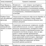
Rice. 5-16. Sarcoidosis of the eye.
A. Sebaceous precipitates on the cornea in ocular sarcoidosis.
B. Color photograph of the fundus with clutches around the retinal veins (arrow) at the periphery of the retina.
B. Corresponding fluorescein angiogram shows staining of vessels with fluorescein and slight leakage of the dye.
D. Optic disc granuloma in sarcoidosis. Vitreitis and the formation of a "macular star".
E. Partial regression of optic disc granuloma was shown 3 months after treatment with systemic glucocorticoids. Associated clinical signs
Damage to the posterior segment. cystic macular edema; clutches around the veins; peripheral chorioretinal white spots; "wax drops" or uneven nodular granulomas around venules; yellow-gray nodular granulomas of the retina, choroid and optic nerve; retinal neovascularization (Fig. 5-16, B-D).
Skin lesion. Granulomas of the orbit and skin of the eyelids.
Other eye manifestations. Granulomas of the bulbar and palpebral conjunctiva; nodules of the iris; cataract; dry keratoconjunctivitis and secondary glaucoma.
Differential Diagnosis
Other inflammatory chorioretinal diseases (eg syphilis, tuberculosis, toxoplasmosis). DiagnosticsSarcoidosis of the eye should be suspected in all patients with uveitis. The examination includes determining the levels of serum lysozyme and angiotensin-converting enzyme; chest x-ray; limited scanning of the head and neck with gallium, as well as biopsy of suspicious areas of skin, conjunctiva, or lacrimal gland formations. Fluorescein angiography reveals leakage of the dye from the vessels (see Fig. 5-16, B).
Prognosis and treatment
The prognosis is variable. To improve the condition, as well as to prevent the formation of synechia, corticosteroids are used locally, parabulbar or systemically, as well as cycloplegia. In some cases, systemic antimetabolites such as methotrexate are used.S.E. Avetisova, V.K. Surguch