Materials of congresses and conferences. Treatment of metastatic germ cell tumors High-dose chemotherapy for testicular cancer and stem cell transplantation
Chemotherapeutic treatment of testicular cancer most often done after surgical intervention. Chemotherapy is carried out in accordance with international protocols. A prerequisite for chemotherapy is the simultaneous appointment of accompanying therapy, which allows you to reduce side effects, and, accordingly, it is well tolerated (for example, antiemetic drugs are prescribed).
Most often, chemotherapy treatment for testicular cancer is carried out with the help of several drugs. This principle significantly increases the effectiveness of the therapy. For the treatment of testicular cancer, the main chemotherapy drugs are:
- Bleomycin.
- Cisplatin.
- Ifosfamide.
- Cisplatin.
- Etoposide, etc.
The most effective combination chemotherapy regimens in the treatment of patients with testicular tumors
- bleomycin 30 mg on the second, ninth and sixteenth days of the course.
- cisplatin 20 mg / m 2 on the first - fifth day of the course.
- etoposide 100 mg/m 2 on the first - fifth day of the course.
All chemotherapy drugs are administered intravenously by stream or drip. The subsequent course of chemotherapy is carried out on the twenty-second day after treatment.
- etoposide 100 mg/m 2 from the first to the fifth day from the start of treatment.
- cisplatin 20 mg/m 2 on the first to fifth day of the course.
The next course begins on the twenty-second day. The drugs are administered intravenously.
- etoposide 75 mg/m 2 on the first to fifth day of treatment.
- mesna 400 mg / m 2 before the introduction of ifosfamide, then 400 mg / m 2 IV after eight hours from the first to the fifth day.
- cisplatin 20 mg/m 2 administration on the first to fifth day of the course.
The introduction of drugs is carried out intravenously. The next course is carried out on the 22nd day.
- ifosfamide 1.2 g/m 2 from the first to the fifth day.
- mesna at 400 mg/m 2 15 minutes before the administration of ifosfamide, then 400 mg/m 2 every eight hours from the first to the fifth day.
- cisplatin 20 mg/m 2 from the first to the fifth day.
- vinblastine 0.11 mg/kg on the first and second days
Chemotherapy drugs are administered intravenously by drip or jet. The second course begins on the twenty-second day.
Side effects of chemotherapy
Complications of chemotherapy treatment are related to the fact that, in addition to tumor cells, it has a detrimental effect on healthy cells of the body, and they are even more sensitive to the effects of treatment. Cells most affected by drugs: brain, gastrointestinal tract, cells that form the basis of hair follicles and brain cells.
The main manifestations of the influence of chemotherapy drugs
- Anemia. The concentration of hemoglobin and erythrocytes decreases.
- Decreased platelet count (thrombocytopenia).
- Leukopenia - a decrease in the number of leukocytes.
- Inhibition of the spermatogenic function of the testis.
- Hair loss.
- Nausea, vomiting.
- Weakness.
This scheme involves the use of two medications: Cyclophosphamide (an alkylating cytostatic with chlorethylamine affiliation) and Adriamycin, the analogue of which is the commonly used Doxorubicin.
Cyclophosphamide is injected into a vein in an amount of 0.6 g per m² in isotonic or glucose solution. The duration of treatment is once every 21 days.
Doxorubicin is administered in an amount of 0.06 g per m², once every 21 days.
The degree of nausea (emetogenicity) of the treatment is quite high.
The most common side effects:
- bouts of nausea and vomiting;
- baldness;
- neutropenia.
The AC regimen is used primarily for the treatment of malignant diseases of the mammary glands.
Chemotherapy according to the XELOX scheme (CapeOx)
The regimen includes the use of Capecitabine and Oxaliplatin, a combination of an antimetabolite and an alkylating agent.
The use of 0.085-0.13 g per m² of Oxaliplatin in 5% glucose solution and 1 g per m² of Capecitabine (twice a day) is envisaged. Treatment is carried out for every 3 weeks.
Possible side effects:
- diarrhea;
- bouts of nausea and vomiting;
- neutropenia;
- irritated palms and soles syndrome.
The XELOX regimen is often prescribed for cancerous tumors of the intestine and esophagus.
Chemotherapy regimens for lymphoma
With lymphoma - a malignant lesion lymphatic system- usually use combination therapy with the introduction of a short chemotherapy course, which is carried out before radiotherapy.
Currently, the standard regimen for lymphoma is considered to be two or three courses of the ABVD protocol - a combination of drugs such as Adriamycin (0.025 g / m), Bleomycin (0.01 g / m), Vinblastine (0.006 g / m) and Dacarbazine ( 0.375 g/m). Injection regimen - 1 and 15 days.
Possible side effects:
- pain in the head;
- baldness;
- lowering blood pressure;
- anorexia;
- leukocytopenia.
With Hodgkin's lymphoma, an extended chemotherapy regimen can be prescribed, which is indicated by the abbreviation BEACOPP escalated.
The extended regimen includes the following drugs: Bleomycin, Etoposide, Adriamycin, Cyclophosphamide, Vincristine, Procarbazine and Prednisolone. This combination allows you to increase the chances of a cure and increase the survival rate of patients. However, when introducing more medicines the degree of toxicity to the body also increases.
FAC Chemotherapy
The FAC regimen is used in the treatment of breast cancer, especially in the early stages.
The protocol includes the use of the following drugs:
- Fluorouracil - 0.5 g per m per day intravenously, on the first and eighth day;
- Adriamycin - 0.05 g per m2 intravenously on the first day;
- Cyclophosphamide - 0.5 g per m2 intravenously on the first day.
Possible side effects include:
- oppression of the hematopoietic function;
- performance degradation digestive system;
- baldness;
- infertility;
- liver damage.
As an analogue, it is possible to prescribe mirror chemotherapy regimens - CAF and CAF extended.
Chemotherapy according to the FOLFOX scheme
There are several similar types of FOLFOX schemes, including an extended version of the protocol. Used chemotherapy drugs:
- 5-fluorouracil - I day: 1.5-2 g for 22 hours in a glucose solution; II day: repeat;
- Leucovorin - 0.5 g for 2 hours, repeated on the second day;
- Oxaliplatin - 0.1 g per m2 on the first day simultaneously with the introduction of Leucovorin.
The course is held once every two weeks.
The scheme is used mainly for the treatment of malignant lesions of the intestine.
Of the probable side effects can be distinguished:
- diarrhea
- neutropenia;
- thrombocytopenia.
Currently, the most commonly used chemotherapy regimen is FOLFOX 7, the course of which is designed for one day.
Chemotherapy regimens for stomach cancer
For chemotherapy of a cancerous tumor in the stomach, several schemes are suitable with different combination drugs. The choice of the scheme remains with the doctor, who takes into account the characteristics of clinical symptoms and the general condition of the patient. The following combinations of cytostatic drugs are most often used:
- ECF - a combination of Epirubicin, Cisplatin and Fluorouracil;
- ECX - a combination of Epirubicin, Cisplatin and Capecitabine;
- FEMTX is a combination of Fluorouracil, Epirubicin and Methotrexate.
Before surgery, Capecitabine or Cisplatin with 5-fluorouracil may be prescribed in combination with radiation therapy.
For the treatment of patients with advanced stages other protocols can be used for gastric cancer:
- DCF - a combination of Docetaxel, Cisplatin and 5-fluorouracil;
- a combination of cisplatin and irinotecan;
- Oxaliplatin and Capecitabine.
Most specialists try to limit the number of chemotherapy drugs in the protocols in order to reduce the degree of side effects. As you know, unwanted side effects are a frequent consequence of chemotherapy.
Mayo chemotherapy regimen
Mayo's regimen is a standard program of adjuvant chemotherapy treatment, that is, such treatment that is prescribed in addition to the main therapy.
The scheme involves the use of Leucovorin in the amount of 0.02 g per m² from days 1 to 5, as well as 5-fluorouracil in the amount of 0.425 g per m², from days 1 to 5. The course is alternated every 4 weeks, and starting from the third course - 5 weeks. The number and names of drugs used in the regimen may vary, but the frequency of administration remains the same.
Side effects of the proposed scheme do not differ from those that can be observed with other combinations of drugs. The protocol is characterized by diarrhea and stomatitis, inhibition of hematopoiesis, dermatitis.
Due to its therapeutic efficacy, the Mayo scheme is actively used in most well-known cancer clinics. This is a convenient and simple program that can be used to treat patients with different stages cancer processes.
Chemotherapy according to the CAF scheme
The CAF regimen is a mirror image of the similar FAC program and is used primarily for the treatment of breast cancer. Chemotherapy drugs for this protocol are as follows:
- Cyclophosphamide - 0.1 g per m² per day (from the first to the 14th day);
- Adriamycin - 0.03 g per m² per day (on the first and 8th day);
- 5-fluorouracil - 0.4-0.5 g per m² per day (on the first and 8th day).
Repeat treatment - every 28 days.
Another CAF scheme also applies:
- Cyclophosphamide - 0.5 g per m² on the first day;
- Adriamycin - 0.05 g per m² on the first day;
- 5-fluorouracil - 0.4-0.5 g per m² on the first day.
This course should be repeated every 28 days.
In addition, there is an extended high-dose CAF protocol supplemented with a granulocyte colon-stimulating factor: this treatment is more effective, but represents a considerable burden on the body. Therefore, high-dose treatment can only be prescribed to patients with good general health indicators.
2303 0
Malignant tumors of the ovaries
World Organization Health (WHO) And International Association of Obstetricians and Gynecologists (FIGO) adopted a unified morphological classification malignant tumors ovaries, which secretes epithelial tumors, sex cord stromal tumors and germ cell tumors.Most malignant tumors (80-90%) are epithelial.
Among them, serous cystadenocarcinoma - 42%, mucinous cystadenocarcinoma - 12%, endometrioid carcinoma - 15%, undifferentiated carcinoma - 17%, clear cell carcinoma - 6%.
Borderline (potentially low malignancy) tumors were identified in the main types. They make up about 15% of epithelial tumors. In addition to the morphological type of tumor, the most important independent prognostic factor in the effectiveness of treatment and survival of patients is the degree of cellular differentiation of epithelial tumors, which determines the degree of its malignancy. The Broders histological assessment system is used, and the I degree of differentiation is more favorable prognostically and the III degree is the least favorable (G1 - highly differentiated, G2 - medium, G3 - low-differentiated).
Of all tumors of stromal origin, including granulosa-, thecacollagen-producing, as well as Sertoli / Leydigo-stromal cells or their embryonic precursors, the granulosa cell tumor is the most common.
Germ cell tumors account for less than 5% of all malignant ovarian tumors, but are important because they occur in young girls and women and require special treatment, different from other ovarian tumors. The most common of these tumors are dysgerminoma, similar to testicular seminoma (tumor of endodermal origin), and embryonic cancer, in which there is an increase in the level tumor markers(serum and a-fetoprotein).
Borderline tumors, or tumors with low malignancy potential, account for approximately 15% of all epithelial ovarian tumors.
Obligatory morphological confirmation of the diagnosis of such a tumor is necessary, since its prognosis and treatment are completely different from others. malignant neoplasms.
A review of 22 studies (953 patients) with a mean follow-up of 7 years showed a survival rate of 92% for advanced disease with the exception of invasive tumor implants.
The method of treatment of borderline tumors is an operation, the volume of which is determined by the stage of the process, the age of the patient and her desire to preserve reproductive function. Patients with a common process perform radical operations in the amount of extirpation or supravaginal amputation of the uterus with appendages, removal of the greater omentum and all tumor nodes in the form of the so-called aggressive cytoreduction.
Patients with residual borderline tumors do not undergo chemo- and radiation therapy, since numerous studies (including those at the N.N. Blokhin Russian Cancer Research Center of the Russian Academy of Medical Sciences) do not show its significance. Patients without residual tumors who do not receive adjuvant treatment have the same or better survival results when compared with the treatment group.
In cases of rapid growth of residual tumors and their repeated removal, some authors use melphalan or cisplatin.
ovarian cancer
Ovarian cancer is one of the most common malignant gynecological tumors and ranks 5th in cancer mortality among women. 50% of all cases occur in people over 65 years of age. The 5-year survival rate improves significantly over time, from 36% in the mid-1970s to 45% in 2002. Approximately 5-10% of ovarian cancer is familial in the three most common variants: ovarian cancer alone, ovarian cancer, and cancer breast, ovarian and colon cancer.First of all, heredity is traced in relatives of the first degree (mother, daughter, sister). Less at risk for women of the second degree of kinship (grandmother, aunt). Genetic studies reveal BRCA1 mutations at the 17q21 locus. The BRCA2 gene, also responsible for familial ovarian cancer and breast cancer (BC), located on chromosome 13q12.
Prophylactic oophorectomy may be considered in women at increased risk over 35 years of age with children, but its relevance has not yet been fully established. Cases of the disease after prophylactic surgery are described, starting with peritoneal tumor growths similar to ovarian cancer.
A feature of ovarian cancer is spread into the abdominal cavity by cell implantation and local invasion into bladder and intestines. The incidence of lymph node involvement is 24% in stage I, 50% in stage II, 74% in stage III, and 73% in stage IV. The pelvic lymph nodes are involved as often as the para-aortic ones. The tumor, by transdiaphragmatic spread, can block diaphragmatic lymphatic drainage, which causes ascites and pleurisy.
The most informative prognostic factors for ovarian cancer include the following (Table 9.23).
Table 9.23. Main prognostic factors in ovarian cancer
Note. "+" - favorable; "-" - unfavorable, "±" - intermediate
For patients with stage I, the degree of morphological differentiation of the tumor is most important. Flow cytometric analysis of DNA in stages I and IIA can identify an increased risk group.
After optimal operations in stage III, the median survival is 52-63 months.
In table. Figure 9.24 shows the FIGO classification of ovarian cancer.
Table 9.24. Classification of ovarian cancer (FIGO)

The survival of patients directly depends on the stage of the process (Table 9.25).
Table 9.25. Survival of patients according to FIGO stages

To diagnose and monitor the effectiveness of treatment in epithelial tumors, tumor markers such as cancer embryonic antigenREA) and tumor-specific antigen CA-125. There is a high correlation of the level of CA-125 one month after the 3rd course chemotherapy (XT) at III and IV stages and survival. In cases of normalization of this marker during treatment, its repeated increase determines the activation of the process, although it does not mean the need for immediate treatment.
An elevated CA-125 level indicates a high probability of ovarian cancer, while a negative response does not rule out the presence of a residual tumor. The level of CA-125 can be elevated both in other malignant tumors and in various diseases genital organs, such as endometriosis.
Treatment methods depend on the stage of the process. Surgery is the key to treatment. Unlike other tumors of the female genital organs, the stage of the process with ovarian cancer is established after surgery. Despite the fact that only a small number of patients can be cured by one operation, the success of therapy is determined by the amount of initial intervention. The possibility of achieving subsequent complete remission, confirmed morphologically, depends on the size of the residual tumors.
Radical operation in ovarian cancer, bilateral ovariosalpingectomy with hysterectomy and removal of the greater omentum is considered. In young women who insist on preserving reproductive function, with stage I and grade I (G1), unilateral oophorectomy is possible.
During the operation, to clarify the stage and morphological variant, a biopsy is taken from the lateral canals, the pelvic peritoneum and diaphragm, the ligament that suspends the ovary, para-aortic, common iliac, external and internal iliac lymph nodes, serosa of the rectum and bladder.
Studies have not shown improvement in long-term outcomes with neoadjuvant XT. Currently, aggressive operative tactics as initial therapy are considered to be preferred for best survival. However, in case of doubtful success of the operation in patients with potential complications and comorbidities, neoadjuvant XT is possible.
Treatment tactics
Stage IPatients with tumors in stage IA-IB with a high or medium degree of differentiation (i.e. I-II degree of malignancy, G1-G2) do not require additional treatment after surgery.
At III degree malignancy (G3) stage 1C, there is a high probability of recurrence (up to 20%), which requires additional methods treatment.
Possible systemic chemotherapy, intraperitoneal (ip) administration of radioactive phosphorus 32P, or radiation abdominal cavity and small pelvis. However, the administration of 32P turned out to be more toxic with the same efficiency when compared with 6 courses of cisplatin.
Stage II
After surgical treatment adjuvant XT is performed according to the TS scheme.
Stage III
Extirpation or supravaginal amputation of the uterus with appendages with resection of the greater omentum and removal of all or most of the tumors. In the absence of visible tumors, multiple biopsies and washings from the abdominal cavity are performed.
Further treatment includes the following:
1. With minimal residual tumors (
Perhaps total irradiation of the abdominal cavity and small pelvis (only if there are no macroscopic manifestations of the disease in the abdominal cavity and there are minimal residual tumors less than 0.5 cm in diameter in the pelvic cavity) or intravenous administration of 32R (only if residual tumors less than 1 cm) or colloidal radioactive gold.
2. In case of macroscopic residual tumors more than 2 cm in diameter in the pelvic cavity, combined chemotherapy is performed in the TC, TP, CP or CC mode.
The effectiveness of XT is assessed clinically, radiologically and by marker level. Increasingly important for confirmation of complete remission is positron emission tomography (PAT).
Research programs have demonstrated a statistically significant improvement in relapse-free survival in patients with minimal residual tumors treated with ip cisplatin and ip and iv paclitaxel compared to those who received only iv cisplatin with paclitaxel. These data open up prospects for intraperitoneal chemotherapy in patients with minimal residual tumors.
Stages III and IV. Operations in full and cytoreductive to remove the largest volume of tumor masses, after which a combined XT is performed.
Therapeutic approaches for stage III and IV ovarian cancer are the same, despite the fact that the prognosis for patients with stage IV is worse. In patients with stage IV, the main manifestation is usually large tumors in the abdominal cavity and cytoreductive surgery, if possible, should be carried out in order to minimize the volume of tumor masses as much as possible.
The volume of residual tumors is a prognostic factor that significantly affects survival. Median survival in patients after optimal cytoreductive surgery is 39 months, and after suboptimal cytoreduction - only 17 months. In case of technical impossibility of performing the operation, treatment can be started with chemotherapy in order to re-evaluate the possibility of cytoreductive surgery after 3 courses. The value of repeated cytoreductive operations has not been proven.
Chemotherapy
Platinum derivatives form the basis of first-line XT combinations for advanced ovarian cancer. The standard dose is cisplatin 75 mg/m2 and carboplatin AUC-6.0~7.5.Cisplatin and carboplatin are equivalent in efficacy in ovarian cancer. Few studies have shown superiority of carboplatin (AUC 7.5) + paclitaxel (175 mg/m2) 3-hour infusion over cisplatin (75 mg/m2) + paclitaxel (135 mg/m2) 24-hour infusion.
An alternative to the paclitaxel regimen is the docetaxel and carboplatin regimen, which has shown similar efficacy in a comparative study with greater hematologic and lower neurotoxicity. Survival at 2 years of follow-up remains the same. The TC regimen (paclitaxel and carboplatin) is considered to be the best for initial XT in terms of efficacy, toxicity, and patient quality of life. Cisplatin is associated with greater neuro-, nephro-, oto-, and gastrointestinal toxicity but less myelosuppression than carboplatin.
Despite anecdotal evidence of equivalent efficacy for HT, ATS, and carboplatin monotherapy (ICON-3) regimens, most authors consider HT to be the preferred regimen.
Docetaxel may replace paclitaxel in cases where neurotoxicity needs to be reduced. Adding a third agent to such combinations is not justified.
Starting regimen: paclitaxel 175 mg/m2 3-hour infusion and carboplatin AUC 6.0-7.5 (high dose for patients in good general condition) every 3 weeks for a total of 6 cycles. Chemotherapy should be started after 4-6 weeks. after operation.
Intraperitoneal XT in a comparative study showed a significant improvement in median progression-free survival (29.8 vs 18.3 months) and overall survival (65.6 vs 49.7 months).
This type of treatment may be considered for patients with minimal residual tumors, as it is for this category of patients that it has an advantage: the median survival for minimal tumors is 66 months, and for large residual tumors - 26 months.
The preferred regimen studied is as follows: paclitaxel 135 mg/m2 IV 24-hour infusion on day 1. Sequentially cisplatin 100 mg/m2 i.p. on day 2 and paclitaxel 60 mg/m2 i.p. on day 8. There are six 21-day courses of treatment in total.
This approach should be discussed in detail with the patient as is associated with more significant toxicity than intravenous XT. In addition to catheter-related complications (infection, prolapse, blockage), it may be accompanied by grade III-IV fatigue, neutro- and thrombocytopenia, as well as gastrointestinal toxicity, abdominal pain, metabolic disorders, and neuropathy. Intraperitoneal therapy should be carried out only in clinics with relevant experience.
New drugs such as gemcitabine (Gemzar), oxaliplatin, topotecan, and triple regimens including epirubicin (Pharmorubicin) and altretamine continue to be studied with encouraging results.
Maintenance and consolidation chemotherapy, as well as high-dose XT, is not justified due to the lack of data on improving overall survival.
Relapses of ovarian cancer. Second line chemotherapy
Most important factors predict ovarian cancer recurrence clinical stage and size of residual tumors (Table 9.26).Table 9.26. Predictive factors for ovarian cancer recurrence

The age of patients also matters: 5-year survival in women younger and older than 40 years correlates as 65 and 20%. Other negative factors include clear cell or mucinous histology, poor differentiation, poor general condition, non-platinum first line XT regimens, presence of ascites. The overall recurrence rate is 62%.
The choice of second-line chemotherapy is based on tumor sensitivity to first-line XT.
Allocate:
Platinum-sensitive tumors - the first line with platinum derivatives is effective, the relapse-free interval is more than 6 months;
platinum-resistant - relapse-free interval is less than 6 months;
refractory cases - patients progress in the process of the first line XT.
Recurrent ovarian cancer may be clinically manifested by new symptoms or radiographic findings. computed tomography(CT), as well as an increase in the level of CA-125, which may precede other symptoms for 6 months. and more.
For women with asymptomatic relapses, the advisability of starting treatment immediately should be carefully considered and discussed.
The goal is palliative treatment with long-term remissions, since a cure in this situation is unlikely. Immediate initiation of treatment is justified for patients with symptoms of the disease, as well as in the presence of a small tumor volume that responds better to chemotherapy. The greatest efficacy is likely in patients with a platinum-sensitive relapse and a relapse-free interval of 12-24 months. and more. It is up to 60% with a median survival of up to 2-4 years. These patients are subject to immediate treatment.
For patients with platinum-resistant recurrence and a short relapse-free period, treatment can be delayed until a certain point (appearance of symptoms, etc.), and only the growth of the CA-125 marker requires further monitoring.
For platinum-sensitive relapses, resumption of platinum-containing regimens, primarily TC or TR, is the treatment of choice. The exception is clear cell adenocarcinoma (mesonephroid), which is relatively resistant to these regimens.
Other regimens may be: liposomal doxorubicin + carboplatin or carboplatin + gemcitabine. The latter regimen is preferred for patients with residual neurotoxicity after first line XT.
Combined XT showed better results compared to monotherapy with one of the platinum derivatives. Success depends on the duration of the relapse-free interval: if it is 5-12 months. - effect 27%, s pathomorphological complete remission (pPR)- 5%, 13-24 months. - 33% and PPR - 11%, more than 24 months. - 51% and PPR - 22%.
Platinum-resistant relapses
Paclitaxel should be used if it has not been used in first-line chemotherapy.The drug of choice for platinum- and taxane-resistant relapses is liposomal doxorubicin (Doxil in the US, Kelix in Europe). Oral etoposide, topotecan, gemcitabine, vinorelbine, 5-fluorouracil (5-FU) with leucovorin and ifosfamide have some efficacy. Altretamine (Hexalen) and oxaliplatin may also be used.
Tamoxifen gives 9.6% objective effects.
For the second line XT, weekly regimens of paclitaxel and carboplatin or docetaxel and carboplatin are more effective.
An active and relatively well tolerated regimen is the combination of gemcitabine 650 mg/m2 on days 1 and 8 and liposomal doxorubicin 30 mg/m2 on day 1. Gemcitabine can be used in combination with cisplatin and oxaliplatin.
Topotecan is used in different dose regimens: standard 5-day dose of 1.5 mg/m2/day (grade IV neutropenia is 70-80% and requires dose reduction to 1 mg/m2/day). To reduce haematological toxicity, topotecan may be supplemented with amifostine.
A weekly regimen of topotecan 4 mg/m2 on days 1.8 and 15 of a 28-day cycle is less toxic. In practice, the 15th day of administration must often be skipped. A 24-hour infusion of 8.5 mg/m2 every 3 weeks is being studied, and oral form topotecan 2.3 mg/m2 daily for 5 days every 3 weeks. Myelosuppression is lower. There are literature data on the effectiveness of irinotecan in platinum-resistant or refractory patients (250-300 mg / m2 90-minute infusion every 3 weeks.).
Efficacy in refractory cancer is: ifosfamide - 12-20%, altretamine (Hexamethylmelamine) - 12-14%, fluorouracil with calcium folinate (Leucovorin) - 10-17%, etoposide (oral) - 6-26%, epirubicin (Pharmorubicin) - 16-30%.
The effectiveness of docetaxel is 24-41%, vinorelbine - 15%, topotecan - 14-37%, irinotecan (Campto) - 21%, gemcitabine (Gemzar) - 15-28%, oxaliplatin (Eloxatin) - 29% (46% - at potentially platinum-sensitive tumors, 17% - with resistant ones), liposomal doxorubicin - 19.7%.
Several studies have demonstrated the effectiveness of thalidomide and lenalidomide either alone or in combination with other agents.
Promising is new drug trabectedin (Yondelis), isolated from the marine product Ecteinascidia turbinate and then synthetically produced, which has a unique mechanism of action.
For platinum-sensitive relapses, trabectedin 1.3 mg/m2 as a 3-hour infusion every 3 weeks. caused an objective effect in 43% of patients with a median to progression of 7.9 months.
The predominant toxicity was asthenia, neutropenia, and increased aminotransferase activity. Other studies have confirmed a 28.3% efficacy with the 1.3 mg/m2 3-hour infusion every 3 weeks regimen. and 29.6% for the regimen of 1.5 mg / m2 24-hour infusion every 3 weeks.
Efficacy, according to 3 phase II studies, was 34% with a median to progression of 5.8 months. in patients with platinum-sensitive tumors and 8% and 2.1 months. - with platinum-resistant. The combination regimen of trabectedin with doxorubicin is considered promising as a second line XT for recurrent ovarian cancer.
Bevacizumab (Avastin) 15 mg/kg IV every 3 weeks showed encouraging results. It can be used in combination with paclitaxel (3-week or weekly regimen) or with endoxan (50 mg/day orally on a long-term basis with blood counts monitored). The side effects of bevacizumab should be borne in mind, especially the risk of intestinal perforation when it is involved in the process or after irradiation of the abdominal cavity.
Therapy regimens
MonochemotherapyPaclitaxel (Taxol) - 175-250 mg / m2 ± granulocyte colony stimulating factor (G-CSF) 3-hour IV infusion once every 3 weeks. with premedication with corticosteroids, antihistamines and blockers of H2 receptors: 20 mg of dexamethasone orally or intramuscularly for 12 and 6 hours, 300 mg of cimetidine or 50 mg of ranitidine and 50 mg of diphenhydramine (diphenylhydralin hydrochloride) intravenously 30-60 minutes before administration. It is necessary to use special infusion systems that do not contain polyvinyl chloride (PVC).
Paclitaxel 70-80 mg / m2 in a solution of 0.9% sodium chloride or 5% glucose to a concentration of 0.3-1.2 mg / ml IV 60-minute infusion weekly for 6 weeks. or on days 1, 8 and 15 every 28 days. Premedication: dexamethasone 20 mg IV by bolus over 30 minutes, diphenhydramine 50 mg IV over 30 minutes and ranitidine 50 mg IV in 20-100 ml of 0.9% sodium chloride solution or 5% glucose 30 minutes before administration paclitaxel.
Docetaxel - 75-100 mg / m2 1-hour IV infusion 1 time in 3 weeks. with pre- and postmedication with corticosteroids: 32 mg of methylprednisolone or 8 mg of dexamethasone orally 13, 7 and 1 hour before administration and then 2 times a day for 3-4 days.
Cisplatin - 75-100 mg / m2 IV drip with hyperhydration and forced diuresis every 3 weeks.
Carboplatin - 400-450 mg/m2 IV drip once every 4 weeks. Given the significant difference in AUC and creatinine clearance in patients with normal and impaired renal function, it is recommended to calculate the dose using the Calvert formula.
Doxorubicin liposomal (Doxil, Kelix) - 40-50 mg / m2 IV infusion in 250 ml of 5% glucose for a dose of up to 90 mg and in 500 ml - for a dose of more than 90 mg every 3-4 weeks. The initial rate of administration is 1 mg / min for 10-15 minutes. In the absence of reactions, the rate is increased and the entire dose can be administered in 60 minutes.
Altretamine (Hexamethylmelamine, Hexalene) 6-8 mg/kg po daily for 21-28 days, or 65 mg/m2 po 4 times a day after meals and at bedtime daily for 14 days of a 28-day cycle (total dose per cycle - 3640 mg / m2), or 65 mg / m2 orally 4 times a day after meals and at night daily for 21 days of a 28-day cycle (total dose per cycle - 5460 mg / m2).
Oxaliplatin - 135 mg / m2 IV 2-hour infusion every 3 weeks, diluted in 5% glucose solution.
Vinorelbine (Navelbin) - 25-30 mg/m2 IV weekly for 8-10 weeks.
Gemcitabine (Gemzar) - 800-1250 mg / m2 IV on days 1, 8 and 15 of a 28-day cycle.
Topotecan -1.5 mg/m2/day IV 30-minute infusion for 5 days, or 2.3 mg/m2/day orally for 5 days, or 2.25-4 mg/m2 30-minute infusion in 50-250 ml of 0.9% sodium chloride solution or 5% glucose on days 1.8 and 15 of a 28-day cycle.
Irinotecan - 250-350 mg / m2 30-minute IV infusion once every 3 weeks; in case of diarrhea, reduce the dose to no more than 250 mg/m2.
Epirubicin (Farmorubicin) - 75-100 mg / m2 IV 1 time in 3 weeks.
Etoposide (Vepezid, Lasted) 50 mg/day orally for 21 days every 4 weeks. (total dose per cycle - 1050 mg).
5-FU + LV: leucovorin - 500 mg / m2 in 25-100 ml of 0.9% sodium chloride solution or 5% glucose IV 30-minute infusion daily on days 1-5 of a 21-day cycle. After 1 hour, 5-FU - 375 mg/m2 i.v.
Trabectedin (Yondelis) - 1.3 mg / m2 3-hour infusion or 1.5 mg / m2 24-hour infusion every 3 weeks.
Combination chemotherapy TS
Paclitaxel (Taxol) - 175 mg / m2 3-hour IV infusion with premedication.
Carboplatin - AUC 5.0-7.5 IV. Repeat cycle every 3 weeks.
Paclitaxel (Taxol) 175 mg/m2 3-hour IV infusion with premedication
Cisplatin - 75 mg/m2 intravenously with hydration. Repeat cycle every 3 weeks.
Paclitaxel (Taxol) 135 mg/m2 IV 24-hour infusion on day 1. Cisplatin - 75 mg/m2 IV on the 2nd day.
Docetaxel (Taxotere) - 75 mg/m2 on day 1 with pre- and post-medication.
Carboplatin - AUC 6 IV or cisplatin - 75 mg/m2 IV on day 1. Repeat cycle after 3 weeks.
Cisplatin - 75 mg/m2 on day 1 or 20 mg/m2/day for 5 days.
Cyclophosphamide - 600-750 mg / m2 on the 1st day. Repeat cycle after 3 weeks.
Cyclophosphamide - 600 mg / m2 IV on the 1st day.
Carboplatin - AUC 5-6 IV on day 1. Repetition of the cycle after 3-4 weeks.
Cisplatin - 75 mg/m2 IV on the 1st day.
Doxorubicin - 40-50 mg/m2 IV on the 1st day.
Cyclophosphamide - 600 mg / m2 IV on the 1st day. Repeat cycle after 3 weeks.
Ifosfamide - 3000-4000 mg/m2 IV (+ mesna) on day 1 or 1500 mg/m2 IV on days 1-5 (+ mesna).
Cisplatin - 60 mg/m2 IV on the 1st day. Repeat cycle every 4 weeks.
Gemcitabine (Gemzar) - 1000 mg/m2 IV on days 1, 8 and 15.
Cisplatin - 75 mg / m2 on the 1st or 8th day. Repeat cycle after 2 weeks.
Gemcitabine - 750 mg/m2 IV on days 1 and 8. Cisplatin - 30 mg/m2 IV on days 1 and 8. Repeat cycle every 21 days.
Gemcitabine - 650 mg/m2 IV on days 1 and 8.
Liposomal doxorubicin - 30 mg/m2 IV on the 1st day. Repeat cycle every 21 days.
Vinorelbine (Navelbin) - 25 mg/m2 IV on days 1 and 8.
Cisplatin - 75 mg/m2 IV on days 1 or 8. Repeat cycle every 21 days.
Liposomal doxorubicin (Doxil, Kelix) - 30 mg / m2 90-minute infusion, then Trabectedin - 1.1 mg / m2 3-hour infusion. Repeat cycle every 3 weeks.
During treatment exudative pleurisy and ascites, platinum derivatives are effective, as well as the following drugs administered intraperitoneally or intrapleurally after exudate evacuation: thiotepa - 20-40 mg, fluorouracil - 0.75-1 g (or a combination thereof), bleomycin - 30-60 mg, mitoxantrone - 25 -50 mg. A large dose of thiotepa, 60-100 mg, can also be administered intramuscularly. Effective intravenous administration of cisplatin (100-200 mg in 200-1000 ml of saline with intravenous hydration) or carboplatin (600-750 mg), as well as IFN-a2, 5-50 million units.
Stromal and germ cell tumors of the ovaries
These tumors account for 5 to 10% of all malignant ovarian tumors.They can be divided into three groups:
Ovarian stromal tumors are associated with increased estrogen secretion and concomitant endometrial cancer in 7.8% of patients. 43% of tumors are thecacellular, 24% are granulosa cells, and 33% are mixed theca and granulosa cells. The worst prognosis for granulosa cell tumors with metastases. In the case of residual tumors after surgery, use radiation therapy at a dose of 50-60 Gy to the pelvic area. For widespread metastases, alkylating agents, doxorubicin, a combination of PVB, and combinations used in ovarian cancer are used.
Experience in the treatment of Sertoli/Leydigo cell tumors is limited due to their rarity. The efficacy of combinations of VAC (vincristine, dactinomycin, cyclophosphamide) and CAP (cyclophosphamide + doxorubicin + cisplatin) has been described.
In malignant mixed ovarian tumors, tumor size and histological structure serve as the main factors determining the prognosis. The prognosis is generally poor in large tumors in which more than Y3 are elements of an endodermal sinus tumor, choriocarcinoma, or grade III immature teratoma.
For germ cell tumors, which most often occur in adolescence and adolescence, the operation of choice for lesions of one ovary is unilateral ovariosalpingectomy and biopsy of the second ovary. With bilateral lesions, a panhysterectomy is performed.
Many tumors produce proteins and enzymes that can be detected in serum as tumor markers: alphafetoprotein (AFP), chorionic gonadotropin (CG), lactate dehydrogenase (LDH).
5-year survival depends on the stage: at stage 1C - 100%, stage II - 85%, stage III - 79%, stage IV - 71%.
For dysgerminomas less than 10 cm in diameter without disruption of the capsule and invasion of other organs and without ascites, the 10-year survival rate after conservative surgery was 88.6% in one series of studies; at the same time, many women had one or more normal pregnancies that ended in childbirth after unilateral ovariosalpingectomy. Even in the case of non-radical operations after subsequent chemotherapy according to the BEP or PVB scheme, good long-term results are possible.
All patients, except those with stage I and grade I (G1) immature teratoma and stage IA dysgerminoma, require postoperative XT.
Patients after operations with incomplete removal of tumors (cytoreductive) also undergo 3-4 courses of XT according to the BEP or PVB scheme (Table 9.27).

In patients with multiple extraperitoneal lesions or who are not subject to surgical treatment according to the general condition, chemotherapy is carried out at the first stage of treatment. Patients who do not respond to the BEP regimen receive XT as a second line under the VAC or VIP regimen. The issue of a subsequent operation is decided after a thorough examination and control of the level of markers.
Combination XT includes a set of drugs and treatment regimens used for testicular germ cell tumors. In order to reduce the pulmonary toxicity of bleomycin for young patients, some modification of the treatment regimens according to the PVB and BEP regimens has been proposed.
Can carboplatin replace cisplatin in the combinations used? Carboplatin is associated with less oto- and neurotoxicity. For many tumors, but not all, carboplatin can replace cisplatin without compromising efficacy. However, this does not apply to testicular germ cell tumors. In ovarian germ cell tumors, carboplatin may be a substitute for cisplatin.
In the treatment of children with extracranial germ cell tumors, 5-year survival and recurrence-free survival were 91% and 88%, respectively, when using a combination of carboplatin, etoposide, and bleomycin.
Therapy regimens
First line chemotherapy regimensBleomycin - 30 mg IV or IM once a week for 12 weeks.
Etoposide (VP-16) - 100 mg/m2 IV drip daily on days 1-5.
PVB or UVS
Vinblastine - 3 mg/m2 IV on days 1 and 2.
Bleomycin - 15 mg / m2 (maximum 20 mg) continuous IV 24-hour infusion daily on days 1-3.
Cisplatin - 20 mg/m2 IV drip on days 4-8. Repeat cycles every 3 weeks.
Etoposide (Vepezid) - 100 mg/m2 IV drip on days 1-3.
Cisplatin - 20 mg/m2 intravenously daily on days 1-5. Repeat cycles every 3 weeks.
Etoposide (Vepezid) - 100 mg/m2 IV drip on days 1-3.
Ifosfamide - 1500 mg / m2 intravenously daily on the 1-5th day with mass in the standard mode.
Vinblastine - 0.11 mg/m2/day IV on days 1 and 2.
Ifosfamide - 1200 mg/m2/day IV on days 1-5.
Cisplatin - 25 mg/m2/day IV on days 1-5.
Paclitaxel (Taxol) 250 mg/m2 IV 24-hour infusion on day 1
Ifosfamide - 1500 mg/m2/day IV on days 2-6.
Cisplatin - 20 mg/m2/day IV on days 2-6.
Carboplatin - 600 mg/m2 IV on the 2nd day.
Etoposide - 1 20 mg/m2 IV on days 1-3.
Bleomycin - 15 mg/m2 IV on the 3rd day. Repetition of cycles every 3-4 weeks.
Second line chemotherapy regimens
VAC (vincristine, dactinomycin, cyclophosphamide)
For immature grade II and III teratomas, a VAC regimen or similar combination with vinblastine is considered best: Vinblastine 3 mg/m2 IV on days 1 and 2. Dactinomycin - 0.5 mg/m2 IV on days 1-3. Cyclophosphamide - 800 mg / m2 IV on the 3rd day.
V.A. Gorbunova
Germ cell tumors (GO)- a relatively rare pathology, accounting for about 1% of all malignant neoplasms in men, but in the age group from 17 to 35 years, this is the main variant of tumors. Over 90% of HOs are localized in the testis, primary extragonadal tumors of the mediastinum and retroperitoneum are rare. Over the past decades, there has been a significant increase in the incidence of HO.
The results of HO treatment clearly demonstrated the success of chemotherapy in disseminated solid tumors. Prior to the advent of effective chemotherapy, HO with distant metastases was an absolutely fatal disease. Survival of patients did not exceed 1 year. Only after creation efficient schemes chemotherapy, the basic drug for which was cisplatin, patients with HO got a chance for a cure. Currently, more than 80% of patients with a disseminated process are cured, and HO can be called one of the most “favorable” options for the treatment of solid tumors. At the same time, it should be noted that such progress has been achieved due to the creation of rational chemotherapy regimens, selected on the basis of an individual patient's prognosis, as well as as a result of strict adherence to the treatment algorithms that will be outlined in this article.
Orchiofuniculectomy
Even if the patient has a disseminated process at the time of detection of the primary testicular tumor, then at stage I, an orchiofuniculectomy is performed for diagnostic and therapeutic purposes. At the same time, it should be remembered that the prognosis of the disease in accordance with international recommendations is based on the determination of the level of tumor markers (see below) before orchiofuniculectomy and the results of relevant blood samples taken before surgery. Only in a severe general condition of the patient, due to a widespread tumor process, treatment for health reasons begins with chemotherapy, and orchiofuniculectomy is performed later.
Examination before starting chemotherapy
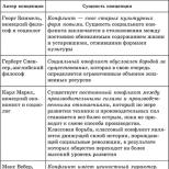
Routine examination has the following main goals: staging in accordance with the IGCCCG classification (Table 1), clarification of the localization of metastases for planning post-chemotherapeutic cytoreductive interventions, and determination of concomitant clinically significant pathology. X-rays of the organs are required chest, Ultrasound of the abdominal cavity, retroperitoneal space and small pelvis (or CT of these areas).
| Nonseminoma GO | Semin |
| Good prognosis: 56% of patients, 5-year overall survival 92% | |
| And And AFP< 1000 нг/мл, ХГ < 5000 мЕ/л и ЛДГ < 1,5 х ВГН |
And Absence of non-pulmonary visceral metastases And Normal level AFP, any levels of hCG and LDH |
| Intermediate prognosis: 28% of patients, 5-year overall survival 80% | |
| Localization of the primary tumor in the testicle or retroperitoneum or Absence of non-pulmonary visceral metastases or AFP 1000-10,000 ng/mL, CG 5000-50,000 mU/L or LDH 1.5-10 x ULN |
Any localization of the primary tumor And Presence of non-pulmonary visceral metastases |
| Poor prognosis: 16% of patients, 5-year overall survival 48% | |
| Localization of the primary tumor in the mediastinum or Presence of non-pulmonary visceral metastases or AFP > 10,000 ng/mL, CG > 50,000 mU/L, or LDH > 10 x ULN |
No poor prognosis option for seminoma |
| Note. VGN - the upper limit of the norm. | |
Table 1. International Germ Cell Cancer Collaborative Group (IGCCCG) classification
For the purpose of staging and subsequent monitoring of the effectiveness of therapy, the levels of a-fetoprotein (AFP) and human chorionic gonadotropin (CG) must be examined. It is desirable to determine the p-subunit of CG, which significantly increases the specificity. It is also mandatory to determine the level of lactate dehydrogenase (LDH).
GO rarely metastasizes to the bones, so skeletal bone scintigraphy is indicated only if the patient complains or elevated level alkaline phosphatase. MRI (CT) of the brain is recommended for high levels of hCG (more than 10,000 IU / ml) and extensive metastatic lung disease, or in the presence of neurological symptoms.
Choice of chemotherapy regimen
Patients with a good prognosis are shown 3 courses of chemotherapy according to the BEP scheme (bleomycin, etoposide, cisplatin) or 4 courses according to the EP scheme (etoposide, cisplatin) (Table 2). Four courses of the EP regimen are associated with a higher incidence of acute and mainly delayed toxicity, and therefore this regimen is prescribed mainly to avoid the pulmonary toxicity of bleomycin in patients with chronic diseases lungs or because of the characteristics of the profession (for example, athletes).
In patients with intermediate and poor prognosis, 4 courses of the VER scheme are indicated. The PEI regimen (cisplatin, etoposide, ifosfamide) is as effective as VER, but has a greater hematological toxicity (see Table 2). It is an alternative to VER when bleomycin is contraindicated.
| Mode | A drug | Introduction | day of treatment | Indications |
| EP | Etoposide 100 mg/m2 Cisplatin* 20 mg/m2 | IV 40 min IV 1 hour |
1st-5th 1st-5th |
4 courses - good forecast (with contraindications to bleomycin) |
| VER | Bleomycin 30 mg Etoposide 100 mg/m2 Cisplatin* 20 mg/m2 |
In / in 2-20 minutes IV 40 min IV 1 hour |
1st, 3rd,5th 1st-5th 1st-5th |
3 courses - good forecast, 4 courses - intermediate / bad prognosis |
| PEI | Etoposide 75 mg/m2 Ifosfamide 1200 mg/m2 Cisplatin* 20 mg/m2 Uromitexan** 800 mg |
IV 40 min In / in 1-2 hours IV 1 hour In/in jet* |
1st-5th |
4 courses - intermediate / bad prognosis (with contraindications to bleomycin as an alternative to VER) |
| Note. *Cisplatin is administered against the background of intravenous hydration with saline sodium chloride solution (total daily volume of 2.5 l), necessary to maintain a diuresis of more than 100 ml/h during the administration of cisplatin and for the next 3 hours. **Uromitexan is administered at a dose of 800 mg immediately before infusion ifosfamide and then 4 and 8 hours after its onset. | ||||
In the treatment of patients with HO, adherence to the regimen of administration and maintenance of the planned intensity of treatment are extremely important. Any delay in chemotherapy and/or reduction of doses of cytostatics lead to a significant deterioration in patient survival. On the other hand, maintaining the planned dose intensity is associated with an increased risk of severe side effects (most often due to hemosuppression), which requires experience in the accompanying treatment of such patients. A retrospective analysis showed that in centers that included less than 5 patients with a poor prognosis in one of the EORTC studies, patient survival was significantly worse (L. Collette et al., 1999). In this regard, it is extremely important that the therapy of patients (especially those with a poor prognosis) be carried out in centers with sufficient experience in its implementation.
First course of chemotherapy
The first course of induction chemotherapy is a crucial step, especially in patients with a poor prognosis and a large tumor mass. In order to prevent rapid tumor lysis syndrome (tumor lysis syndrome) and associated uric acid nephropathy 12-24 hours before the start of chemotherapy, it is necessary to start taking allopurinol at a dose of 600 mg / day. In addition, in patients with a large tumor mass, prehydration is recommended to maintain a urine output of more than 100 ml/h before and during chemotherapy.
In patients with large tumor mass and high level CG (metastatic choriocarcinoma) the first course of chemotherapy may be complicated by the development of bleeding due to tumor decay. With severe respiratory failure associated with multiple lung metastases, their decay may be accompanied by perifocal edema, followed by the development of total pneumonia and death. Prevention of this is a prolonged course of chemotherapy: for example, the course of VER instead of the prescribed 5 consecutive days is carried out for 7-10 days (for example, cisplatin 20 mg/m2 and etoposide 100 mg/m2 in the 1st, 3rd, 5th, 7th, 8th days). Our experience shows that this approach can significantly reduce the incidence of these severe complications.
Note. * Monitoring the number of neutrophils and platelets on the 4th day of chemotherapy to reduce doses in case of further reduction
Intensity of therapy and dose reduction
As mentioned above, in the conduct of induction chemotherapy for HO, the cornerstone is dose intensity, which consists of two components: strict adherence to adequate single doses of drugs and adequate intervals between cycles (21 days from the start of the previous course of chemotherapy). In contrast to the classical indications for the beginning of the next course of therapy for other solid tumors (the absolute number of neutrophils is more than 1500 in 1 μl and platelets is more than 100,000 -109/l), HO treatment is started with almost any blood count. In our clinic, we adhere to the following algorithm.
If before the start of the next course of VER, the number of neutrophils is< 0,5 -109/л или количество тромбоцитов составляет < 50 -109/л, то начало курса откладывается на 4 дня. Если количество нейтрофилов варьирует от 0,5 -Ю"/л до 1,0 -109/л, а количество тромбоцитов - от 50,0 -109/л до 100,0 -109/л, то используют схему модификации доз, представленную в табл. 3. При развитии на предыдущем курсе фебрильной нейтропении/инфекции в дальнейшем показано профилактическое применение гранулоцитарного колониестимулирующего фактора. Все это позволяет проводить адекватную химиотерапию в нужные сроки у подавляющего большинства больных.
Metastatic brain injury
Metastases to the brain in previously untreated patients are rare. This explains the fact that no randomized trials have been conducted on the therapy of this category of patients, and all recommendations are based on observations of small groups of patients.
If a brain metastasis is suspected, to resolve the issue of the number and localization of metastases, it is recommended to perform MRI with gadolinium, which allows planning further treatment. When metastases are detected at stage I, dexamethasone is prescribed at a dose of 12–24 mg/day in order to stop perifocal cerebral edema. With the number of metastatic foci less than 5, good general condition and potential resectability of at least the largest of them, neurosurgical intervention is possible at stage I. In the future, radiation therapy is indicated for the remaining metastases, as well as for the bed of remote foci, preferably in the radiosurgical version (gamma-knife apparatus, Gamma-Knife). If it is impossible to perform neurosurgical intervention, radiation therapy is indicated (in the radiosurgical version, irradiation of part or the entire brain up to a total total dose of 30-50 Gy) against the background of chemotherapy. There is no evidence of benefit from any chemotherapy regimen, with 4 courses of PEP being the standard. It should be emphasized that with adequate treatment, even patients with brain metastases have a fairly high chance of a cure.
Monitoring the effectiveness of treatment during chemotherapy
Before each course of chemotherapy, it is mandatory to control the level of tumor markers. Instrumental assessment of the effectiveness of treatment (ultrasound, chest x-ray, etc.) is usually performed every 2 cycles of chemotherapy or more often if clinically indicated. It should be remembered that in patients with a large tumor mass after the 1st course of chemotherapy, there is often an increase in the level of markers associated with tumor lysis. In this situation, it is necessary to conduct a 2nd similar course, followed by an assessment of the effect.
Growing mature teratoma syndrome
A unique feature of HO is the so-called growing mature teratoma syndrome. Its essence lies in the fact that under the influence of chemotherapy the malignant component of the tumor dies and a mature teratoma remains insensitive to therapy. It is also possible that chemotherapy promotes differentiation (maturing) of the tumor. Clinically, this is manifested by an increase in the size or, much less frequently, in the number of metastatic foci (often with the formation of cystic structures) against the background of a decrease in the level of tumor markers during chemotherapy. A huge mistake is the interpretation of this situation as the progression of the disease! It is necessary to complete the induction stage of chemotherapy with subsequent removal of the residual tumor.
Post-chemotherapeutic cytoreductive interventions
Seminoma
Residual tumor in patients with seminoma after completion of chemotherapy in more than 90% is represented by necrosis. In addition, due to the invasive growth of metastases in the retroperitoneal lymph nodes, radical intervention can be performed in no more than half of the patients. Prophylactic radiotherapy is not accompanied by improved results, so the standard tactic is the dynamic monitoring of the residual tumor. In recent years, data have appeared that allow recommending positron emission tomography. It should be carried out no earlier than 4 weeks after completion of chemotherapy with persistent foci with a diameter of more than 3 cm. At the same time, the prognostic significance of a positive or negative result of the study is unclear, and additional studies are required.
Nonseminoma Tumors
After the completion of induction chemotherapy, many patients remain with a residual tumor against the background of a normalized level of tumor markers. Unfortunately, there are no reliable criteria to predict its morphology. Approximately 35% of cases it is represented by necrosis, 50% by mature teratoma, and 15% by viable malignant tumor. Postoperative cytoreductive interventions are indicated for residual tumors larger than 1 cm and within 4-6 weeks after completion of drug therapy. Most often, retroperitoneal lymphadenectomy is performed, less often - resection of the lung, liver, removal of a mediastinal tumor. In the presence of residual tumor masses in different anatomical regions, the largest mass is usually removed at the first stage - as a rule, this is retroperitoneal lymphadenectomy. When planning further treatment, it should be remembered that the morphological structure of the retroperitoneal lymph nodes in 30-50% of cases does not correspond to that of pulmonary metastases, and therefore, even in the presence of necrosis, cytoreduction in other anatomical areas is justified.
After the end of first-line chemotherapy, removal of the residual tumor is also indicated in patients with a persistent slightly elevated level of tumor markers. With an increase in the level of markers after the first line of chemotherapy, the situation is regarded as a relapse of the disease and second-line chemotherapy is prescribed. The question of the need for postoperative chemotherapy after radical removal of residual foci containing a viable tumor still remains unclear. In one retrospective analysis, it was shown to improve only disease-free survival. In our clinic, in this situation, we usually recommend 2 cycles of chemotherapy (EP or VAB-6).
Conclusion
Advances in chemotherapy have made HO a prime example of curable disseminated solid tumors. These achievements are due not only to the development of chemotherapy, but also to more "intensive" surgery, increased diagnostic capabilities, and rational tactics for treating patients depending on prognostic factors.
Treatment algorithm for common germ cell tumors
Once again, I would like to remind you that the maximum chance of curing a patient can be achieved only with strict adherence to the recommendations for the treatment of such patients and the ability to cope with the side effects of therapy. Currently, in patients with a good prognosis, most of whom can be cured with adequate chemotherapy, it is promising to develop methods to reduce the toxicity of treatment. Against this background, the results of treatment of patients with an unfavorable prognosis are unsatisfactory, and new approaches to therapy are being sought. Its improvement is seen in the emergence of new drugs, possibly intensification of therapy, as well as in the identification of modern molecular biological factors that make it possible to individualize the treatment of patients with HO.
"Together against cancer. Doctors of all specialties" No. 1, 2006
LITERATURE
1. International Germ Cell Cancer Collaborative Group. International Germ Cell Consensus Classification: A prognostic Factor-Based Staging System for Metastatic Germ Cell Cancers. J Clin Oncol 1997, 15: 594-603.
2. de Wit R., Roberts J.T., Wilkinson P.M. et al. Equivalence of Three or Four Cycles of Bleomycin, Etoposide, and Cisplatin Chemotherapy and of a 3- or 5-Day Schedule in Good-Prognosis Germ Cell Cancer: A Randomized Study of the European Organization for Research and Treatment of Cancer Genitourinary Tract Cancer Cooperative Group and the Medical Research Council. J Clin Oncol 2001:1629-40. Pure sodium chloride.
3. Nichols C.R., Loehrer P.J., Einhorn L.H. et al: Phase III study of cisplatin, etoposide and bleomycin or etoposide, ifosfamide and cisplatin in advanced stage germ cell tumors: An intergroup trial. Proc Am Soc Clin Oncol 1995;14:239.
4. Albers P., Weinknecht S., Krege S. et al. Prediction of necrosis after chemotherapy of advanced germ cell tumors - results of a prospective multicenter trial of the GTCSG. J Urol 2002;167(Suppl.):172.
5. De Santis M., Bokemeyer C., Becherer A. et al. Predictive impact of 2-18fluoro-2-deoxy-D-glucose positron emission tomography (FDG PET) for residual postchemotherapy masses in patients with bulky seminoma. J Clin Oncol 2001;19:3740-4.
6. Aprikian A.G., Herr H.W., Bajorin D.F. et al. Resection of postchemotherapy residual masses and limited retroperitoneal lymphadenectomy in patients with metastatic testicular nonseminomatous germ cell tumors. Cancer 1994;74:1329-34.
7. Hartmann J.T., Candelaria M., Kuczyk M.A. et al. Comparison of histological results from the resection of residual masses at different sites after chemotherapy for metastatic non-semino-matous germ cell tumors. Eur J Cancer 1997;33:843-7.
8. Fizazi K., Tjulandin S., Salvioni R. et al. Viable malignant cells after primary chemotherapy for disseminated nonsemi-natous germ cell tumors: prognostic factors and role of postsurgery chemotherapy -results from an international study. J Clin Oncol 2001;19:2647-57.