Features of the choice of orthoses for the knee joint. Devices for immobilization of the knee joint: the best orthoses, bandages and splints Diagnosis of hemarthrosis with elements of treatment
The creation of immobility of nearby joints in the conservative treatment of fractures of the bones of the extremities by the method of fixation bandages is an indispensable condition for optimal consolidation and is recommended in all guidelines for traumatology and orthopedics. For example, immobilization of all three large joints of the lower limb in fractures of the femur with a circular plaster cast is the oldest method of treatment.
At the same time, all specialists understand that the longer the joints do not function, the more often contractures form and muscle hypotrophy develops. So, back in 1936, R.R. Vreden wrote that one of the main defects of the “circular bandages” is the long-term immobilization of the muscles and joints of the leg. Turning off all, even minimal active muscle contractions, leads to weak arterialization of the limb and stagnation of venous blood and lymph. The conditions for resorption of exudate and cellular decay products worsen, and thereby the regenerative abilities of damaged muscles, tendons and ligamentous apparatus decrease.
Full immobilization of the joints causes their stiffness and hypotrophy of the muscles of the limb, which have to be fought for a long time, and sometimes unsuccessfully, after the bandage is removed. He believed that, for example, the treatment of hip fractures by immobilization with "circular bandages" often does not provide a satisfactory anatomical recovery and at the same time prevents the functional recovery of the affected limb. A major disadvantage of immobilization treatment should be considered that the damage to the function of the limb is not so much the result of the fracture itself, but rather the result of this method of treatment.
Therefore, not to the detriment of the process of consolidation, for a long time they have been trying to determine the moment when it is possible to start a motor function in previously fixed joints. If this is not possible, other methods are used for treatment. One of the optimal solutions to achieve maximum release from immobility of the joints closest to the fracture was and still is the creation of a rigid bandage, both in its design and with the help of the materials used.
At the end of the 19th century, for fractures in the middle third of the bones of the lower leg, Professor Volkovich applied a cardboard-gypsum or plaster splint 6-7 cm wide to the limb in the form of a stirrup, starting from the level of the knee joint, along the outer surface of the lower leg through the sole to the inner surface as well as to knee lines.
on thus located on the anterior-inner surface of the leg along the tibia and on the posterior-outer along the fibula and was fixed with soft bandages. After the final hardening of the bandage, patients were allowed to load the damaged surface. Volkovich attached great importance to the possibility of independent movement in the joints of the lower limb and early functional loading. The same type of dressing was proposed in 1920. in Germany Brunn. in France in 1910. Delba was also offered a bandage similar to Volkovich's bandage. In the 30s of the XX century, Beler's splint-gypsum bandages (3 splints) were widely used. All these dressings were united by the desire to achieve maximum rigidity of fixation of fractures, the possibility of movements in the joints and early function.
In the future, the design of dressings used in the treatment of bone fractures of both the upper and lower extremities was constantly improved with the advent of new technological possibilities.
Interesting solutions in the use of "functional" dressings with partial release of the ankle joint and foot joints in ankle fractures were proposed by S.N. Khoroshkov (2006).
Sarmiento A et all (2000) on a large group of patients (922 patients participated in the study) with diaphyseal fractures humerus used specially made orthoses for the shoulder segment without immobilization of the shoulder and elbow joints. Moreover, in 87% the fractures healed. Less than 16% of them had slight varus deformity or angular deformity with an anterior open angle.
Comparative analysis results of treatment of fractures of the shaft of the shoulder in a similar brace with the results of treatment after surgical treatment with a lockable pin (n=89) are given by Wallny Tetall (1997) and Campbell J.T. et all (1998). Thus, 44 patients were treated conservatively in a brace, and 45 patients were treated with an operatively lockable pin. 86% of the patients in the conservative group and 47% in the operative group did not experience any restrictions in movement in the joints after the end of treatment. Functional results in the conservative group were significantly better.
Gypsum bandage is still widely used for the manufacture of fixing dressings in traumatology. But today, plaster casts are being replaced different kinds orthoses, in the manufacture of which modern materials are used: polyurethane bandage; low temperature or high temperature plastics.
Already today, many manufacturers in this area are establishing and expanding the arsenal of mass-produced orthoses made of various elastic materials such as neoprene or other multilayer dense elastic fabric consisting of elastic and cotton fibers, with additional stiffening ribs made of metal or polymer plates, depending on the location and purpose. applications. This makes it possible in some cases to use a finished orthopedic product instead of plaster, which allows you to maintain control over the fit of the fixator around the limb and, if possible, maintain movement in adjacent joints.
In this regard, it is necessary to understand exactly for what purpose, and for what indications, this or that group of products is used.
Of particular interest to practicing traumatologists, in our opinion, is the "polyurethane bandage", which, again, not in all cases, replaces the plaster bandage.
Bandages made of "plastic plaster" have a number of differences from traditional plaster bandages:
However, the possibilities of its use, and, accordingly, the presence of indications and contraindications for the appointment, as a rule, are little known to doctors working in a wide clinical practice.
Synthetic polymer bandages are produced in the USA - "Scotchcast", "Softcast" (firm "ZM"); in Germany - "Cellacast" (firm "Lohmann & Rauscher"), "Rhena®therm", "Rhena®cast" (firm "Hartmann") and in Russia - "Super-cast" (for rigid immobilization) and "Super-cast- elast" (to create an elastic sleeve) (firm "Novomed", Moscow).
The fabric base of the bandage consists of a fiberglass or polyester mesh impregnated with polyurethane resin. It is made in the form of a bandage or longuet. Form of release of bandages: individual packaging for each bandage in a hermetically sealed foil bag.
When the bandage is exposed to water, the polymerization reaction of the resin is activated, as a result, the bandage hardens. Full strength of the material occurs after 30 minutes. The bandage is applied quickly and easily. Thanks to its stretchability, it precisely follows the contours of the body, which ensures an excellent fit and optimal fixation. Bandages are intended for the manufacture of immobilizing dressings in traumatology and orthopedics, as well as other orthopedic removable devices.
In order to compare the elasto-mechanical properties of plaster and polymer bandages, we conducted special studies of the elasticity, elasticity and rigidity of standardized samples in the laboratory of polymers of GNU CITO.
Identical samples of longet and circular rings (imitation of "circular dressings") were prepared from plaster and polymer bandages (Fig. 1).
Fig.1. Appearance of the prepared samples of longet and "circular dressings" from different layers of plaster and polymer bandages

It can be seen from the table that a splint made of 4 layers of a polymer bandage is 3 times stronger than a 12-layer analogue of a plaster bandage. When comparing the weight characteristics of samples with an equal number of layers and sizes, gypsum samples are 2 times heavier than polymer samples.
Technique for the manufacture of "express orthosis".
The method of applying a bandage differs little from the application of conventional plaster bandages. Although differences exist and lie in the need for even more careful and careful attitude to the application of such bandages with strict observance of all the rules for applying a hard (gypsum) bandage, since irregularities on the inner surface of the bandage due to its high rigidity can lead to skin damage.
The following materials and equipment are required for the manufacture of an express orthosis:
1. Synthetic bandage, consisting of specially woven glass fibers impregnated with polyurethane resin. Under the influence of water or a humid environment, a reaction occurs, leading to the hardening of the material.
2. Seamless knitted tubular bandage with a high degree of stretch in the longitudinal and transverse directions. Used as lining material.
3. Lining bandage made of soft synthetic cotton.
4. Metal rivets, velcro tape, hinged devices
5. Vibrating saw for cutting polymer dressings.

Fig.2. Appearance of a splint for the knee joint made of a polymer bandage
In the manufacture of orthoses, we used the following technique for their manufacture:
1. When acute injury limbs, at the first stage, a plaster splint lining bandage was applied. A cotton lining layer, especially in case of an acute injury, helps to prevent the development of phlekten and additional damage to the skin. After subsidence of soft tissue edema and relief of pain syndrome, the plaster cast was changed to the necessary one made of synthetic material.
2. PREPARATION for the imposition of a synthetic bandage. Skin, clothing and work surfaces must be protected from contact with the super-cast bandage. The patient's extremity is preliminarily put on a cushioning (synthetic or cotton tight stocking) and lining material (a special thin cotton bandage that allows you to protect the skin, especially in the area of \u200b\u200bbone protrusions).
The doctor and his assistant must wear gloves. Open packages of super-cast bandage as needed (when in contact with air moisture, it begins to harden).
3. SOAKING. Only when moistened does the material acquire excellent adhesive properties. The water temperature should not exceed 20-24°C (otherwise, the patient may get burned due to the heat generated during the hardening of the bandage). When immersing the bandage in water, it is necessary to lightly press on it 3-4 times, for a more complete soaking with water. After that, squeeze out excess water, as carefully as when working with a plaster bandage. If the package of the "super-cast" bandage is opened, but not immersed in water, then the polymerization process will begin from interaction with air containing moisture. The time for the complete hardening of the dressing will increase to 10-15 minutes, which gives more time for the reposition of bone fragments and modeling of the dressing.
4. OVERLAY TECHNIQUE. Super-cast bandages are applied in circular rounds without tension and so that each subsequent round of the bandage overlaps the previous half and overlaps the edge of the underlying round. Due to the special weaving, the “super-cast” bandage is easily applied in difficult places, while there are no folds and bends. The simulation lasts 2.5-3 minutes. At this stage, it is possible to mount various devices into the bandage, such as hinges, staples, etc.
To this end, the legs of metal hinges (or staples) are treated with a specially prepared urethane resin with a brush and applied to the already applied layers of the dressing. Three additional layers of bandage are applied over the legs to secure the hinges in a given position.

Fig.3. The appearance of the patient in a circular non-removable "apparatus on the knee joint"
5. READY BANDAGE. The bandage hardens within 5-8 minutes. The polymerization process is accelerated by wetting the dressing surface with water using a sponge. After 20-30 min. the bandage can be given a partial load. Full polymerization occurs within a day, after which it is recommended to give a full load. Processing, formation of holes, removal of the dressing is possible with conventional tools or an oscillating saw.
The advantage of bandages from the bandage "super-cast" is:
- high strength and reliable stabilization, since, based on our research, a four-layer bandage made of polyurethane bandage has 5 times higher operational strength than a 12-layer plaster bandage.

If we enter the numbers we received into the formula, then for similar dressings from the “super-cast” bandage, 4 units will be required, and from plaster bandages - 12 of the same size.
- 4 - 6-layer circular bandage eliminates the use of reinforcing splints and withstands a weight load suitable for long-term use;
- moisture resistance and moisture permeability;
– breathability (excludes maceration of the skin);
- slight radiopacity;
- the possibility of staged use of the imposed circular bandage for further rehabilitation (the bandage can be cut, form "windows", used as the basis for the manufacture of a removable orthosis, splint).
NOTE: If the super-cast bandage comes in contact with the doctor's or patient's skin, wipe the area with alcohol or acetone. Dressings made of "super-cast" synthetic bandage do not get wet.
Further, during operation, regular bathing or showering is not recommended, because. wet cushioning material can cause maceration of the skin, while at the same time the quality and strength of the dressing itself does not suffer. However, if the patient still resorted to water procedures dry the dressing with a towel and hair dryer.
In the course of treatment, if necessary, the circular bandage can be easily converted into a removable longet. With the help of a special vibrating saw, cuts are made along the lateral and medial surfaces of the dressing, and the front “cover” is removed.

Fig.4. Converting a circular dressing to a splint
Then the bandage was completely removed and the sharp edges of the cuts were processed. Along the edges of the back splint, using a hole punch and metal rivets, from 1 to 5 elastic Velcro tapes were fixed to ensure the fixation of both parts of the product to each other, thus obtaining a circular split splint. Lining material was added to the inner surface, if necessary, and a bandage was tried on.
Fig.5. Removable ankle splint
A synthetic circular hard bandage has the same scope as a plaster bandage, but, it must be noted, contraindications to its application are:
- cases associated with a rapid significant change in the volume of a limb segment with an increase and decrease in edema in the first week after injury;
- the planned repeated manual repositions of the fracture through the bandage, which cause deformation of the inner wall of the applied bandage and cause severe skin damage in the form of bedsores and deep deposits.
The indication for applying this bandage is the need for the patient to achieve high mobility for a long time. This is ensured by its elasto-mechanical properties and a wider possibility of built-in various combinations hinges at the level of the joint, which, while providing the necessary rigidity of immobilization, will create the possibility of dosed movement for the prevention of contractures.
Analysis of the results of the use of solid polymer "foot splints" for non-displaced metatarsal fractures and metatarsal fractures showed an important positive economic effect of the proposed treatment. This method treated 15 patients, 12 of them had a fracture of the 5th metatarsal bone with a slight displacement, 2 patients had a fracture of the base of the 3rd-4th metatarsal bones and 1 patient had a fracture of the cuboid bone. The "foot splint" was made from a synthetic polymer bandage as a one-piece construction. With careful modeling, the bandage provides a high degree of rigidity of fixation of the foot with the talocalcaneal joint and provides partial mobility in the ankle joint, which makes it possible to allow dosed walking at the stages of treatment on the 5th day after the injury in sports shoes. This allowed patients to return to normal life within 2 weeks after the injury.

Fig.6. The appearance of the patient and the function of the limb a week after the injury in a "shortened bandage" for a fracture of the IV metatarsal bone
By the end of immobilization, the patients had no pain and stiffness of the ankle joint. When analyzing the results of a survey of patients on the Hauser Walk Index (I.X.H.) test (Hauser Ambulation Index,) which was developed by Hauser S., 1983. Patients treated with traditional technique(control group with plaster immobilization) had a level of “4”, and patients treated with the use of a “foot splint” had a level of “1 or O”, which characterizes more high level patient activity.
However, not in all cases with fractures of the bones of the extremities, short bandages provide the necessary immobilization of the segment.
In difficult situations, you can use combined bandages with the inclusion of hinged devices in the design.
For a correct understanding of the terms, we list all possible types of devices for the lower limb based on the localization for which it is intended:
Devices for the lower limb:
1. Apparatus for the ankle joint;
2. Apparatus for the knee joint;
3. Apparatus for the knee joint with a block for the ankle joint;
4. Devices for the knee and ankle joints (or "Apparatus for the whole leg"):
5. Apparatus for the whole leg with unloading under the tuber;
6. Device for the whole leg with unloading under the tuber and with a stirrup;
7. Full leg device with double track;
8. Apparatus for the hip joint;
9. Apparatus for the hip and knee joints;
10. Apparatus for hip, knee and ankle joints;
11. Devices for two hip, knee and ankle joints connected through a lumbosacral corset (“tee”)
Regardless of the localization of the distribution of apparatuses, in their manufacture, hinges of different functions are required, which are used for specific applications. pathological conditions in the same joints:

are used to implement the full range of motion in the joint of the same name when walking, but strictly along the specified axes. To approach the physiology of movement in the knee joint, the hinge is made as biaxial.

is used to implement a dosed range of motion in the joint of the same name when walking along strictly specified axes. To approach the physiology of movement in the knee joint, the hinge can be made as a biaxial one.
It is used for functional deviations associated with instability in the joints or for partial unloading of the joint while maintaining the full range of motion.

A discrete change in the hinge of the angle of fixation - 8 degrees creates the possibility of using it to hold the joint in a given position.
It is used in orthoses designed to develop joint contractures.

The hinge is equipped with a special spring and an adjusting screw to create forced flexion-extension movements.
It is used in orthoses designed to create forced walking conditions in myoneurotrophic diseases leading to muscle weakness of the segment, in post-traumatic manifestations such as "equinovarus foot", "hanging foot" to develop or maintain specified movements.

hinges for the knee joint, having a special device that provides a dosed fixed movement in the sagittal plane, are used to correct the varus or valgus installation of the knee joint. Model - "TRASTER".

Structurally, there is a falling lock that provides rigid fixation at the level of the joint at the moment of full extension, moreover, the lock is released manually, that is, there is no possibility of spontaneous opening of the lock at the moment of walking.
It is recommended in the manufacture of orthoses used for walking with eliminated flexion contractures, with paresis and paralysis of the muscles of the limb, for the treatment of intra-articular and peri-articular fractures of the joint or in the postoperative period.
In the manufacture of an orthopedic device for the knee joint, sleeves from three layers of a "super-cast" synthetic bandage were applied to adjacent segments of the limb. Then, according to the method developed by us, hinges of the same name to the joint were attached to the sleeves.
Since we still do not have mass-produced special models of joints for this purpose, in order to improve the kinematics of movement in the orthosis at the level of the knee joint, a knee joint joint was developed for functional deviations associated with instability in the knee joint to partially unload the joint and maintain maximum volume movements.

Fig.7. Appearance of the patient in a non-removable "apparatus for the knee joint"
The results of clinical, physiological, biomechanical examinations of patients with the consequences of diseases, injuries of the musculoskeletal system, equipped with various designs of orthoses, suggest that training weakened and paretic muscles when walking in lockless devices contributes to the restoration of motor function.
In isolated fractures of the tibial condyles without displacement (18 patients), after the phenomenon of hemarthrosis had subsided (at this stage, "splints for the knee joint" were used), we used express devices individually made directly on the patient from polymer bandages with hinges for the knee joint.
I would like to note that the replacement of plaster immobilization in this group of patients with modern orthopedic products made it possible in all cases to begin active development of movements in the joint not after the termination of immobilization, but in parallel with it, that is, usually a full course of exercise therapy began in the second week after the injury.

Fig.8. The volume of passive movements in the knee joint after the imposition of a non-removable "apparatus for the knee joint"
This allowed patients treated by this technique to return to normal life without an additional period of rehabilitation, which, on average, reduced the total period of disability by 2–4 weeks. By the end of immobilization, the patients had no pain and stiffness of the knee joint.

Fig.9. Function of the lower limb in a non-removable “knee joint apparatus” by the end of 4 weeks after damage to the lateral ligament of the knee joint
When analyzing the results of the questionnaire according to I.Kh.Kh. patients treated according to the traditional method (the control group was treated with plaster immobilization) had a level of "4", and patients treated with orthopedic apparatus had a level of "1 or O", indicating a higher physical activity of this group.
In conclusion, I would like to answer the question posed in the title of the article, that modern possibilities (when used) create ample opportunities in many cases to comply with the principles of combining the conditions necessary for consolidation with early development of movements in adjacent joints.
www.cito-pro.ru
Joint immobilization
Most often, the cause of pain and damage in the wrist joint is an injury caused by sudden movements or their large amplitude, which is usually obtained when falling on the hand, less often with a sharp jerk or impact.
Another physiological feature of this joint is the passage through it of the endings of the median nerve. Therefore, quite often the pain can be localized in the area of the phalanges of the fingers, and not in the damaged area.
One of the mandatory components of the treatment of injuries is the use of various orthopedic bandages for immobilization. However, the wrist brace is not only used in the case of injuries, it is also necessary:
- With inflammation of the joint and surrounding tissues with arthritis, tendonitis and myositis.
- To prevent the development of flexion contractures of the hand that disrupt the normal mobility of the joint.
- At various neuropathies developing as a result of compression of the median nerve due to overstrain of the ligaments and tendons, such as carpal tunnel syndrome.
- AT complex treatment various osteochondropathy arising from circulatory disorders as a result of injuries or other diseases and leading to microfractures.
Types and features of orthoses
Bandage on the wrist joint may differ in the degree of rigidity and the ability to limit mobility. Usually, the whole variety of models is usually divided into several types, depending on the rigidity of the product and design features.
Soft orthoses

Such products are made of breathable elastic fabrics. They are often called sports bandages or calipers. They do not restrict the movement of the hand and fingers, but at the same time protect the joint from excessive stress.
Often used to prevent injury by athletes, especially those involved in weightlifting, tennis or basketball and people who prefer outdoor activities. Sometimes these dressings are prescribed for such conditions:
- at the last stage rehabilitation period after operation;
- joint instability;
- tunnel syndrome;
- a mild form of inflammation of the ligaments of the hand;
- arthrosis or arthritis.
Depending on the properties of the material, such a wrist brace can additionally have a light, massaging and warming effect.
Semi-rigid orthosis
This orthosis is made of a soft elastic material, but with the addition of stiffeners, which are thin plates made of metal or polymer materials. It moderately limits the movement of the hand in the wrist joint. Most often assigned:
- in the early period after surgical interventions;
- for fixing the wrist after removing the cast;
- with bruises, sprains or torn ligaments.
Rigid orthosis

It is a dense plastic frame, which can sometimes be additionally reinforced with metal inserts. It is attached to the hand and fingers with the help of special straps that allow you to adjust the degree of fixation. Completely excludes movement in the joint. Applies to the following conditions:
- in the early period of rehabilitation, after operations associated with complicated fractures and torn ligaments;
- at the last stage of inflammatory and degenerative diseases.
There are models that fix not only the wrist, but the entire hand with fingers, which allows you to do without plaster even with the most complex fractures.
Appointment of orthoses
Semi-rigid or rigid orthoses prevent the development of contractures in the wrist and fingers - pathological processes, limiting passive movements in the joint, in which the arm cannot normally bend and unbend.
Most orthoses tend to combine several functions, such as relieving excess tension and helping to restore mobility. Also, fixing dressings are usually divided, depending on their purpose, into:
- Prophylactic, which must be used when playing sports, outdoor activities or any other activity related to constant loads, as well as on early stages development of joint deformity.
- Therapeutic fixatives are used temporarily for injuries and in the postoperative period.
- Constants are appointed with a complete loss of form or function of the wrist joint.
The difference between an orthosis and other dressings

Sometimes an orthosis is confused with a splint or splint. Both are orthopedic products that serve to protect, relieve stress and, if necessary, ensure complete immobility of the joints.
However, the orthosis differs in that it is a device fastened with hinges, and the splint looks like a sleeve or shoe connected by tires.
As for the splint, it is a long strip of plaster or quick-hardening plastic, which is usually used for fractures as a fixing bandage on the wrist joint.
How to choose?
Today on sale you can find a huge number of the most diverse models of bandages and among such an assortment it can be very difficult to find exactly the one that is needed.
First of all, it all depends on the disease, the age of the patient and his physiological characteristics. In addition to the wrist itself, orthoses can additionally fix the thumb or the entire hand.
In practice, each manufacturer has its own size grid of orthopedic products. All you need to do before buying is to measure the circumference of the arm in the area of the joint.
It is also worth considering for which hand a wrist brace is needed, since not all models are universal. Some manufacturers produce products for both the left and right limbs. As for the material from which it will be made, the main requirement here is the absence of allergies.
Application results

With the help of an orthosis, excess tension is removed from a fixed limb, which helps to avoid injuries. And in case of diseases or after operations, the resting joint and ligaments recover faster. If there is a fracture with a displacement, then the fixator will help prevent the development of deformation.
The effectiveness of the use of an orthosis depends on the correctness of its choice. The doctor should select the degree of fixation, mode and duration of use.
There is an opinion that wearing an orthopedic fixator can cause the development of muscle atrophy. However, this is nothing more than a myth. The cause of atrophy often lies in an improperly fitted bandage or in ignoring exercises to restore joint mobility.
A properly selected orthosis does not compress the tissues surrounding the joint and does not interfere with blood circulation in them. In addition, wearing an orthopedic fixator must be combined with physiotherapy and physiotherapy exercises.
medotvet.com
Immobilization of the lower limb
1. Immobilization in case of a fracture of the lower leg is carried out in a straight position of the leg or slight flexion at the knee joint. The foot is fixed in the position of dorsal flexion at a right angle with respect to the lower leg. An exception to this situation may be an injury calf muscle, where in order to reduce pain, you can save a slight flexion of the foot. It is advisable to use at least 2 splints applied in 2 planes for immobilization. Wooden tires are located both on the outer and inner surfaces of the leg, and ladder tires - one on the back, the second on the outer surface. In the case of using 3 splints, the latter is placed along the back surface of the leg, preferably a ladder one (Fig. 8).
|
|

Immobilization with 3 splints is desirable for severe, especially gunshot fractures of the diaphysis of the leg, severe pathological mobility of fragments and bleeding from the wound. Modeling requires a rear tire. Curves must be created for the foot, heel, Achilles tendon, calf and knee. The length of immobilization: in case of damage to the foot - from the fingers to the upper third of the lower leg; ankle joint and lower leg - up to the upper third of the thigh; knee joint, hip and hip joint - to the level of the shoulder blade and armpit. With mild closed injuries of the knee joint, immobilization is limited to the level of the hip joint. Side wood splints require thicker padding at the ankles and knees.
2. Transport immobilization for injuries of the knee and hip joints and thigh is usually carried out with a Dieterichs splint, in addition, there are other splints (Goncharov, Thomas-Vinogradov, etc.)
|
|

Stages of applying the Dieterichs bus (Fig. 9):
1. Before applying, the splint is adjusted in height, while the lower ends of the crutches should protrude beyond the “sole” by 15-20 cm.
2. Fitted crutches at the level of the pegs are tied with bandages.
3. The plantar part of the tire is fixed to the foot with an eight-shaped bandage, carefully strengthening the heel area.
4. The lower ends of the crutches are passed through the metal eye of the plantar part of the tire and applied to the side surfaces of the limb and torso.
5. In the area of the protrusions of the greater trochanter and the knee joint, cotton is placed.
6. The tire is attached to the body with scarves or straps threaded through the crutches on the lower leg, thigh, abdomen and chest.
7. The ends of the twist laces are threaded through the hole cross bar inner branches and inserted into the rings of the sole, brought back through the hole of the strap and tied around the twist.
8. The leg is pulled by the foot until the transverse bars of the branches rest against the groin and armpit.
9. After stretching, the splint is fixed along the entire length of the limb with circular tours of the bandage.
To improve fixation under rear surface legs and pelvis enclose a ladder or plywood splint with thick linings in the region of the hamstring and Achilles tendon. Under favorable conditions, the Dieterichs tire can be strengthened with plaster rings.
Transport immobilization for fractures of the spine in the cervical and upper thoracic regions is carried out on the back with a roller under the neck. The most reliable immobilization for severe, especially multiple fractures can be performed using vacuum immobilizing stretchers (Fig. 11,12).


Fig.11. Preparation for immobilization Fig.12. Case lacing
using NIV-2
Transport immobilization in case of damage to the thoracic and lumbar spine and transportation must be carried out on a rigid stretcher. The victim is placed on a stretcher and fixed together with a solid pad to the stretcher. A small roller is placed under the knees, and in the presence of paraplegia, an inflatable rubber or cotton-gauze circle is placed under the sacrum.
If the victim has to be transported on a conventional soft stretcher, then he should be laid on his stomach, which provides some extension of the spine. Some kind of roller (coat, etc.) is placed under the chest. At gunshot wounds the spine should not be created lordosis, but it is better to put the victim flat on his stomach.
In case of pelvic fractures, the victim can be transported on a regular stretcher, but it is better on a hard stretcher. The legs should be bent at the knee and hip joints, for which a roller is placed under the knees of the victim. The victim must be fixed to a stretcher.
Currently, at the pre-hospital and early hospital stages, an anti-shock pneumatic suit "Kashtan" is used (Fig. 13).
Pneumatic anti-shock fixing suit "Kashtan" is intended for emergency use in order to prevent and relieve hypovolemic shock at the pre-hospital and resuscitation stages. The action of the suit is based on the principle of controlled circular external pressure. When inflated, controlled pressure in the suit (up to 100 mmHg) redistributes blood from the lower extremities and abdomen to the heart and vital organs of the upper half of the body. Simultaneously with In this way, external pneumatic compression often helps to stop sludge, significantly reduce internal and external bleeding, and also provides stable immobilization of fractures of the lower extremities and pelvis.
Indications for use are:
1. Systolic blood pressure of 100 mm Hg accompanied by symptoms of shock (pallor, cyanosis, cold clammy sweat, tachycardia, tachypnea) or systolic pressure below 80 mm Hg, regardless of the cause, are absolute indications for the use of the suit, in the absence of contraindications.
2. traumatic shock II - IV degree with multiple fractures and amputations of the lower extremities, pelvic fractures.
3. Internal and external bleeding of the lower half of the body: intra-abdominal bleeding as a result of blunt or penetrating abdominal trauma; postpartum, uterine, gastrointestinal bleeding; bleeding or ruptured aneurysms of the abdominal aorta.
Contraindications:
1. Respiratory failure due to pulmonary edema, tension hemopneumothorax.
2. Massive unstopped bleeding of the upper half of the body.
3. Prolapse of internal organs.
4. Cardiac tamponade, acute heart failure, cardiogenic shock.
5. Pregnancy (because of the threat of miscarriage).
If there are contraindications, only the abdominal section cannot be inflated on the suit, but the leg and pelvic sections can be inflated.
Sticks, boards, skis and any similar items can be used as improvised means for transport immobilization. When immobilized with these objects, it should be borne in mind that they are hard, inflexible and cannot be modeled on the surface on which they are applied. Therefore, improvised means should be applied only from the outer and inner surface of the limb, always with soft pads in the area of the ankles and knee joint. Improvised means, like standard ones, should immobilize 2 joints - above and below the fracture.
If there are no means for transport immobilization at hand, then the injured arm can be immobilized with a jacket, bandaged to the chest, and the leg is fixed to the other, healthy leg (Fig.). Foot-to-foot immobilization is a last resort and is not very reliable for hip fractures, especially in the middle and upper third.
STOP BLEEDING (HEMOSTASIS).
In almost any injury, blood vessels are injured. In this case, bleeding is of varying intensity and depends on the type and nature of the damaged vessel.
Anatomically distinguish:
arterial bleeding characterized by intense blood loss. The blood is bright red (scarlet) in color, beats with a pulsating jet under great pressure. In case of damage to large vessels (aorta, femoral artery, etc.), blood loss that is incompatible with life can occur within a few minutes.
Venous bleeding. The blood is dark cherry in color, flows out slowly, evenly, in a continuous stream. This bleeding is less intense than arterial, and therefore less likely to lead to irreversible blood loss. However, it must be borne in mind that when injured, for example, the veins of the neck and chest air can enter into their lumen at the moment of inspiration. Air bubbles entering the heart with blood flow can cause an air embolism and cause death.
capillary bleeding observed with superficial wounds, shallow skin cuts, abrasions. Blood from the wound flows slowly drop by drop, and with normal clotting, the bleeding stops on its own.
mixed bleeding occurs with simultaneous injury of arteries and veins, most often with deep wounds.
Parenchymal bleeding in case of damage to parenchymal organs (liver, spleen, kidneys), which have a developed network of arterial and venous vessels, the walls of which do not collapse when damaged.
By time of occurrence:
1.primary
2.secondary
- early (from several hours to 5 days)
- late (after 5 or more days)
In relation to the external environment:
1. external (if blood is poured outside the body)
2. internal (if blood accumulates in cavities and tissues)
- open - if the cavity has an anatomical connection with the environment (nasal, pulmonary, uterine, gastric, intestinal)
- closed - if the cavity has no anatomical connection with the environment (hemothorax, hemoperitoneum, hemarthrosis, hematoma)
3.interstitial
- petechiae - small hemorrhages in the skin
- ecchymosis - pinpoint hemorrhages in the skin
- hematomas - accumulations of blood in tissues and organs.
By clinical course:
- acute
- chronic
By intensity:
- profuse
- moderate
- weak
Distinguish temporary and final stop of bleeding.
Temporary stop of bleeding used in the provision of first medical and first aid. It can be achieved by pressing the damaged vessel in the wound or along the length, maximum flexion and fixation of the limb in this position, applying a pressure bandage, giving an elevated (raised) position to the damaged part of the body, applying a hemostatic tourniquet (twisting) and clamping the vessel.
Pressing the vessel throughout is carried out by squeezing the bleeding vessel above the site of bleeding when an artery is injured and below it when a vein is injured. Pressing with a finger (fingers) to the underlying bone formations is carried out in case of damage to large arterial or venous vessels, when it is necessary to immediately stop the bleeding and gain time to prepare for stopping the bleeding in other ways that allow the victim to be transported. Besides, manual pressing of the bleeding vessel demands application of considerable efforts; even a physically strong person can perform this procedure for no more than 15-20 minutes.
|
For each large arterial vessel, there are typical places where it is digitally pressed (Fig. 10). However, stopping bleeding with finger pressure should be replaced as soon as possible by pressing the bleeding vessel in the wound with tight tamponade, clamping it with a clamp or applying a tourniquet. If finger pressure on a bleeding vessel can be performed in a mutually beneficial manner, then tight tamponade of the wound should only be performed by a physician. A tampon that has tightly filled the wound must be fixed on top with a pressure bandage. It should be remembered that tight tamponade is contraindicated for wounds in the popliteal fossa, as it often leads to gangrene of the limb. |

Fig.10 (1-temporal, 2-mandibular, 3-carotid, 4-subclavian, 5-axillary, 6-humeral, 7-ulnar, radial, 8-femoral, 9-popliteal, 10-rear foot)
Most fast way a temporary stop of arterial bleeding is the imposition of a hemostatic tourniquet. This manipulation is indicated only for massive arterial (not venous!) bleeding from the vessels of the limb. In the absence of an elastic rubber band, you can and should use the material at hand: a rubber tube, a towel, a belt, a rope. A tourniquet is applied above the (central) site of bleeding and as close as possible to the wound (Fig. 11).
The harness is applied as follows:
the place of the alleged application of the tourniquet is wrapped with a towel, a piece of cloth, several layers of a bandage;
the tourniquet is stretched and 2-3 turns are made around the limb along the specified substrate, the ends of the tourniquet are fixed either with a chain and hook, or tied in a knot;
the limb must be tightened until the bleeding stops completely;
the time of applying the tourniquet must be indicated in a note attached to the victim’s clothing, as well as in medical documents accompanying the victim.
|
With a properly applied tourniquet, bleeding from the wound stops and the peripheral pulse on the limb is not determined by palpation. You should know that the tourniquet can be kept for no more than 2 hours on the lower limb and no more than 1.5 hours on the shoulder. In the cold season, these periods are reduced. A longer stay of the limb under the tourniquet can lead to its necrosis. It is strictly forbidden to apply bandages over the tourniquet. The tourniquet should lie so that it is conspicuous. After applying a tourniquet, the victim must be immediately transported to medical institution to stop bleeding completely. If the evacuation is delayed, then after the critical time for the tourniquet to partially restore blood circulation, it must be removed or loosened for 10-15 minutes, and then reapplied slightly above or below the place where it was located. For the period of release of the limb from the tourniquet, arterial bleeding is prevented by finger pressure of the artery throughout. Sometimes the procedure for loosening and applying the tourniquet has to be repeated: in winter every 30 minutes, in summer after 50-60 minutes. |

Fig.11 Places of overlap
hemostatic tourniquet to stop bleeding from the arteries. 1-foot; 2-shin and knee joint; 3-hands and forearms; 4-shoulder and elbow joint; 5-neck and head; 6-shoulder joint and shoulder; 7-hips.
To stop arterial bleeding, you can use the so-called twist from improvised means (belt, scarf, towel). When applying a twist, the material used should be loosely tied at the required level and form a loop. A stick is inserted into the loop, and, rotating it, twist until the bleeding stops. After that, the specified stick is fixed. It must be remembered that the twist overlay is quite painful procedure and possible skin injury. To prevent infringement of the skin during twisting and reduce pain, some kind of dense gasket is placed under the knot. All the rules for applying a twist are similar to the rules for applying a tourniquet.
To temporarily stop bleeding at the scene, it is sometimes possible to successfully apply a sharp (maximum) flexion of the limb, followed by its fixation in this position. This method of stopping bleeding is advisable to use in case of intensive bleeding from wounds located at the base of the limb. The maximum flexion of the limb is performed in the joint above the wound and the limb is fixed with bandages in this position. So, in case of injury to the forearm and lower leg, the limb is fixed in the elbow and knee joints; in case of bleeding from the vessels of the shoulder - the arm should be brought to failure behind the back and fixed; when the thigh is injured - the leg is bent in the hip and knee joints and the thigh are fixed in the position given to the stomach.
Often bleeding can be stopped with a pressure bandage. Several sterile napkins are applied to the wound, over which a thick roll of cotton wool or bandage is tightly bandaged.
To temporarily stop venous bleeding, in some cases it is effective to create an elevated position as a result of placing a pillow, rolling up clothing or other suitable material under the injured limb. This position should be given after applying a pressure bandage to the wound. It is advisable to put an ice pack and a moderate load such as a sandbag on top of the bandage on the wound area.
final stop bleeding carried out in the operating room, tying the vessel in wound or throughout, stitching the bleeding area, applying a temporary or permanent shunt.
ANESTHESIA
Anesthesia for bone fractures and associated injuries has the following goals:
eliminate pain impulses;
minimize the negative effects of psycho-emotional stress;
prevent or normalize neuroendocrine disorders that occur in response to severe mechanical damage.
Methods and means of prehospital anesthesia have a number of specific features and the following requirements must be imposed on them:
high analgesic and hypnotic activity of the drugs used;
fast-onset and soon-to-be-passing action;
sufficient simplicity and reliability of the applied methods;
a large therapeutic latitude and the absence of pronounced side effects.
It is important that the duration of any method of pain management used for trauma on prehospital stage, did not go beyond the time required to complete the evacuation from the scene and deliver the patient to a medical facility. This is due to the fact that the presence of spontaneous reflex activity remains the basis for making the correct diagnosis.
For anesthesia in an ambulance, in addition to immobilization and rational laying of the patient, analgesics, hypnotics, inhalation and intravenous anesthetics are fundamentally applicable.
Most often, narcotic (opioid) analgesics are used for pain relief in prehospital injuries.
M is traditionally considered the reference opioid. orfin. Its main effect - painkiller - develops against the background of preserved consciousness. The average dose is 1-2 ml of a 1% solution, however, morphine has a number of side effects, such as dose-dependent depression of the respiratory center, nausea, and vomiting. They try to avoid respiratory depression by observing the recommended dosages of the drug, nausea and vomiting are stopped by the introduction of metoclopramide.
Widespread and available in ambulance settings romedol. In terms of analgesic activity, the drug is inferior to morphine by about 10 times, but to a lesser extent it depresses the respiratory center. The average dose is 1-2 ml of a 2% solution. Preference is given to the intravenous route of administration of the drug, since in conditions of shock, absorption from subcutaneous tissue and muscles slowly.
Quite widely used drugs from the group of opioid agonist-antagonists or partial agonists of opioid receptors. The main distinguishing feature of this group of drugs is that the analgesic effect and respiratory depression increase with increasing dose to a certain level, and then change little (the “plateau” effect). A prominent representative of the agonist-antagonist group is Nalbufin(Nubain). The drug is characterized by a distinct analgesic, sedative effect and a limited depressant effect on breathing. Nalbuphine can be combined, if necessary, with midazolam or etomidate for ultrashort anesthesia in manual simultaneous reposition of bone fragments.
Convenient to use stadol, which is 5 times superior to morphine in analgesic activity (used at a dose of 2-4 mg). Stadol is not included in the official list of drugs subject to strict accounting and is an opioid that can be prescribed for traumatic brain injury.
For minor injuries, use is indicated Tramalol(tramal) at a dose of 50-100 mg. The analgesic effect persists for 2.5-3 hours, the drug does not depress external respiration, does not have a significant effect on central and peripheral hemodynamics.
It must be remembered that any analgesic used at the prehospital stage is able to mask the clinic of intracavitary injuries. Therefore, before deciding on their introduction, it is necessary to reliably exclude an intra-abdominal catastrophe.
In cases of excessive pain certain types injuries (burns of the face, hands) to narcotic analgesics add Diazepam (relanium) at a dose of 5-10 mg, midazolam(flormidal, dormicum) at a dose of 0.15 mg/kg or non-narcotic analgesic (analgin, ketorolac).
Inhalation anesthetics are not used so often in pre-hospital care, but they have one important advantage - their action is easily dosed and controlled, which makes it possible to correct the diagnosis when delivering the victim to the hospital at a minimum level of analgesia.
Previously, the most commonly used in ambulances was 3 nitrous oxide. In a mixture with oxygen (1:2, 1:3), nitrous oxide has a slight negative effect on hemodynamics, but often causes strong excitation, which is extremely undesirable in injuries due to the risk of displacement of bone fragments, secondary damage to large vessels and nerves. In addition, this anesthetic has a small breadth of therapeutic action, which implies a certain experience of the anesthesiologist when working with him.
Fluorotan has properties that are valuable for anesthesia precisely at the prehospital stage: a powerful anesthetic effect, a quick loss of consciousness, and the absence of a masking effect on the clinic of abdominal injuries. However, its use requires a special evaporator, which must be carefully calibrated. In addition, the use of halothane has several more negative aspects: a small breadth of therapeutic action, the need for prior administration of atropine, the risk of serious heart rhythm disturbances (tachycardia, fibrillation).
Methoxyflurane (pentran, inhalan) has a good analgesic effect in injuries. For its inhalation, a special evaporator (Analgizer, AP-1) was designed, which is convenient for prehospital anesthesia. The device is used for autoanalgesia. The method is extremely simple (the principle of "smoking pipe"), safe and associated with a small consumption of anesthetic (15 ml for 2-2.5 hours). The evaporator is fixed to the patient's wrist with a loop of ribbon. With the onset of anesthesia sleep and relaxation of the muscles, the hand, together with the apparatus, goes down and self-analgesia is interrupted until the moment of awakening. With this technique, an overdose of methoxyflurane is excluded. After the cessation of inhalation of anesthetic vapors, pain sensitivity remains reduced for 8-10 minutes. The main disadvantage of autoanalgesia with methoxyflurane for pre-hospital pain relief is its late development - 5-12 minutes after the start of inhalation.
The method of inhalation autoanalgesia can be used when extracting a victim from a rubble or from a damaged vehicle, when performing transport immobilization of fractures and applying bandages to burnt surfaces, less often during transportation.
Of the intravenous anesthetics at the prehospital stage, they use Ketamine, which is used here not as an anesthetic agent, but as an analgesic, therefore, doses of ketamine should not exceed 0.5 mg / kg at intravenous administration and 1.5 mg/kg intramuscularly. The introduction of ketamine at recommended doses for bone fractures, closed injuries, wounds and burns is accompanied by either complete disappearance or a sharp decrease in pain without a noticeable effect on the state of consciousness. Sometimes drowsiness, disorientation develops, which, as a rule, disappear by the time of delivery to the hospital. Ketamine is the drug of choice for hypovolemic conditions, because it does not lower blood pressure, and often even slightly increases it. In small doses (up to 0.5 mg/kg), ketamine does not increase intracranial pressure, therefore, it can also be used for traumatic brain injuries. Relative contraindications to its use are alcohol intoxication and concomitant hypertonic disease. Sometimes, when using ketamine, psychomotor agitation develops, which is stopped by diazepam at a dose of 0.15-0.3 mg / kg.
AT last years has become widespread in the prehospital stage hypnotic Etomidat (hypnomidat), which is characterized by rapid action and a slight effect on hemodynamics. It is administered once at a dose of 0.2 - 0.3 mg.
Specifically and reliably suppresses pain reactions local anesthesia in its various versions: superficial, infiltration, regional.
Sometimes used for local anesthesia novocaine blockades fracture sites (40 - 80 ml of a 0.5% solution of novocaine in the area of \u200b\u200beach fracture).
Intercostal nerve block indicated for fractures of the ribs and severe contusions of the chest. It is performed in the position of the patient on the back or on the healthy side. After anesthesia of the skin, the needle is inserted until it comes into contact with the surface of the lower edge of the rib. With a slight advance in depth, the end of the needle enters the zone of the neurovascular bundle, where 10-30 ml of a 0.25% solution of novocaine is injected.
Blockade brachial plexus indicated for trauma to the upper extremity. It is performed with the patient in the supine position. The left forefinger is pressed outwards from the middle of the clavicle downwards and backwards in order to push the subclavian artery. Anesthesia of the skin is carried out at the upper edge of the clavicle, after which the needle is advanced backwards, down and inwards at an angle of 30 degrees towards the first rib. Enter 30 - 60 ml of a 0.25% solution of novocaine. Then the end of the needle is brought to the lateral edge of the first rib and an additional 20-30 ml of 0.25% novocaine solution is injected.
Pelvic ring block carried out in the position of the patient on the back or on the side with knees pulled up to the stomach. In the area between the coccyx and the anus, the skin is anesthetized, then a long needle is inserted along the midline parallel to the anterior surface of the sacrum. Enter 100 - 200 ml of a 0.25% solution of novocaine.
In case of fractures and associated injuries, DO NOT:
DO NOT administer central (opioid) analgesics for traumatic brain injury (except stadol) and signs of organ damage abdominal cavity. It is not recommended to enter diphenhydramine.
DO NOT lift an injured person lying on the ground, on the road, or on the floor until the nature of the injury is determined.
DO NOT tilt the head of the victim and turn it if you suspect a fracture of the spine in the cervical region; lift and place an adult patient alone or together with a fracture of the cervical or thoracic spine; only 3-4 people can put such a victim on a hard stretcher and fix it.
It is IMPOSSIBLE to transfer and transport the victim with obvious and possible fractures of large bones without transport immobilization.
It is IMPOSSIBLE to transport the victim with signs of shock without initial compensation of blood loss by jet infusion of 1-1.5 liters of crystalloids; when installing a plastic cannula in a peripheral vein or catheterization of the subclavian vein, infusion therapy (colloidal solutions) can be continued during transportation.
DO NOT transport an unconscious victim without an inserted airway or endotracheal tube.
|
Introduction………………………………………………………………………… |
|
|
Injuries of the bones of the extremities………………………………………………………. |
|
|
Transport immobilization…………………………………………………. |
|
|
Stop bleeding (hemostasis).……………………………………………… Elbow bandage |
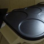
Knee brace- an orthopedic device designed to immobilize the knee in the treatment of injuries or diseases of the musculoskeletal system. Knee pads are made from different materials, providing a wide range of applications and a degree of protection. Such devices can be used not only for therapeutic, but also for preventive purposes. They are included in the equipment of professional athletes in order to protect the knee joint in the event of traumatic situations.
Purpose of knee brace
The main purpose of the knee brace is to fix the joint in the correct position, reduce the load on the knee and reduce pain associated with articular pathologies. Such devices not only unload the joint, but also provide a compression, massage and warming effect. Clamps (bandages) on the knee are recommended to be worn in the following cases:
- diseases of the ligamentous apparatus of the knee joint (arthrosis, gonarthrosis);
- knee injuries (stretching or rupture of ligaments, meniscus damage, dislocation, fracture);
- joint instability associated with weakness of the ligamentous apparatus;
- intense pain syndrome degenerative changes in the tissues of the joint;
- rheumatoid arthritis;
- the rehabilitation period after the surgical intervention on the knee joint.
More prone to knee injuries professional athletes and people leading an active lifestyle. Therefore, they are advised to wear knee pads as a preventive measure, which allows them to evenly distribute the load on the joint and prevent its injury.
In addition, orthopedists advise the use of knee braces for certain groups of patients. For example, women in late pregnancy, people suffering from obesity or representatives of certain professions who experience high loads when lifting weights.
Properties of knee braces

Orthopedic products for the knee joint bring tangible benefits, which are as follows:
- wearing a knee brace can reduce the severity of edema and pain;
- provides a warming effect, improves blood circulation and nutrition of joint tissues;
- allows you to lead full life, improving the mobility of the knee joints;
- stimulates metabolic processes in the affected area;
- evenly distributes the load, securely fixes the patella, without restricting the freedom of movement of the limb;
- accelerates recovery after surgeries and injuries;
- prevents re-injury to the joint.
Almost all diseases affecting the musculoskeletal system require an integrated approach. Treatment of arthrosis, arthritis and other inflammatory and degenerative joint lesions is long-term, requiring patience and strict adherence to medical recommendations from the patient. In the treatment regimen, in addition to medications, physiotherapy and physiotherapy exercises necessarily include the wearing of orthopedic structures, which allows you to restore the function of the joint and speed up recovery.
Types of knee pads
Orthopedic products designed to fix the knee joint are divided into several types:
Knee pads with a light degree of fixation (trapes, bandages, calipers)

They are made from natural elastic materials, or from fabrics combined with high-quality synthetics.Elastic knee braceare used for minor knee injuries (bruises, sprains), after surgical operations, or are recommended for wearing in professional sports to reduce excessive stress on the joint.
Such knee pads are characterized by good air and moisture permeability. They are made of modern, durable and high-quality materials that provide orthopedic products with a long service life, a high degree of wear resistance and do not cause allergies. Today, a wide range of elastic bandages are produced to provide stability and fixation of the knee joint. Natural materials additionally have medicinal properties- warm, improve blood circulation in the diseased joint, preventing further development of the inflammatory process.
The most popular elasticknee bracefrom neoprene. It is easy and reliable fixation, long service life and ease of operation. It can be washed by hand and dried naturally. The most convenient models are detachable bandages, which are interconnected with Velcro. Such elastic bandages can be direct fixation, lateral (located on the sides of the knee) or provided with stiffeners that run along the kneecap in a spiral.
Semi-rigid knee orthoses

Such orthopedic structures consist of a splint, metal hinges and fasteners that allow you to adjust the degree of fit of the brace to the knee. Orthoses are used for lateral and direct fixation of the joint, they provide reliable protection of the knee, without interfering with the free movement of the leg.
Semi-rigid orthoses help to quickly recover from surgery, fractures (after removal of the cast), dislocations of the patella and other knee injuries. Such designs are recommended to be worn for arthritis, bursitis, gonarthrosis, Osgood-Schlatter disease. In addition, semi-rigid orthoses perfectly protect the joint during intense physical exertion.
Semi-rigid knee pads can have a fabric or neoprene base, silicone side plates and inserts, and other additional devices and straps that allow you to securely fix the joint. They can be washed in cold water, after removing the removable elements and fastening the fasteners. Dry the product away from heating devices.
Rigid fixation knee pads (braces)

Rigid orthopedic structures securely fix the knee, simultaneously covering the upper part of the thigh and lower leg. The knee pad made of polymer fabrics or leather is equipped with silicone rings, side plates or metal hinges, and may have different shape, size and configuration. The fixation of the structure on the leg is carried out with the help of special belts or lacing. Tutor performs the following functions:
- securely fixes the knee joint during rehabilitation and treatment;
- in case of injuries, replaces the plaster splint or splint;
- due to immobilization of the joint relieves pain;
- prevents further progression of the disease;
- prevents recurrence of articular pathologies.
Rigid structures should be lightweight, made of hypoallergenic, durable materials with a high degree of wear resistance, provide reliable fixation of the joint during round-the-clock use. Such products are designed for long-term wear, so they are made detachable so that the splint can be removed when performing medical or hygiene procedures.
Other types of knee pads
Heated knee pads are a separate group. These products are in high demand, despite the rather high cost. Now they produce bandages equipped with sources of infrared or halogen light, which provide deep heating of the diseased knee, remove pain and improve joint mobility. Knee pads made of animal hair (dog, sheep, camel) have a good warming effect.
In case of arthrosis of the knee joint, it is recommended to wear magnetic knee pads, which help to restore the working capacity of the diseased knee by improving blood supply and activating metabolic processes. Magnets inside the knee pad relieve overload from the muscles and ligaments and help to properly distribute the load on the knee joint.

Choice sports knee bracedepends on the intensity of the loads, the type of sport, the reliability and ease of use of the structure. With average physical activity, it is recommended to choose elastic bandages, calipers, neoprene knee pads. With regular sports and increased loads on the knee joint, professional bandages are preferred.
For athletes and extreme sportsmen, knee bandages, supplemented with a patellar ring, are best suited. This design does not slip even under the most intense loads, since the silicone parts placed inside the knee pad are responsible for reliable grip. The lateral fixation of the joint in the products is provided by a spring design, and the silicone ring protects the patella from injuries and strong impacts. Additional fixation is achieved through a special compression bandage.
The materials from which light fixing devices are made are characterized by increased strength and elasticity, well remove moisture, preventing skin irritation. It is recommended to wear such knee pads no more than 8 hours a day.
Sports enthusiasts prefer Fosta neoprene braces with silicone inserts under the knee, knee padsVariteks, which are suitable for surfing and swimming, or orthoses with a warming effect from Pharmacels, which securely fix the joint and prevent sprains during active training.
In professional sports, special sports fixators are used to protect the knee from injuries to the meniscus and ligamentous apparatus; when doing power sports, it is recommended to wear orthoses with side inserts that immobilize the knee joint while maintaining its mobility.
Material for knee brace
Bandages used in the treatment of joint diseases are made from the following materials:
- Elastane or polyester. Modern materials, which are distinguished by strength, elasticity, good breathability, ease of use. However, the synthetic base of such knee pads does not provide a warming effect.
- Neoprene - bandages based on it are distinguished by the highest wear resistance, elasticity and long service life. They are easy to care for - just wash by hand in cool water and dry in the open air. Neoprene knee pads have only one drawback - the skin under them does not breathe, since the material does not allow air to pass through. Therefore, it is recommended to wear such bandages only during sports training, that is, 2-3 hours a day.
- Cotton is a natural material that does not cause allergic reactions, perfectly passes air and removes moisture, but does not differ in durability. To ensure elasticity, stretching fibers are included in the composition of the material. This is a lightweight version of the clamps, which are preferable to use in the warm season. Cotton retainers are easy to wash, dry quickly, but are not as durable as products made from modern synthetic materials.
- Wool - orthopedic fixators made from the wool of dogs and sheep have a healing effect, since they provide deep warming of the joint. Such bandages pass air and moisture well, but from frequent washing they quickly lose their original appearance and some of their healing properties.
How to choose a fixative?

The specialist selects an orthopedic fixator for the knee joint individually, taking into account the specific situation and the purpose of the bandage. The product must be selected according to size, degree of density, structural rigidity and type of material. The retainer should not interfere with the required range of motion, be comfortable to wear, but at the same time securely fix the knee.
It is best to seek help from a specialist - an orthopedist or traumatologist, who will select the optimal type of fixator. The size of the brace is determined by measuring the circumference of the joint above the knee, in the center of the patella and in the popliteal area.
The density of the bandage is selected taking into account the type of articular pathology or the degree of load during sports activities. After an injury, it is recommended to wear rigid fixatives that can replace a plaster cast. Such rigid knee pads are designed for long-term wear.
For arthrosis, elastic knee pads or semi-rigid orthoses are usually used to fix the joint in a certain position. It is recommended to wear them for several hours a day. It is very important that the orthopedic design for joint diseases meets all the requirements and ensures an even distribution of the load. Otherwise, an incorrectly selected fixator can aggravate the course of the disease and accelerate the course of irreversible processes leading to disability.
How much do fixators cost?
Knee braces, braces, orthoses and other devices for fixing the knee joint can be purchased in specialized departments of pharmacies or stores selling orthopedic goods. Mediumpricesfor simple fixators and soft bandages range from 800 to 2500 rubles. The cost of structures with silicone tabs varies from 4,000 to 10,000 rubles. The most expensive and complex orthoses cost about 40,000 rubles.
Causes: falling on the knee or hitting it with a hard object.
Signs: complaints of pain in the joint, difficulty walking. The damaged joint is enlarged in volume, its contours are smoothed, a bruise is sometimes visible under the skin on the anterior surface. Joint movements are difficult and painful. The accumulation of blood in the joint is determined by balloting the patella. If the amount of blood in the joint is insignificant, then by squeezing the joint with the palms of the hands from the sides, the symptom of patella balloting can be made more distinct. Hemarthroses of the knee joint sometimes reach a significant size (100-150 ml). In this case, the limb is half-bent, since only in this position the joint cavity reaches its maximum size. Be sure to produce x-rays of the joint in two projections.
Treatment. Patients with bruises of the knee joint with the presence of hemarthrosis are subject to treatment in a hospital. For mild bruises without accumulation of blood, it is possible to carry out ambulatory treatment with fixation of the joint with a tight bandage bandage. If fluid appears in the joint a few days after the injury, the limb should be fixed with a splint cast from the ankle joint to the upper third of the thigh until the fluid disappears.
In the presence of hemarthrosis, which sometimes develops several hours after the injury, first aid is to immobilize the limb transport bus from the toes to the upper third of the thigh. The victim is taken to the hospital in the supine position on a stretcher. Treatment of hemarthrosis of the knee joint is to puncture the joint and remove the accumulated blood in it. After that, the limb is fixed with a plaster splint. It can be removed after 4-5 days if fluid does not accumulate in the joint again. The patient can walk with crutches. After the termination of immobilization, exercise therapy and thermal procedures, massage are prescribed.
Sometimes, with a sharp twisting of the leg in the knee joint, the same hemarthrosis can develop, as with a bruise, although there was no bruise of the joint as such. In these cases, probably due to uncoordinated tension of the quadriceps femoris and the displacement of its tendon relative to the condyles, ruptures of the synovial membrane of the joint occur. Symptoms of damage to the ligamentous apparatus of the joint in such cases are absent. Treatment of such injuries is the same as for bruises of the joint.
KNEE MENISKI INJURY
Causes: a direct blow with the knee on a hard object or crushing the meniscus between the articular surfaces when jumping from a height. More often observed indirect mechanism damage. With a sharp uncoordinated flexion or extension of the leg in the knee joint with simultaneous rotation of it inward and outward, the meniscus does not keep up with the movement of the articular surfaces and is crushed by them. The meniscus associated with the joint capsule, when the articular surfaces are abruptly moved, breaks away from it, torn along or across, sometimes shifting into the intercondylar space (Fig. 1 1 4). Damage to the medial meniscus is observed 10 times more often than the lateral one.
Signs: pain and dysfunction of the knee joint. The leg in the joint is often bent and it usually cannot be straightened. In the future, hemarthrosis joins, and the clinical picture resembles a bruised joint. Typical circumstances of injury sharp pain in the area of the joint space, blocking of the joint in the bent position of the limb, recurrence of blockades make it possible to establish the correct diagnosis with a significant degree of certainty.
X-ray examination in case of suspected meniscus injury is mandatory to rule out other diseases and injuries of the knee joint. For more accurate X-ray diagnostics, air, liquid contrast agents, or both are injected into the joint. The development of deforming arthrosis, especially pronounced on the side of damage, can serve as an indirect sign of meniscus damage.
The use of arthroscopy in recent years has greatly improved the diagnosis and treatment of meniscal injuries.
Treatment. Puncture of the joint and removal of accumulated blood, followed by immobilization of the limb with a plaster splint bandage from the toes to the gluteal fold. The blockade is removed under local anesthesia with novocaine, which is injected into the joint cavity. The meniscus, pinched between the articular surfaces or displaced in the intercondylar space, is reduced by bending the leg at a right angle at the knee joint, pulling the lower leg along its length while simultaneously rotating it and moving it to the healthy side. Under these conditions, a gap is formed between the articular surfaces, and the meniscus is set into place.
Immobilization of the limb continues until the disappearance of hemarthrosis and subsidence of the phenomena of secondary synovitis, which takes an average of 10-14 days. Then thermal procedures, muscle massage and exercise therapy are prescribed. Usually after 3-4 weeks the patient can start working.
Early surgical treatment for fresh meniscal injuries is rarely performed and only in cases where the diagnosis is not in doubt. More often it is performed with repeated blockades of the joint. The operation is performed under conduction, local or intraosseous anesthesia. The damaged meniscus is removed completely or partially (only the torn part). After the operation, a plaster splint is applied for 7-10 days, followed by exercise therapy, massage and thermal procedures. Ability to work is restored after 6-8 weeks. With the help of arthroscopic technique, the traumatism of the intervention and the terms of disability are significantly reduced.
The most common combinations are: damage to the anterior cruciate ligament and one or two menisci (up to 80.5%); damage to the anterior cruciate ligament, medial meniscus and tibial collateral ligament ("ill-fated triad" - up to 70%); damage to the anterior cruciate ligament and tibial collateral ligament (up to 50%). The frequency of damage to the anterior cruciate ligament - 33-92%; posterior cruciate ligament - 5 - 12%; tibial collateral ligament - 1 9 - 7 7%; peroneal collateral ligament - 2 - 1 3%.
Causes: simultaneous flexion, abduction and external rotation of the lower leg (sharp, uncoordinated); flexion, abduction and internal rotation; hyperextension in the knee joint; direct blow to the joint.
Signs. General manifestations: diffuse pain, limitation of mobility, reflex muscle tension, effusion into the joint cavity, swelling of the periarticular tissues, hemarthrosis.
Diagnosis of injuries of the lateral ligaments. The main techniques are abduction and adduction of the lower leg. The position of the patient is on the back, the legs are slightly apart, the muscles are relaxed. The test is first carried out on a healthy leg (determination of individual anatomical and functional features). The surgeon places one hand on the outer surface of the knee joint. The other covers the foot and ankle area. In the position of full extension in the knee joint, the doctor gently abducts the lower leg, while slightly rotating it outward (Fig. 1 1 5). Then the technique is repeated in the position of leg flexion up to 150-160°. Change in the axis of the damaged limb by more than 10-15° and expansion of the medial articular
gaps (on radiographs) of more than 5 - 8 mm are signs of damage to the tibial collateral ligament. Expansion of the joint space by more than 10 mm indicates concomitant damage to the cruciate ligaments. Double testing (in the position of full extension and flexion to an angle of 150-160°) allows you to navigate in the predominant damage to the anteromedial or posteromedial part of the medial collateral ligament.
Identification of damage to the peroneal collateral ligament is carried out similarly with the opposite direction of the load forces. In the position of full extension, the peroneal collateral ligament and the biceps tendon are examined, in the position of flexion up to 160°, the anterolateral part of the articular capsule, the distal part of the ilio-tibial tract. All these formations provide stability to the knee joint, which is disturbed if even one of them is damaged.
Diagnosis of cruciate ligament injuries.
Drawer front test: position of the patient on the back, the leg is bent in hip joint up to 45 ° and in the knee - up to 80-90 °. The doctor sits down, presses the patient's forefoot with his thigh, covers the upper third of the shin with his fingers and gently jerks several times in the anteroposterior direction (Fig. 116): first without rotation of the shin, and then with external rotation of the shin (behind the foot) up to 15 ° and internal rotation - up to 25-30°. In the middle position of the lower leg, the stabilization of the knee joint is mainly (up to 90%) carried out by the anterior cruciate ligament. A displacement of 5 mm corresponds to I degree, by 6-10 mm - II degree, more than 10 mm - III degree(i.e., complete rupture of the anterior cruciate ligament). During rotation of the lower leg, additional damage to the lateral ligamentous structures of the knee joint is determined.
Lakhman test (1976): the position of the patient on the back, the leg is bent at the knee joint up to 160 °. The doctor covers the lower third of the thigh with his left hand, palm right hand, brought under the upper third of the lower leg, gently and smoothly pulls the lower leg anteriorly. At positive test in the area of retraction of the own ligament of the patella, a bulge appears from excessive displacement of the lower leg relative to the condyles of the thigh.
I degree - the displacement of the lower leg is felt only by the patient ("proprioceptive feeling").
II degree - visible displacement of the lower leg anteriorly.
III degree - passive subluxation of the lower leg posteriorly in the position of the patient on the back.
IV degree - the possibility of active subluxation of the lower leg
(the occurrence of subluxation with muscle tension).
Makintosh test (1972)- detection of excessive rotation of the lower leg in case of damage to the anterior cruciate ligament. The position of the patient on the back, the leg at the knee joint is extended. The doctor grabs the foot with one hand and rotates the tibia inwards, with the other hand exercises a load from the lateral side on the upper third of the tibia in the valus direction, while slowly bending the tibia at the knee joint. When the anterior cruciate ligament is damaged, subluxation of the lateral condyle occurs; when the lower leg is bent to 160-140°, this subluxation is suddenly reduced due to the posterior displacement of the ilio-tibial tract. Valgus load on the knee joint accelerates the reduction of dislocation. In this case, the doctor has a feeling of push. The absence of such a sensation indicates a negative test result (the cruciate ligament is not damaged).
The diagnostic capabilities of tests are most effective in chronic injuries of the cruciate ligaments. The Lachman test is the most sensitive, and with fresh injuries of the knee joint, its diagnostic efficiency reaches 90%.
When the posterior cruciate ligament is damaged, the symptom of the "posterior drawer" is revealed, which is more pronounced in acute period and may disappear over time.
Joint effusion is an important symptom of ligament injury. It is necessary to specify the rate of formation and the severity of the effusion. Hemorrhagic effusion indicates damage to the ligaments, paracapsular part of the meniscus, synovial membrane. The appearance of effusion after 6-12 hours or on the 2nd day is more often associated with the development of post-traumatic synovitis and indicates a predominant damage to the menisci. With the development of hemarthrosis in the first 6 hours and its volume of more than 40 ml, a diagnosis of serious intra-articular damage to the capsular-ligamentous apparatus should be made, even without pronounced symptoms of knee joint instability. Clarifies the diagnosis of arthroscopic examination (up to 96%).
Treatment. With conservative treatment, after puncture of the joint and removal of accumulated blood, the limb is fixed with a deep plaster splint from the fingers to the upper third of the thigh for 3 weeks. After the plaster bandage has dried, UHF therapy is prescribed, then, after immobilization has ceased, massage, exercise therapy and thermal procedures are prescribed. In the future, if the failure of the ligamentous apparatus is revealed, surgical treatment is undertaken.
Surgical treatment in the early stages is indicated for complete damage to the ligaments. Several U-shaped sutures are applied to the torn capsule and ligament. When the ligament is torn from the bone, a transosseous suture is used. In case of defibration, defect, chronic damage, auto- or alloplasty of the ligaments is performed (Fig. 117).
After the operation, the limb is fixed with a circular plaster bandage with a flexion angle in the knee joint of 140 - 160° for 4-6 weeks, followed by thermal procedures, exercise therapy and muscle massage. Ability to work is restored after 3 months,
117. Variants of plasty of the anterior cruciate and collateral ligaments of the knee joint.
INJURY OF THE BUCKET AND PATELLET LINING TENDON
Causes. The extensor apparatus of the knee joint (quadriceps femoris tendon, patella and its ligament) is damaged as a result of a sharp tension of the thigh muscle or from direct trauma when hitting or falling on one or both knees.
Signs: pain on the anterior surface of the thigh and knee joint, instability of the damaged limb, which, as it were, gives way due to loss of function of the quadriceps femoris muscle. Active extension of the leg in the knee joint is impossible. When pressing with the ends of the fingers along the extensor apparatus, one can feel a drop above or below the patella (especially with active tension of the quadriceps femoris muscle). On radiographs of the knee joint, if the tendon of the quadriceps muscle is damaged, the patella remains in place or slightly shifts downward, and with complete damage to the patellar ligament, the latter is significantly shifted upward.
Treatment. Partial injuries of the extensor apparatus are subject to conservative treatment. The limb is fixed with a circular plaster splint from the ankle joint to the gluteal fold with full extension of the leg in the knee joint. After 4 weeks, the bandage is removed, exercise therapy and thermal procedures are prescribed.
With complete damage to the extensor apparatus, surgical treatment is indicated: the imposition of strong U-shaped silk sutures on the damaged tendon, auto- or alloplasty with the wide fascia of the thigh or tendon grafts. After the operation, the limb is fixed with a plaster splint from the ankle joint to the gluteal fold for 2 months. Subsequently, thermal procedures, muscle massage, active and passive exercise therapy are carried out. Ability to work is restored in 3-3 1/2 months after the operation.
FRACTURES OF THE PATELLUM
Causes: hitting the knee or falling on it. Almost all patella fractures are intra-articular. Only fractures of its lower pole can be extra-articular. The degree of divergence of fragments depends on damage to the lateral tendon sprain of the extensor apparatus of the knee joint. With significant ruptures, the proximal fragment is displaced upward by the traction of the quadriceps femoris muscle. If the extensor apparatus was not significantly damaged, then there may be no displacement of the fragments or it may be insignificant (Fig. 1 1 8).
Signs: the contours of the joint are smoothed, in its cavity a free fluid is determined - hemarthrosis. Expressed instability in the knee joint.
With simultaneous damage to the lateral extensor apparatus, active extension of the lower leg is impossible, the patient cannot keep the extended leg on weight. At the same time, she slides along the plane of the bed without breaking away from it (symptom of "stuck heel"). On palpation of the patella, it is usually possible to feel the fracture gap or the ends of the dispersed fragments. It should be remembered that sometimes the impression of failure is created even with an intact patella, when blood accumulates in the prepatellar bag.
An x-ray examination of the knee joint in two projections is necessary even with a clear clinical picture of a patella fracture in order to exclude other injuries. Be sure to make an additional x-ray in axial projection. At the same time, the patient is laid on his stomach, the injured leg in the knee joint is bent at a right or sharp angle. The cassette is placed under the knee, and the central beam is directed obliquely at an angle of 45° to the cassette from the side of the lower pole of the patella. At the same time, longitudinal fractures of the patella are revealed, which are invisible in the pictures in ordinary projections.
Treatment. For fractures without displacement or displacement of fragments by several millimeters (which indicates the preservation of the integrity of the extensor apparatus), treatment should be conservative. It consists in puncturing the joint and removing the accumulated blood, followed by immobilization of the limb with a deep plaster splint from the fingers to the gluteal fold.
The puncture of the joint should be performed in the first hours after the injury, since the blood in the joint with fractures, unlike bruises, quickly coagulates. Cold is prescribed locally, and on the third day - UHF therapy. 5-7 days after the edema subsides, the longet bandage is replaced with a circular plaster
a splint from the ankle joint to the upper third of the thigh, in which the patient can walk with support on the diseased limb. Further treatment carried out in the clinic. After 3-4 weeks, the splint is removed. Exercise therapy, massage, thermal procedures are prescribed.
In fractures with displacement of fragments, surgical treatment is indicated. It should also be undertaken in cases where the congruence of the articular surfaces of the fragments is disturbed, although the fragments themselves may not be displaced along the length. The operation is performed under local anesthesia or anesthesia. To connect the bone fragments, a double semi-purse-string suture is used (Fig. 119). It is necessary to impose additional sutures on the lateral extensor apparatus. Thick silk threads are used as suture material. At comminuted fractures, especially when crushing one of the fragments, it is permissible to remove the crushed part of the patella with the restoration of the extensor apparatus of the joint. Screws, knitting needles, wire cerclages, external fixation devices are also used to fasten fragments (Fig. 120-121).
After the operation, the limb is fixed with a splint plaster bandage up to the upper third of the thigh. After 10-12 days, the sutures are removed and the longet bandage is replaced with a plaster splint, in which the patient can walk with full load on the sore leg. After 4-5 weeks after the operation, the plaster bandage is removed, exercise therapy, massage and thermal procedures are prescribed. Ability to work is restored after 2-2 3/2 months.
118. Variants of fractures of the patella. a - the norm; 6 - subaponeurotic fracture; c - fracture with partial damage to the extensor structures; d - fracture with complete
119. Sh about in the patella.
120. Internal (fixation of fractures of the patella.
121. External fixation of fractures by rupture of the extensor apparatus. patella.
DISTRUCTIONS OF THE PATELLA
Causes: a fall on the knee joint or a sharp tension of the quadriceps femoris muscle with simultaneous abduction of the lower leg outward. The inner part of the fibrous capsule of the joint is torn, and the patella is displaced to the outer surface of the joint by the force of impact or traction of the extensor apparatus. Dislocation of the patella is facilitated by the valus installation of the lower leg of a congenital nature, as well as underdevelopment of the external condyle of the femur. Sometimes dislocations become habitual, arise from a little violence and are easily reduced by patients without the help of medical workers.
Signs: typical displacement of the patella on the outer surface of the joint, half-bent position of the lower leg, movement in the joint is impossible. The patella is palpated to the side of the lateral condyle of the thigh, the tendon of the quadriceps muscle and the patellar tendon are sharply strained. The diagnosis is confirmed by X-ray examination.
Treatment. The reduction of the dislocation is performed under local anesthesia. The leg is fully extended at the knee joint and
the patella is shifted into place with the fingers. After that, the limb is fixed for 2-3 weeks with a splint plaster bandage in the extension position in the knee joint. Subsequently, exercise therapy, massage and thermal procedures are prescribed. Ability to work after traumatic dislocation is restored after 4-5 weeks.
With frequent habitual dislocations of the patella, surgical treatment is indicated.
122. Scheme of dislocations of the lower leg, a - anterior; 6 - back.
123. Immobilization of the knee joint with a plaster bandage.
2431 0
Various diseases and injuries of the knee joints bring a lot of problems. Not only do they require long-term treatment, but the knees also take a very long time to recover.
For the speedy recovery of the knee joints after injuries and during diseases, doctors recommend wearing special fixing devices - orthoses, splints and bandages.
These products provide reliable fixation of the diseased joint and accelerate the process of restoring its structure.
Applied clamps - their capabilities
All fixators for the knee joint are divided into several types, but the purpose of their use is common. They are designed to support and fix the damaged joint for preventive or therapeutic purposes.
The knee joint has a rather complex structure. It consists of three bones, three synovial bags, menisci and tendons. Due to the well-coordinated workflow of all these components, a person can perform all the necessary flexion and extension functions. However, sometimes as a result of injury or disease, the structural units of the joints can be damaged.
In these cases, various fixators are required to maintain the joints. These immobilizing devices include:
- bandages;
- orthoses;
- splints.
All these fixing structures have some distinctive features that you should pay attention to:

Varieties of designs
Consider the types of devices for joint immobilization.
By appearance
On sale there are the following types of fixing devices for the knee joint:
- Bandages- an elastic support device that can have different compression and density. It is used to improve the structure of the joint, normalizes blood circulation, relieves swelling, inflammation.
- Orthoses- high-strength structures to support the knee joint. They provide a secure fixation of the knee. Applicable for various diseases joints and traumatic injuries.
- Splints- These are rigid devices made in the form of sleeves. They are attached to the knee joint with straps, lacing or hinged elements. Used for injuries and diseases of the knee joint.
The choice of product according to the degree of rigidity
According to the degree of rigidity, the products are of different types:
- elastic knitted material;
- light rigidity, made of metal plates;
- medium hardness, based on plastic or iron material;
- hinge structures.

Articulated orthosis Orliman knee
Features of each group of clamps:
- The knee brace has the most low rigidity, most often used for preventive purposes. They also produce bandages for the knee joint, which are additionally equipped with stiffeners.
- Orthoses with weak rigidity are based on metal plates, which are covered with natural knitwear. Additionally, products of this type are fitted with a mesh, so when worn, the skin does not feel discomfort and breathes completely.
- Products with medium hardness made of plastic or iron. The dense frame is covered with synthetic material on the outside. These designs allow you to fix the knee joint after severe injuries or with complications of articular diseases -,. Often these devices are made to order.
- Articulated orthoses and splints. This is the toughest products. They are used to completely immobilize the joint. These devices are used during severe injuries, in which case they replace the cast. In the design, in addition to a strong frame, there are belts, rollers, hinges.
By type of fixation
According to the type and strength of fixation, products are divided into the following types:

Knee soft orthosis GenuTrain
- Functional. These devices provide complete fixation and immobilization of the joint.
- dynamic or compression products with this type of fixation are used in periods after surgical interventions, injuries and for prevention during exercise.
- Stabilizer. Products with this type of latch have plastic and metal inserts. Often these devices replace tires.
- Stage-functional orthosis on the knee joint. This device has a rigid design, which is used to gradually increase the range of motion during joint rehabilitation.
Other types
Depending on the design features, the following types of products are distinguished:
- Articulated. This tire, the design of which is connected by special hinges. During use, provides full mobility of the knee joint.
- Hingeless. They look like bandages.
When certain products are used
Bandages with a soft elastic structure of the material are usually used as a preventive measure to restore the structure of the joint tissue and to maintain a fixed position of the knees in cases of heavy load on them.
These products can be worn after mild disorders, they are also used during various physical exercises to relieve increased stress on the joint.
The main indications for wearing orthoses and splints are the following conditions:

Hard knee orthosis Medi
- during the rehabilitation period after traumatic injuries of the knee joint;
- after the procedure for;
- in the period after traumatic injuries and surgical interventions in the lower femur and upper leg bones;
- after traumatic injuries of the ligamentous apparatus of the knee joint;
- with hemorrhages in the joint area;
- during and after , ;
- with inflammation of the articular bag, synovial membranes, menisci;
- in the presence of congenital and acquired deformities of the lower leg and knee joint - with valgus or, with recurvation of the knee joint;
- in the presence of paralysis, including cerebral palsy, as well as paralysis after a stroke or sclerosis.
Rigid orthoses and splints are installed on the knee in such cases:
- after severe traumatic injuries, fractures, in the postoperative period. In these cases, these devices are used as plaster;
- with deformity of the knee joint and bones of the lower leg;
- can be used to ensure complete rest during acute and progressive articular diseases;
- with paralysis.
Structures with a rigid structure can be used as fixation devices when transporting a patient who has a suspected knee fracture.
Fixation devices, which have a moderate degree of rigidity, are usually used to support joints in the following conditions:
- during the rehabilitation period after traumatic injuries or illnesses;
- at .
Application for fractures during the rehabilitation period
During this period, fixing devices are used that provide complete restriction of movements. The use of orthoses during the rehabilitation period after fractures completely immobilizes the patient, thereby providing complete rest to the damaged area. Fixing the device is provided by special straps and hinges.
In any case, it is necessary to use any fixing devices strictly according to indications. pick up necessary product the attending physician should, in accordance with the degree of damage to the joint and the severity of the disease or injury.
Features of choice
Regardless of the type of design of the fixing product for the knee joint, a number of features must be taken into account during its selection, from which determine the quality and durability of the immobilizer.
What features you need to pay attention to first of all:
- good degree of fixation;
- ease of construction;
- high wear resistance;
- convenient adjustment in length and width;
- the base must be made of safe materials;
- high hygiene;
- size range, it is best to measure the product immediately, it must exactly fit the size of the foot;
- the structure of the structure must be breathable.
Use fixing products for about three weeks. Usually the course of application is determined by the attending physician. Also, a specialist can choose a course of exercises that will help develop the joint.
Operating rules:
- before putting on products, you should check the skin - it should not have wounds and damage;
- orthoses, bandages and splints can be worn directly on bare skin;
- wear products of this type should be on the lower limb, then fix on the required area of \u200b\u200bthe leg;
- sometimes doctors may recommend wearing thin leggings or underpants under the retainer.
With regard to care, each orthopedic product is accompanied by special instruction, which gives detailed description operating rules. Its observance will significantly extend the life of the product. 
Basic care rules:
- knee pads are not recommended to be washed in a washing machine, these products should only be washed by hand;
- dry in the unfolded state on a flat surface;
- it is not recommended to stretch, hang on batteries or in the sun;
- when washing, do not use various chemical detergents, only baby soap;
- ironing is not recommended.
Editor's Choice
After conducting a market review, as well as having studied numerous reviews, we have selected the 10 best orthoses, bandages and splints for the knee joint:

Word to consumers
The point of view of people who used the knee splint and brace.
After a severe knee injury, the doctor recommended wearing a splint. He said that this device would help the quick recovery of the joint structure. I chose the Fosta articulated splint. I liked the product, the device is firmly fixed on the knee, it does not cause difficulties during movements. There was no swelling or irritation during wearing. My knee recovered quickly and there were no complications.
Maria, 29 years old
Often have to perform heavy exercises in the gym. Lately, I've been noticing pain in my knees. I immediately decided to consult a doctor, you never know.
The doctor recommended using special bandages for the knee joint. I chose the Genumedi model. The product is excellent - it is firmly fixed, relieves stress and overvoltage. The main thing is all the pain and discomfort gone!
Oleg, 48 years old
price issue
The cost of devices for immobilization is different, it all depends on the degree of rigidity and fixation. Bandages for prevention can be purchased for a price of 600 to 5,000 rubles, orthoses and splints for the knee joint are much more expensive, on average, such products cost from 5,000 to 50,000 rubles.