Vestibular reflexes and their characteristics. Vestibular pathways and reflexes. The structure of pathways and centers of the vestibular system
Neurons of the vestibular nuclei provide control and management of various motor reactions. Vestibulospinal influences change the impulse of neurons at segmental levels of the spinal cord. This is how the skeletal muscle tone is dynamically redistributed and the reflex reactions necessary to maintain balance are activated. The vestibulo-vegetative reactions involve the cardiovascular system, gastrointestinal tract and others internal organs. With strong and prolonged stress on the vestibular apparatus, motion sickness occurs (for example, motion sickness). Vestibulo-oculomotor reflexes (ocular nystagmus) consist of a slow rhythmic movement of the eyes in the opposite direction to rotation, followed by a jump back. The occurrence and characteristics of rotational ocular nystagmus are important indicators of the state of the vestibular system and are widely used experimentally and clinically.
End of work -
This topic belongs to the section:
Fundamentals of psychophysiology: Textbook / Ed. ed. Yu.I. Alexandrov
On the website read: 075 basics of psychophysiology: textbook / resp. ed. Yu.I. Alexandrov. - M.: infra-m, 1997. - 349 pp...
If you need additional material on this topic, or you did not find what you were looking for, we recommend using the search in our database of works:
What will we do with the received material:
If this material was useful to you, you can save it to your page on social networks:
| Tweet |
All topics in this section:
General information
Traditionally, since the time of the French physiologist Bichat (early 19th century), the nervous system has been divided into somatic and autonomic, each of which includes structures of the brain and spinal cord.
Neuron. Its structure and functions
The human brain consists of 1012 nerve cells. An ordinary nerve cell receives information from hundreds and thousands of other cells and transmits it to hundreds and thousands, and the number is connected
Size and shape
The sizes of neurons can range from 1 (the size of a photoreceptor) to 1000 μm (the size of a giant neuron in the marine mollusk Aplysia) (see [Sakharov, 1992]). The shape of the neurons is also exceptional
Neuron color
Next external characteristic nerve cells are their color. It is also varied and can indicate the function of the cell - for example, neuroendocrine cells are white. Yellow
Synapses
The biophysical and cell biological approach to the analysis of neural functions, the possibility of identifying and cloning genes essential for signaling, revealed a close connection between
Electrical excitability
All functions inherent in the nervous system are associated with the presence of structural and functional features in nerve cells that provide the ability to generate under the influence of external
Pacemaker
One of the surprising types of electrical activity of neurons recorded by an intracellular microelectrode is pacemaker potentials. A. Arvanitaki and N. Chalazonitis. Chemoreceptors inform the central nervous system about changes in chemistry
Pathways and centers of the visceral sensory system
The pathways and centers of the visceral sensory system are represented mainly by the vagus, splanchnic and pelvic nerves. The vagus nerve transmits afferent signals to the central nervous system through thin
Visceral sensations and perception
Excitation of some interoceptors leads to the emergence of clear localized sensations, i.e. to perception (for example, when stretching the walls Bladder or rectum). IN
Basic quantitative characteristics of human sensory systems
Near point of clear vision 10 cm Diameter of the retinal macula about 0.5 mm (1.5–2 angular degrees) Accommodative power about 10 diopters (D)
Motion Control
Movement (including speech and writing) is the main means of interaction between the human body and the environment. In this interaction, reflex responses, stimulated by environmental stimuli, with
General information about the neuromuscular system
It is impossible to understand the principles of operation of the control system without knowing the structural features of the control object. In relation to the movements of animals and humans, the object of control is the
Proprioception
For the successful implementation of movements, it is necessary that the centers that control these movements at any time have information about the position of the body parts in space and about
Central motion control devices
Almost all parts of the central nervous system are involved in controlling movements - from the spinal cord to the cerebral cortex. In animals, the spinal cord can carry out a fairly extensive function
Motor programs
Movement control is unthinkable without activity coordination large quantity muscles. The nature of this coordination depends on the motor task. So, if you need to take a glass of water,
Coordination of movements
The concept of coordination of movements arose on the basis of observations of patients who, for various reasons, are not able to smoothly and accurately carry out movements that are easily accessible to healthy people.
Types of movements
Human movements are very diverse, but all this diversity can be reduced to a small number of basic types of activity: ensuring posture and balance, locomotion and arbitrariness
Development of motor skills
Improvement of motor function in ontogenesis occurs both due to the maturation of innate mechanisms involved in the coordination of physical activity, which continues in the first years after birth.
Psychophysiology of memory
Learning can be viewed as a sequence of complex processes involved in the acquisition, storage and reproduction of information (see Chapter 15). As a result of learning,
Temporal organization of memory
The temporal organization of a memory trace implies a sequence of development over time of qualitatively different processes leading to the recording of acquired experience. Basic concepts to
Gradient of retrograde amnesia
The dependence of the efficiency of memory modulation on the time interval between training and the use of an amnestic agent characterizes the gradient of retrograde amnesia. hail
Stages of memory fixation
Hypothesis of two sequentially developing traces. According to the hypothesis, the formation of an engram is carried out in two stages: the first is characterized by an unstable form
Short-term and long-term memory
The development of the concept of temporary organization of memory is hampered by the vagueness and vagueness of the basic concepts on which the theoretical structure is based. The concept of brief
Spontaneous memory recovery
Facts about spontaneous memory recovery after amnestic electric shock were known back in the 50s. (see in [Grechenko, 1979]). Skill recovery was reported after
Restoring an engram with a second electric shock
Many researchers have reported the restorative effect of a secondly presented combination of “punishment (stimulus used during learning) - electric shock” (see in)
Restoring memory using the reminder method
In the work of R.J. Koppenaal et al. were the first to use the “reminder” method. It consists in the fact that before testing the retention of a skill, animals are presented
Memory restoration using the familiarization method
If, before training, the animal is placed in an experimental chamber and given the opportunity to move freely around it, then after the use of an amnestic agent, retrograde amnesia will occur.
Retrograde amnesia for reactivated memory traces
It has been discovered that retrograde amnesia can be caused after memory has passed into long-term storage. The experiments studied the effect of electric shock on a skill formed several days ago.
The main provisions of the theory of active memory
The main provisions of the concept of active memory are as follows. Memory acts as a single property, i.e. there is no division into short-term and long-term
Engram Distribution Hypothesis
Experiments with local stimulation of the brain have shown that the development of retrograde amnesia upon stimulation of a certain structure depends on the time interval that has passed from the moment the
Engram distribution in experiments with local brain stimulation
Studies performed using electric shocks, which cause the development of electrical convulsive activity, show the complex dynamics of the movement of the active engram along
Distribution of an engram over many brain elements
The idea that a memory trace does not have a specific localization, but is read from neurons in different brain structures depending on the circumstances, has been confirmed by experiments. F
Procedural and declarative memory
Recently, the concept of a multiplicity of memory systems has become increasingly important. This idea was formed on the basis of data obtained during research
Molecular mechanisms of memory
In neuroscience, research into the mechanisms of learning and memory is conducted primarily in the context of plasticity (see Chapter 15). That is why many studies were aimed at identifying
Discreteness of mnemonic processes
Letters of the alphabet, atoms and molecules are all codes for important entities, the significance of whose discoveries cannot be overestimated. The first was the discovery of hieroglyphs and the alphabet. ABC
Constant Livanov
There are many empirical relationships called laws. Examples include the basic psychophysical law, which establishes the dependence of the strength of sensation on f
Memory capacity and speed
If you follow the logic of D. Hartley, A.A. Ukhtomsky, N.G. Samoilova, M.N. Livanov, G. Walter, E.R. John, K. Pribram and other supporters of the idea of dynamic coding perceive
Range of sensations
In psychophysics, Weber's fraction defines the boundary between the sensible and the intangible. This is a barely noticeable subjective increase in sensation, taken in relation to the initial value of the stimulus. I'm leaking
Neural memory codes
Neural memory codes are cyclically repeating waves of impulses generated by neural ensembles. The duration of one cycle is about 100 ms. Note that according to the data
Emotion as a reflection of an actual need and the likelihood of its satisfaction
William James, the author of one of the first physiological theories of emotion, gave his article, published more than 100 years ago, a very expressive title: “What is an emotion?” [
Brain structures that implement reinforcing, switching, compensatory-replacing and communicative functions of emotions
The results of neurophysiological experiments show that needs, motivations and emotions have different morphological substrates. Thus, when stimulating self-irritation zones later
Individual characteristics of the interaction of brain structures that implement the functions of emotions as the basis of temperaments
As methods for diagnosing stable (typological) individual characteristics of behavior, we used two behavioral models: the probability preference test, or value
The influence of emotions on activity and objective methods of monitoring a person’s emotional state
The very fact of generating emotions in a situation of pragmatic uncertainty predetermines and explains their adaptive compensatory value. The fact is that when an emotion arises
Determination of functional status
Most often, the functional state (FS) is defined as the background activity of the nerve centers, during which this or that specific human activity is realized. In classical
The role and place of the functional state in behavior
Functional states regulated by the brain's modulating system are a necessary component of any type of activity and behavior. The relationship between activation level is well studied
Brainstem-thalamo-cortical system
The studies of J. Moruzzi and G. Magun led them to the discovery in the trunk at the level of the midbrain of a nonspecific system, or mesencephalic reticular formation, activating the cortex
Basal forebrain cholinergic system
Recently, it was shown that magnocellular ACh-containing neurons located in the basal forebrain project monosynaptically to the cortex. They are teaching
Caudo-thalamo-cortical system
The basal ganglia are also involved in regulating the level of activity of the body. Their other name is the striopallidal system, which is a complex of neuronal nodes, c
Modulating neurons
In the nervous system, a special group of cells is identified - modulating neurons, which themselves do not cause a reaction, but regulate the activity of other neurons. They form contacts with others
What is attention
Rhetoric occupied a large place in the life of the ancient Romans and Greeks. As an art and as a medium, it was only effective if the speakers were well-executed.
Filter theories
First theoretical model attention, or filter model, was created by D.E. Broadbent. He assumed that the nervous system, despite its many inputs, to some extent
The problem of attention in traditional psychophysiology
The general idea of the previously described models of attention is that the path of nerve impulses from receptors exposed to external stimuli to the cortex has
The problem of attention in systems psychophysiology
This paradox does not arise if we refuse to consider behavior as a reaction to presented stimuli. From the standpoint of systemic psychophysiology [Shvyrkov, 1995], behavior is real
Orienting reflex
Orienting reflex, or reflex “What is it?” was discovered by I.P. Pavlov. He described it as a complex of motor reactions that arose to the unexpected appearance of a new stimulus
Approximate research activities
The orienting reaction (as the tuning of analyzers for better perception of a new stimulus) should be distinguished from exploratory reactions and orienting-exploratory behavior.
Psychophysiology of consciousness
Consciousness is one of the most complex and at the same time mysterious manifestations of brain activity. Although the word “consciousness” is quite widely used in everyday speech
Basic Concepts of Consciousness
Speaking about the brain foundations of the psyche and consciousness, it must be said that these most complex manifestations of the work of the brain cannot be explained by the work of some separate, isolated
Excitation re-entry and information synthesis
The “bright spot” concept described earlier assumes that consciousness is determined by some level of excitability brain structures. However, it can be assumed that this is a disadvantage
Brain basis of sensations
Psychologists since the 20s. it is known that the sensation occurs quite late - after 100 ms from the moment the stimulus is presented (i.e., much later than the arrival of sensory impulses in the cortex). IN
Mechanisms of thinking
Our research over the past ten years has been devoted to answering this question. The aim of the work was to study the structure of cortical connections when solving various mental operations.
Consciousness, communication and speech
The concept of the communicative nature of consciousness was first put forward by P. V. Simonov. Later, similar thoughts were expressed by other authors. According to Oprah
Functions of consciousness
The question of the functional meaning of subjective experiences and their role in behavior is one of the most important problems in brain science. Representing the result of information synthesis, mental functions
The concept of the unconscious in psychophysiology
During human activity in a constantly changing environment, incoming information is processed at different levels of the central nervous system. Switching nervous
Indicators of conscious and unconscious perception
The problem of the experimental study of unconscious perception comes down to attempts to identify the threshold difference between two indicators: one of them is an indicator of awareness of the stimulus; d
Semantic differentiation of unconscious stimuli
For the first time in an experiment, the phenomenon of unconscious perception of verbal stimuli was reproduced by a group of New Look psychologists. In healthy people (students), the threshold is
Temporary connections (associations) at an unconscious level
From the observations of psychiatrists it is known that in certain cases unconscious external signals, if they once or several times coincided with a strong negative emotional
Functional asymmetry of the hemispheres and the unconscious
The classic work of R. Sperry and his colleagues on people with “split brain” opened the way for neuropsychological experimental research into functional
Reverse temporal connections and the unconscious
6.1. The role of temporal feedback in the nervous mechanism of “psychological defense” One of the forms of psychological defense is expressed in an increase
The importance of unconscious feedback stimuli in cognitive activity
Many researchers have written about the influence of unconscious stimuli on cognitive functions [Kostandov, 1983; Velmans, 1991], although unambiguous results were not always described. This effect
The role of the unconscious in some forms of pathology
The formation of a conditioned reflex to unconscious external stimuli explains the nervous mechanism of spatial orientation of blind people [Beritashvili, 1969]. Significant role of sound
Sleep and dreams
1. ACTIVE SLEEP INJECTION OR AWAKE DEPRIVATION? Already in the earliest studies of sleep mechanisms, two main points are clearly identified
Stages of slow-wave sleep and REM sleep
The main findings from years of numerous and varied sleep studies are as follows. Sleep is not a break in brain activity, it is simply a different state. In time
Sleep in relation and phylogenesis
During ontogenesis, the sleep-wake ratio changes. Thus, in newborns, the state of wakefulness constitutes only a small part of the day, and a significant part of sleep is occupied by sleep.
Need for sleep
Many people would like to sleep less, since sleep, in their opinion, is time lost from life. Others, on the contrary, would like to sleep more because they do not feel well enough
Sleep deprivation
Experiments with deprivation (artificial sleep deprivation) suggest that the body has a special need for delta sleep and REM sleep. After prolonged sleep deprivation, the main
Dreams
Dreams have long amazed and worried people. In ancient times, dreams were seen as “the gateway to another world”; it was believed that through dreams contact with other worlds could occur
Two paradigms in the study of behavior and activity
With all the variety of theories and approaches used in psychology, psychophysiology and neuroscience, they can be divided into two groups. In the first of the groups as the main m
Reactivity
Using the principle of reactivity as an explanatory principle in scientific research is based on the ideas of Rene Descartes, outlined by him in the first half of the 17th century. Descartes believed that the body
Activity
Consideration of behavior and activity as activity directed to the future includes an understanding of activity as a fundamental property of living matter; specific form of manifestation a
Eclecticism in psychology and psychophysiology
Recently, the idea of the active, purposeful nature of human and animal behavior has become increasingly widespread. Along with positive consequences this
Functional systems theory
2.1. What is the system? The term "system" is usually used to indicate the collection, organization of a group of elements and
Time Paradox
How can a result (an event that will occur in the future) determine current activity and be its cause? The solution to this “time paradox” was the development of a pre-
Purposefulness of behavior
Already for Aristotle, the purposefulness of behavior was obvious. Thus, the idea of purposefulness cannot in any way be considered new, although a period can be distinguished in history when
Leading reflection
An analysis of the problems of the origin and development of life from the perspective of TPS led P.K. Anokhin to the need to introduce a new category: advanced reflection. Leading
Theory P.K. Anokhin as a holistic system of ideas
So, the first most important advantage and feature that distinguishes TFS from other options systematic approach, – introduction of the idea of the result of an action into the conceptual scheme. So about
System processes
As the key provisions of the reflex theory of P.K. Anokhin identified the following: a) the exclusivity of the trigger stimulus as a factor determining the action, which is its
Behavior as a continuum of outcomes
Until now, for didactic purposes, as well as following the tradition of the original version of TFS, we have used the concept of a trigger stimulus. However, it is clear that the use of this concept within
Systemic determination of neuron activity
3.1. The reactivity paradigm: a neuron, like an individual, responds to a stimulus. As we have already noted, from the standpoint of the reactivity paradigm, the behavior of individuals
Activity as a subjective reflection
Consideration of the relationship between the individual and the environment from the perspective of TPS has long led to the conclusion that the behavioral continuum is entirely occupied by the processes of organization and implementation of functions.
Physical characteristics of the environment and goal-directed behavior
More than 30 years ago, J. Lettvin et al., having studied the connection between the activity of frog retinal neurons and its behavior, formulated in a very vivid form their idea that
Dependence of the activity of central and peripheral neurons on the goal of behavior
Convincing examples of how the subjectivity of reflection manifests itself in the organization of brain activity can be obtained by analyzing the dependence of the activity of neurons “se” on the goals of behavior
The significance of efferent influences
The connection between the activity of retinal ganglion cells and behavior with eyes closed is due to the already mentioned efferent influences. Even at the beginning of this century, S. Ramon y Cajal
Psychophysiological problem and tasks of systemic psychophysiology
In this paragraph we will answer the questions listed below. How do the tasks of psychophysiology depend on methodological settings? Is there a specific task of psychotherapy among them?
Correlative psychophysiology
Traditional psychophysiological studies are carried out, as a rule, from the standpoint of “correlative (comparing) psychophysiology.” In these studies, mental
Systemic solution to a psychophysiological problem
The essence of a systemic solution to a psychophysiological problem lies in the following position. Mental processes characterizing the organism and behavioral act as a whole, and neurophysiologists
The tasks of systemic psychophysiology and its significance for psychology
Using the given solution to a psychophysiological problem in systemic psychophysiology as one of the most important components of the methodology allows us to avoid reductionism
Interaction of correlative and systemic psychophysiology
The philosophy of science affirms the usefulness of the coexistence of alternative theories, facilitating their mutual criticism and accelerating the development of science. A good example of justice
Systemogynesis
In the previous paragraph, when formulating the tasks of systemic psychophysiology, it is no coincidence that the first place was given to the task of studying the formation of systems. We will see further that the history of
Organogenesis and systemogenesis
In contrast to the concept of organogenesis, which postulates the gradual development of individual morphological organs that perform corresponding local “private” functions, the concept of sys
Learning as reactivation of developmental processes
It is now becoming generally accepted that many patterns of modification of functional and morphological properties neurons, as well as the regulation of gene expression lying in the
System specialization and system specificity of neurons
The specialization of neurons relative to newly formed systems – system specialization – is constant, i.e. the neuron is system specific. Currently
Historical determination of level organization of systems
Many authors have developed ideas about patterns of development in connection with the ideas of level organization (see [Anokhin, 1975, 1980; Rogovin, 1977; Alexandrov, 1989, 1995,
The structure of the subjective world and the subject of behavior
The specialization of neurons relative to the elements of individual experience means that their activity reflects not the external world as such, but the individual’s relationship with it (see also the pair
Dynamics of the subjective world as a change in the states of the subject of behavior
From these positions, the dynamics of the subjective world can be characterized as a change in the states of the subject of behavior during the unfolding of the behavioral continuum (see Fig.
Modifiability of the systemic organization of a behavioral act in successive implementations
Even F. Bartlett proposed to completely discard the views according to which “reproduction from memory” is considered as “re-excitation of unchanged “traces”, the appearance of a new element in the individual’s repertoire
Recording stages of learning in the form of elements of experience
Having outlined a fundamental approach to identifying the elements of subjective experience, the theory functional systems formed the basis for experiments studying the subjective fragmentation of behavior and
The influence of learning history on the structure of experience and the organization of brain activity
The subjective continuum, like the behavioral continuum, is a linear sequence of alternating states that correspond to acts of behavior. These changes
Event-related brain potentials
ERPs represent a wide class of electrophysiological phenomena that are isolated from the “background” or “raw” electroencephalogram (EEG) using special methods. The term BSC -
Brief history of the SSP method
The connection between electrical activity of the brain and events in the environment and behavior was first demonstrated and described by the Englishman Richard Caton in 1875–1887. and n
General signal characteristics
ERPs are isolated using special methods from EEG. The frequency range of the SSP includes a band from 0 Hz to 3 kHz and is limited, on the one hand, by the ultra-slow electrical activity of the brain.
Standard methods for obtaining a reproducible BSC configuration
Methodological requirements for EEG recording (installation of electrodes, choice of lead system, amplifier bandwidth, methods for eliminating artifacts) are described in Chapter 2. Note that
Averaging
The basis for isolating ERPs from the EEG signal is the following assumptions: a) in a situation of multiple repetitions of an event, the recorded EEG signal (SUMi (t)) is the sum
Filtration
The random, “noise” component of a single ERP implementation (“raw” EEG) can be eliminated through smoothing. ALGEBRAIC FILTERS
Description of BSC
As a result of the accumulation of EEG segments associated with certain events, their averaging, digital filtering or other procedures, an ERP curve is obtained, which is described as
Features of the SSP method
In recent decades, methods for recording brain activity have been developed that have significant research capabilities (see also Chapter 2). However, even when developing new m
Visual evoked potentials
Visual evoked potentials (VEP, visual evoked potentials - VEP) [Chagas, 1975; Rutman, 1979; Maksimova, 1982; Rockstroh et al., 1982] are recorded in the situation of visual presentation
Auditory evoked potentials
Auditory evoked potentials (AEP) [Chagas, 1975; Rutman, 1979; Rockstroh et al., 1982; Hughes, 1985] are recorded in a situation where rumors are presented
Somatosensory evoked potentials
Somatosensory evoked potentials (SSEPs; somatosensory evoked potentials, SEP) [Chagas, 1975; Rutman, 1979; Rockstroh et al., 1982] are recorded in a fur situation
Potentials associated with the execution of movements
Potentials associated with the execution of movements (PSVP, movement-related potentials - MRP, movement-related brain potentials - MRBP; in Russian-language literature using
Conditional negative wave
Conditional negative wave (CNV, contingent negative variation - CNV, or expectation wave, expectancy wave - E-wave). In a situation where two stimuli are presented, the first
Principles for organizing the phenomenology of BSC
The list of known types of BSC is constantly growing, and there is no reason to consider it close to completion. Let us give as examples the most famous phenomena
The problem of the functional meaning of ssp
Within the framework of correlative psychophysiology (see [Shvyrkov, 1995] and Chapter 14), it is assumed that ERP oscillations (components) reflect specific functions of brain structures that implement
Psychological correlates
The search for psychological correlates of ERP showed that: 1) the same ERP is associated with many psychological processes (functions) and 2) the same mental functions
The search for brain sources of ERP showed the following: 1) any fluctuation of ERP recorded from the surface of the head is a reflection of the activity of many cortical and subcortical
SSP as a reflection of the dynamics of individual experience
ERPs represent the total electrical potential of various components of brain tissue, the contribution of which is made by neurons (soma, dendrites and axons), glial cells, cell membranes
Flexible Configuration Potential
A comparison of ERPs accompanying the behavior of subjects in various experimental situations shows that the implementation and change of a behavioral act corresponds to the potential of a universal
Prospects for using SSP
The effectiveness of using ERP as a method of psychophysiological research is determined by solving the main problem: what is the ratio of ERP parameters, brain activity, physical function?
Nervous system properties concept
The problem of individual psychological differences between people has always been considered in Russian psychology as one of the fundamental ones. Greatest contribution to development
General properties of the nervous system and holistic formal-dynamic characteristics of individuality
To experimentally test the developed ideas about the properties of the nervous system and their psychological manifestations, V.D. Nebylitsyn conducted a study of the physiological foundations of intelligence
Integral individuality and its structure
The approach that was developed by B. S. Merlin formed the basis for the development of the original school for studying the nature of temperament. The starting points on which this approach was based
Individual behavioral characteristics of animals
An important trend that emerged in the development of the problem of the nature of individual psychological differences after B.M. Teplova and V.D. Nebylitsyn and was based on the behavioral model
Integration of knowledge about personality
At the end of the 80s. with the aim of forming a new strategy for studying the nature of individual psychological differences between people. M. Rusalov developed a survey-type methodology for ots
Cross-cultural studies of personality
IN last years in differential psychophysiology, the methodology of cross-cultural research began to be used. Cross-cultural differential psychophysiological research
Psychophysiology of professional activity
On the borders of natural sciences and psychology, a number of special scientific disciplines and areas, including labor psychology, engineering psychology and ergonomics, the object
Theoretical foundations for the use of psychophysiology to solve practical problems in occupational psychology
Understanding the need and prospects for studying psychophysiological processes in professional activities is facilitated by ideas that consider the mental and physiological
Methodological support for the psychophysiological aspect of applied research
In applied research, the complex nature of psychophysiological research is ensured by the use of a polyeffector method, including frequency registration
Psychophysiology of professional selection and professional suitability
The use of psychophysiological methods in occupational psychology was caused by the need to develop objective and quantitative criteria for psychophysiological selection, which is both
Psychophysiological components of performance
A person’s ability to perform specific activities within given time limits and efficiency parameters determines the content of performance as the main component
Psychophysiological determinants of human adaptation to extreme operating conditions
Currently, the main directions in the study of adaptation have become the determination of the stages of formation of the psychophysiological adaptation system, the criteria for its formation, the identification
Psychophysiological functional states (PFS)
The relevance of studying PPS is determined by their contribution to ensuring the efficiency and reliability of a person, as well as the increase in the number of professions and changes in working conditions
Biofeedback (boss)
Interest in biological research feedback(BFB) for the purpose of voluntary control of FS based on objective information about the dynamics of psychophysiological displays
Psychophysiological analysis of the content of professional activity
Psychophysiological analysis of professional activity involves “considering it as a complex, multidimensional and multi-level, dynamic and developing phenomenon” [Lomov,
Comparative psychophysiology
Comparative psychophysiology is a science aimed at establishing patterns and identifying differences in the structural and functional organization brain, behavior and psyche in animals
Emergence of the psychic
The most accepted point of view at present is that the psyche is an attribute of living systems, and inanimate nature, including complex devices created by people, is not about the psyche.
Evolution of species
Modern data on the evolution of animals indicate the divergence of evolutionary lines and the development of parallel lines (Fig. 19.2 A), including among mammals (Fig. 19.2 B), and among
Evolutionary transformations of the brain
Brain structure in animals different types varied. And although, as follows from Fig. 19.3, related species, for example among crustaceans or mammals, have common features in the building
Comparative method in systems psychophysiology
Systemic psychophysiology, the foundations of which were laid by the works of V.B. Shvyrkov and his colleagues, is based on the recognition of: 1) a single psychophysiological reality in which psychological
PHYSIOLOGY OF THE SENSE OF BALANCE, HEARING AND SPEECH
R. Klinke
This chapter is devoted to the physiology of two phylogenetically related sensory organs—hearing and balance. They are not only closely related anatomically, located side by side in the petrous bone and forming inner ear, but also originated in the course of evolution from the same structure. Since the most important means of communication for humans—speech—is mediated by the organ of hearing, physiology of speech also discussed in this chapter.
Speech requires hearing. In addition, verbal communication is the most important means of learning, so deafness or even just hearing loss pose the most serious threat to a child’s mental development. Comparative physiological studies have shown that deafness affects him more strongly than blindness. Therefore, hearing is the most important sense for a person.
12.1. Physiology of the sense of balance
Physiology of the peripheral sensory apparatus
Introductory anatomical comments . The vestibular organ is one of the components membranous labyrinth forming inner ear; its other component is the organ of hearing (Fig. 12.1). The membranous labyrinth is filled with fluid, endolymph, and immersed in another, called perilymph. The vestibular organ consists of two morphological subunits - the otolith apparatus ( macula utriculi and macula sacculi ) And semicircular canals (front And rear vertical And horizontal channels). In the area of maculae (spots) and in the semicircular canals near the ampullae there is a sensory epithelium containing receptors, which is covered with a jelly-like mass formed mainly mucopolysaccharides. In the otolithic apparatus, this mass cushions the sensory cells and contains deposits of calcium carbonate in the form of tiny calcite crystals(otoliths). Due to the presence of these “stony” inclusions, it is called otolith membrane. Literal translation of the Greek term " otolit hus"–"ushnoy stone". In the semicircular canals, the jelly-like mass more closely resembles a membranous septum. This structure cupula, does not contain crystals.
Receptors and adequate stimulus . In the sensory epithelium of the macula and semicircular canals there are two morphologically different types of receptor cells, which apparently do not differ significantly in their physiological properties.
Both types of cells bear submicroscopic hairs on their free surface (cilia), therefore they are called hairy (Fig. 12.2). Using an electron microscope, you can distinguish stereocilia(60–80 on each receptor cell) and kinocilia(one by one). Receptors are secondary sensory cells, i.e. they do not carry their own nerve processes, but are innervated by afferent fibers of neurons vestibular ganglion, forming the vestibular nerve. Efferent fibers also end on receptor cells. Afferents transmit information to the central nervous system about the level of excitation of receptors, and efferents change the sensitivity of the latter, but the significance of this influence is still not entirely clear. Registration of the activity of single afferent fibers of the vestibular nerve showed them
Rice. 12.1.Scheme of the vestibular labyrinth. Its lymphatic spaces communicate with the cochlea

Rice. 12.2.Diagram of two receptor cells of the sensory epithelium of the vestibular organ and their nerve fibers. When the cilia bundle is tilted towards the kinocilium, the frequency of impulses in the afferent nerve fiber increases, and when tilted in the opposite direction it decreases
relatively high regular resting activity, those. impulse and in the absence of external stimuli. If the jelly-like mass is experimentally moved relative to the sensory epithelium, such activity increases or decreases depending on the direction of the displacement. These changes occur as follows. Since the cilia are immersed in a jelly-like mass, when the latter moves, they are deflected. The shift of their bundle serves as an adequate stimulus for the receptor. When it is directed towards the kinocilium (Fig. 12.2), the corresponding afferent fiber is activated: its impulse rate increases. When shifted in the opposite direction, the pulse frequency decreases. A shift in the direction perpendicular to this axis does not change the activity. Information is transmitted from the receptor cell to the ending of the afferent nerve due to the receptor potential and an as yet unidentified neurotransmitter. The most significant thing here is that shift(bending) cilia is an adequate stimulus for vestibular receptors, increasing or decreasing (depending on its direction) the activity of the afferent nerve. Thus, there is a morphological (by the location of the cilia) and functional
(by the nature of the effect on activity) orientation receptor cell.
Natural stimuli for the macula . As already mentioned, the cilia of receptor cells are immersed in the otolithic membrane. In the latter, due to the presence of calcite crystals, the density (approximately 2.2) is significantly higher than that of the endolymph (about 1), which fills the remaining internal cavity of the sacculus (spherical sac) and utriculus (elliptical sac, utricle). This means that, due to the ubiquitous gravitational acceleration, whenever the sensory epithelium of the otolithic apparatus does not occupy a completely horizontal position, gravity causes the entire otolith membrane to slide (over a very short distance) along it. (Imagine what would happen if the jelly-like mass, indicated in red in Fig. 12.2, was very heavy, and you, holding the textbook vertically, tilted it to the side. Naturally, it would slide down at an angle.) This movement bends the cilia, i.e. . an adequate stimulus acts on the receptors. When a person stands upright and his head is in a “normal” position, the utriculus macula is located almost horizontally and the otolithic membrane does not apply shear force to the sensory epithelium it covers. When the head is tilted, the utriculus macula is at an angle to the horizon, its cilia bend and the receptors are stimulated. Depending on the direction of inclination, the frequency of impulses of the efferent nerve either increases or decreases. The situation with the macula of the sacculus is basically similar, but in the normal position of the head it is located almost vertically (Fig. 12.1). Thus, with any orientation of the skull, each of the otolith membranes has its own effect on the sensory epithelium and a specific pattern of excitation of nerve fibers arises. Since each macula contains two populations of receptor cells with oppositely oriented cilia, tilting the head in a given direction cannot be said to activate afferents. On the contrary, in any case, some fibers are activated while others are inhibited. There is no such position of the head in which the activity of all nerve fibers would drop to zero.
The central components of the vestibular system, assessing the type of excitation of the vestibular nerve, inform the body about the orientation of the skull in space. Providing such information is the most important function of the otolith organs. Gravitational acceleration is just one special form of linear acceleration; Naturally, the macula reacts to others. However, the acceleration of gravity is so great that in its presence others

Rice. 12.3.Diagram of the left horizontal semicircular canal (top view). With the exception of the swelling marking the utriculus, other parts of the labyrinth are not shown. Angular acceleration in the direction indicated black arrow(imagine rotating the textbook this way), deflects the cupula as it goes red arrow
linear accelerations encountered in Everyday life(for example, when accelerating a car), play a subordinate role for the vestibular system and can even be incorrectly interpreted by the central nervous system.
Natural stimuli for the semicircular canals . The second type of adequate stimuli for the cilia of the vestibular receptors is perceived in the semicircular canals (Fig. 12.3). Although the actual shape of the latter in the body is not an ideal circle (Fig. 12.1), they act as closed circular tubes filled with endolymph. In the area of the ampulla, their outer wall is lined with sensory epithelium (Fig. 12.3); here the cupula with the cilia of receptor cells deeply recessed in it protrudes into the endolymph. The cupula of the semicircular canals, which does not contain mineral inclusions, has exactly the same density same as endolymph. Consequently, linear acceleration (including gravitational) does not affect this organ; with rectilinear movement and different orientations of the head, the relative position of the semicircular canals, cupula and cilia remains unchanged. A different effect corner(rotational) acceleration. When the head turns, the semicircular canals naturally turn with it, but the endolymph, due to its inertia, at the first moment remains in place. A pressure difference arises on both sides of the cupula, connected to the canal wall and forming a waterproof barrier, as a result of which it deflects in the direction opposite to the movement (Fig. 12.3). This causes a shear force to be applied to the cilia and thus changes the activity of the afferent nerve. In horizontal canals, all receptors are oriented so that the kinocilia face the utriculus, so the activity of afferents increases when the cupula deviates in the same direction (utriculopetal). In the left horizontal semicircular canal, this occurs when rotating to the left. In the vertical canals, afferents are activated when utriculofugal deviation of the cupula (from the utriculus). The impulses of all these fibers, coming from three channels on each side, are also assessed by the central nervous system and provide information about the angular accelerations acting on the head. Precisely because the head can rotate around three spatial axes - tilt forward and backward, left and right and rotate around the long axis of the body - three semicircular canals are needed, lying in three planes almost perpendicular to each other. When rotating around any diagonal axis, more than one channel is stimulated. At the same time, the brain performs vector analysis of information, determining the true axis of rotation. In clinical studies, it is important to take into account that the so-called horizontal semicircular canal is not completely horizontal: its anterior edge is elevated by approximately 30°.
Features of cupular mechanics. Let us first consider what happens to the cupula during short-term angular acceleration, i.e. when we just turn our heads. As follows from Fig. 12.4, A, the deflection of the cupula corresponds not to this acceleration, but to the instantaneous angular velocity. Accordingly, changes in the frequency of neural impulses compared to spontaneous ones approach changes in angular velocity, rather than angular acceleration, although the forces causing deformation of the cupula are caused precisely by acceleration. After this short movement is completed, the cupula returns to its original state and afferent nerve activity decreases to resting levels. In Fig. 12.4, B shows a fundamentally different situation observed during long-term rotation (for example, in a centrifuge), when after the initial acceleration a constant angular velocity is established for a long time. The cupula, having deviated at the first moment, then slowly returns to its resting position. A quick stop of uniform rotation again deflects it, but in the opposite direction (due to inertia, the endolymph continues to move, resulting in a pressure difference on both sides of the cupula, leading to its displacement, the characteristics of which, with the exception of directionality, are the same as at the beginning of the movement). It takes a relatively long time (10–30 s) to return the cupula to its original position.

Rice. 12.4.Deviation of the cupula and activity of the afferent nerve fiber: A – with a short turn (for example, of the head); B – during prolonged rotation (for example, on a chair). Note the difference in time scale in the drawings
The difference between the responses of the cupula to short and long stimulation is associated with the mechanical properties of the cupula-endolymph system, which behaves, to a first approximation, like a heavily damped torsional pendulum. It should be remembered that the forces deflecting the cupula Always are caused by acceleration, although during short-term angular accelerations, the most common in physiological conditions, its deviation is proportional not to them, but to the angular velocity.
Deformations of the cupula are usually very small, but its receptors are extremely sensitive. In animal experiments, a very rapid body rotation of only 0.005° (cupula deflection of the same order) turned out to be a suprathreshold stimulus for them.
Primary afferents of the vestibular nerve terminate mainly in the region of the vestibular nuclei medulla oblongata. There are four of them on each side of the body, differing from each other both anatomically and functionally: top(Bekhtereva), medial(Schwalbe), lateral(Deiters) and lower(Roller). The impulses coming into them from the vestibular receptors themselves do not provide accurate information about the position of the body in space, since the angle of rotation of the head due to the mobility of the neck joints does not depend on the orientation of the body. The central nervous system must also take into account the position of the head relative to the body. Consequently, the vestibular nuclei receive additional afferentation from neck receptors(muscles and joints). With experimental blockade of these connections, the same imbalances occur as with damage to the labyrinth. The vestibular nuclei also receive somatosensory signals from other joints (legs, arms).
The nerve fibers emerging from these nuclei are connected to other parts of the central nervous system, which provides reflexes to maintain balance. Such paths include the following.
A. vestibulospinal tract, the fibers of which ultimately influence mainly the γ-motoneurons of the extensor muscles, although they also terminate on the α-motoneurons.
b. Connections with motor neurons cervical spine spinal cord, in principle related to the vestibulospinal tract.
V. Connections with oculomotor nuclei, which mediate eye movements caused by vestibular activity. These fibers pass as part of the medial longitudinal fasciculus.
Routes heading to vestibular nuclei of the opposite side of the brain, making it possible to jointly process afferentation from both sides of the body.
d. Relations with cerebellum, especially with Archicerebellum (see below).
e. Connections with reticular formation, providing an effect on the reticulospinal tract, another (polysynaptic) pathway to α- and γ-motoneurons.
and. Paths passing through thalamus V postcentral gyrus cerebral cortex, which allows you to process vestibular information, and therefore to navigate in space consciously.
h. Fibers going to hypothalamus, mainly involved in the occurrence of kinetoses. This many connections, only the main ones of which are listed above, enable the vestibular system to play a central role in generating motor efferentation, ensuring the maintenance of the desired body position and appropriate oculomotor reactions. Wherein upright posture and gait are determined mainly by the otolithic apparatus, while the semicircular canals control mainly direction of view. It is the afferentation from the semicircular canals, together with the oculomotor mechanisms, that provides visual contact with environment with head movements. When it rotates or tilts, the eyes move in the opposite direction, so the image on the retina does not change (see statokinetic reflexes). Horizontal compensatory eye movements are controlled by the horizontal semicircular canal, vertical ones by the anterior vertical canal, and their rotation mainly by the posterior vertical canal.
Another important part of the central nervous system involved in these processes is the cerebellum, to which some primary vestibular afferents are sent (the so-called direct sensory cerebellar pathway) in addition to the secondary ones discussed above. All of them in mammals end in mossy fibers in the cells-grains of the nodule ( nodulus) and shred (flocculus ), related to the ancient cerebellum ( archicerebellum ), and partly the tongue ( uvula) and paraflocculus ) old cerebellum ( paleocerebellum ). Granule cells have an exciting effect on Purkinje cells of the same areas, and the axons of the latter are directed again to the vestibular nuclei. This circuit fine-tunes the vestibular reflexes. With cerebellar dysfunction, these reflexes are disinhibited, which manifests itself, for example, in increased or spontaneous nystagmus (see below), imbalance expressed in a tendency to fall, unsteady gait and excessive range of motion, especially when walking (“cock stride”). The listed symptoms belong to the syndrome cerebellar ataxia.
The types of impulses of neurons in the vestibular nuclei are as diverse as their contacts, so we do not consider them in detail. Details can be found in specialized literature.
Vestibular reflexes; clinical tests
Static and statokinetic reflexes . Balance is maintained reflexively, without the fundamental participation of consciousness in this. Highlight static and statokinetic reflexes Vestibular receptors and somatosensory afferents, especially from proprioceptors in the cervical region, are associated with both. Static reflexes ensure adequate relative position of the limbs, as well as stable orientation of the body in space, i.e. postural reflexes. Vestibular afferentation comes in this case from the otolith organs. A static reflex, easily observed in a cat due to its vertical shape. pupil, – compensatory rotation of the eyeball when turning the head around the long axis of the body (for example, with the left ear down). At the same time, the pupils always maintain a position very close to vertical. This reflex is also observed in humans. Statokinetic reflexes- these are reactions to motor stimuli that are themselves expressed in movements. They are caused by stimulation of the receptors of the semicircular canals and otolith organs; their examples are the rotation of a cat's body in a fall, ensuring that it lands on all four paws, or the movements of a person regaining his balance after he has tripped.
One of the statokinetic reflexes is vestibular nystagmus– we will look in more detail in connection with its clinical significance. As discussed above, the vestibular system causes various eye movements; nystagmus as their special form is observed at the beginning of a more intense rotation than ordinary short turns of the head. At the same time the eyes turn against the directions of rotation, in order to maintain the original image on the retina, however, without reaching their extreme possible position, they sharply “jump” in the direction of rotation, and another part of space appears in the field of view. Then follows them slow return movement.
The slow phase of nystagmus is triggered by the vestibular system, and the rapid “jump” of gaze is triggered by the prepontine part of the reticular formation.
When the body rotates around a vertical axis, almost only the horizontal semicircular canals are irritated, i.e., the deviation of their cupulas causes horizontal nystagmus. The direction of both its components (fast and slow) depends on the direction of rotation and, thus, on the direction of cupular deformation. If the body is rotated around a horizontal axis (for example, through the ears or sagittally across the forehead), the vertical semicircular canals are stimulated and vertical, or rotational, nystagmus occurs. The direction of nystagmus is usually determined by its fast phase, those. with “right nystagmus,” the gaze “jumps” to the right.
With passive rotation of the body, two factors lead to the occurrence of nystagmus: stimulation of the vestibular apparatus and movement of the visual field relative to the person. Optokinetic (caused by visual afferentation) and vestibular nystagmus act synergistically. The neural connections involved in this are discussed above.
Diagnostic value of nystagmus . Nystagmus (usually called “post-rotational”) is used clinically to vestibular function testing. The subject sits in a special chair, which rotates for a long time at a constant speed and then suddenly stops. In Fig. Figure 12.4 shows the behavior of the cupula. Stopping causes it to deviate in the direction opposite to that in which it deviated at the beginning of the movement; the result is nystagmus. Its direction can be determined by recording the deformation of the cupula; it must be opposite direction of the previous movement. The recording of eye movements resembles that obtained in the case of optokinetic nystagmus (see Fig. 11.2). It is called nystagmogram.
After testing for post-rotational nystagmus, it is important to eliminate the possibility fixation of gaze at one point, since during oculomotor reactions, visual afferentation dominates over vestibular afferentation and, under some conditions, can suppress nystagmus. Therefore, the subject is put on Frenzel glasses with highly convex lenses and built-in light source. They make him “shortsighted” and unable to fixate his gaze, while allowing the doctor to easily observe eye movements. Such glasses are also required in the test for the presence spontaneous nystagmus is the first, simplest and most important procedure in a clinical study of vestibular function.
Another clinical way to trigger vestibular nystagmus is thermal stimulation horizontal semicircular canals. Its advantage is the ability to test each side of the body separately. The head of a sitting subject is tilted back by approximately 60° (for a person lying on his back, it is raised by 30°) so that the horizontal semicircular canal occupies a strictly vertical direction. Then external auditory canal washed with cold or warm water. The outer edge of the semicircular canal is located very close to it, so it immediately cools or heats up. In accordance with Barany's theory, the density of endolymph decreases when heated; consequently, its heated part rises, creating a pressure difference on both sides of the cupula; the resulting deformation causes nystagmus (Fig. 12.3; the situation depicted corresponds to heating of the left ear canal). Based on its nature, this type of nystagmus is called caloric. When heated, it is directed towards the place of thermal impact, and when cooled, it is directed in the opposite direction. In people suffering from vestibular disorders, nystagmus differs from normal qualitatively and quantitatively. Details of its testing are given in the work. It should be noted that caloric nystagmus can occur in spacecraft under conditions of weightlessness, when differences in endolymph density are insignificant. Consequently, at least one other, as yet unknown, mechanism is involved in its triggering, for example, direct thermal effects on the vestibular organ.
The function of the otolithic apparatus can be tested by observing oculomotor reactions when the head is tilted or during back-and-forth movements of the patient located on a special platform.
Disorders of the vestibular system. Severe irritation of the vestibular apparatus often causes unpleasant sensations: dizziness, vomiting, increased sweating, tachycardia, etc. In such cases we talk about kinetosis(sickness, “sea sickness”), Most likely this is the result of exposure to a complex of stimuli unusual for the body (for example, at sea): Coriolis acceleration or discrepancies between visual and vestibular signals. In newborns and patients with removed labyrinths, kinetosis is not observed.
To understand the reasons for their occurrence, it is necessary to take into account that the vestibular system evolved in conditions of locomotion on the legs, and not based on the accelerations that occur in modern aircraft. As a result, sensory illusions arise, often leading to accidents, for example, when the pilot stops noticing the rotation or its stops, incorrectly perceives its direction and reacts accordingly inadequately.
Acute unilateral disorder functions of the labyrinth causes nausea, vomiting, sweating, etc., as well as dizziness and sometimes nystagmus directed in the healthy direction. Patients tend to fall to the side with impaired function. Very often, however, clinical picture complicated by uncertainty in the direction of dizziness, nystagmus and falling. In some diseases, such as Meniere's syndrome, excess endolymph pressure occurs in one of the labyrinths; in this case, the first result of irritation of the receptors is symptoms that are opposite in nature to those described above. In contrast to the striking manifestations of acute vestibular disorders chronic loss of function of one of the labyrinths compensated relatively well. The activity of the central part of the vestibular system can be restructured so that the response to abnormal arousal is weakened, especially when other sensory channels, such as visual or tactile, provide corrective afferentation. Therefore, the pathological manifestations of chronic vestibular disorders are more pronounced in the dark.
Acute bilateral dysfunctions in humans are rare. In animal experiments, their symptoms are much weaker than with a unilateral disorder, since bilateral interruption of afferentation of the vestibular nuclei does not affect the “symmetry” of the body - Weightlessness (during space flights) does not affect the semicircular canals, but eliminates the effect of gravity on the otoliths and otolithic membranes in all maculas they occupy a position determined by their own elastic properties. The resulting pattern of arousal is never seen on Earth, which can lead to symptoms of motion sickness. As one gets used to the conditions of weightlessness, visual afferentation becomes more important, and the role of the otolithic apparatus decreases.
The common distinction between the physical and biological aspects of hearing is reflected in the terminology. “Acoustic” refers to the physical properties of sound and the mechanical devices or anatomical structures they influence. When talking about the physiological processes of hearing and their anatomical correlates, the term “auditory” is used.
Physical properties of sound stimulus (acoustics)
Sound is vibrations of molecules (we are talking about vibrations superimposed on the Brownian motion of molecules) of an elastic medium (in particular, air), propagating in it in the form of a longitudinal pressure wave. Such vibrations of the medium are generated by oscillating bodies, for example a tuning fork or the bell of a loudspeaker, which transfer energy to it, imparting acceleration to the molecules closest to them. From the latter, energy moves to molecules located a little further, etc. This process propagates around the sound source as a wave with a speed (in air) of about 335 m/s. As a result of vibrations of molecules in the medium, zones appear with a higher or lower packing density, where the pressure is respectively higher or lower than average. The amplitude of its change is called sound pressure. It can be measured using special microphones, recording the effective value (see a physics textbook) and frequency features, which serve as characteristics of sound. Like any other, sound pressure is expressed in N/m 2 (Pa), however, in acoustics a comparative value is usually used - the so-called sound pressure level(SPL), measured in decibels (dB). To do this, the sound pressure px of interest to us is divided by an arbitrarily chosen reference p0 equal to 2–10 –5 N/m2 (it is close to the limit of audibility for humans), and decimal logarithm quotient is multiplied by 20. Thus,
SPL =20lgr x / ro[DB]
The logarithmic scale was chosen because it makes it easier to describe the wide range of sound pressure within audible range. The 20 factor is explained simply: the decimal logarithm of the sound intensity ratio (I), originally called “bel” (in honor of Alexander Bell), is equal to 10 dB. However, sound pressure p is easier to measure than sound intensity. Since the latter is proportional to the square of the pressure amplitude (I ~ p 2) and Igp 2 = 2 lgp , this coefficient is introduced into the equation. This type of measurement is carried out mainly in communications technology. The sound pressure level for a tone with a sound pressure of 2 10 –1 N/m 2, for example, is calculated as follows:
r x / ro= 2▪ 10 –1 /2▪ 10 –5 =10 4, SPL= 20 1g 10 4 =20 4=80.
Thus, a sound pressure of 2–10–1 N/m 2 corresponds to an SPL of 80 dB. It is easy to see that doubling the sound pressure increases the SPL by 6 dB, and an increase of 10 is equal to 20 dB. Ordinates in Fig. Figure 12.8 on the left illustrates the relationship between these parameters.
In acoustics it is usually specified: “dB SPL”, since the dB scale is widely used to describe other phenomena (for example, voltage) or with other conventional standard values. The addition of “Ultrasound” emphasizes that the number is obtained from the above equation with p o = 2 10 –5 N/m 2 .
Forcesound is the amount of energy passing through a unit of surface per unit of time; it is expressed in W/m2. A value of 10–12 W/m2 in the plane of the sound wave corresponds to a pressure of 2 10–5 N/m2.
The frequency of sound is expressed in hertz (Hz); One hertz is equal to one cycle of oscillations per second. The frequency of the sound is the same as that of its source if the latter is stationary.
Sound produced by vibrations of the same frequency is called tone. In Fig. 12.5, A shows the time characteristic of sound pressure for this case. However, pure tones are practically never found in everyday life; most sounds are formed by the superposition of several frequencies (Fig. 12.5, B). Usually this is a combination of the fundamental frequency and several harmonics that are multiples of it. These are musical sounds. Fundamental frequency reflected

Rice. 12.5.Change in sound pressure (p) over time: A- pure tone; B– musical sound; IN- noise. T- period of the fundamental musical frequency; noise has no period
in the period of a complex sound pressure wave (T in Fig. 12.5, B). Since different sources form different harmonics, sounds with the same fundamental frequency can differ, which is what achieves the richness of sound shades when an orchestra plays. A sound consisting of many unrelated frequencies is called noise(Fig. 12.5, IN), in particular, “white noise”, if almost all frequencies in the audibility range are equally represented in it. By recording the sound pressure of noise, periodicity cannot be detected.
Anatomical foundations of hearing; peripheral hearing organ
Sound waves are sent to the auditory system through external ear–external auditory canal–to eardrum(Fig. 12.6). This thin, pearlescent membrane separates the ear canal from middle ear, which also contains air. In the cavity of the middle ear there is a chain of three movably articulated auditory ossicles: hammer ( malleus ), anvils ( incus ) And stirrups ( stages ). The "handle" of the malleus is firmly connected to the eardrum, and the base of the stapes (which actually looks like a stirrup) fits into the opening of the petrous bone oval window. Here the stirrup borders inner ear. The energy of sound is transferred to it from the eardrum through the hammer, anvil and stirrup vibrating synchronously with it. The middle ear cavity is connected to the pharynx by the Eustachian tube. At

Rice. 12.6.Diagram of the outer, middle and inner ear. M—hammer, N—incus, C—stirrup. The arrows indicate the corresponding directions of movement of the tympanic membrane (when it is curved inward), the articulation between the incus and stapes, and the cochlear fluid
When swallowing, this passage opens, ventilating the middle ear and equalizing the pressure in it with atmospheric pressure. During the inflammatory process, the mucous membranes here swell, closing the lumen of the tube. If the external pressure changes (for example, on an airplane) or the air from the middle ear cavity is “pumped out,” a pressure difference arises—“stuffing up the ears.” The pressure in this airspace is also important to consider when diving; the diver must try to equalize it with the increasing external pressure by pumping air into the oral cavity (“blowing out the ears”) or making swallowing movements. If this fails, there is a risk of rupture of the eardrum.
The inner ear is located in the petrous part of the temporal bone along with the organ of balance. Because of its shape auditory organ named snail ( cochlea ). It consists of three parallel channels rolled together - the drum ( scala tympani), middle (scala media ) And vestibular ( scala vestibuli )stairsVestibular And drum ladder are connected to each other through Helicotrema(Fig. 12.6). They're filled perilymph, similar in composition to extracellular fluid and containing many sodium ions (about 140 mmol/l). This is probably plasma ultrafiltrate. The spaces filled by perilymph and cerebrospinal fluid are interconnected, but their functional relationships are unknown. In any case, cerebrospinal fluid and perilymph are very similar in chemical composition.
Middle staircase filled endolymph. This liquid is rich in potassium ions (approximately 155 mmol/l), i.e. resembles intracellular. The peri- and endolymphatic spaces of the cochlea are connected to the corresponding areas of the vestibular apparatus (Fig. 12.6). The base of the stapes in the oval window is adjacent to the perilymph of the scala vestibuli; the hole closes ring ligament, so that fluid cannot leak into the middle ear. It communicates with the base of the scala tympani by another hole - round window, also closed by a thin membrane that holds the perilymph inside.
In Fig. 12.7 shown cross section snails The scala vestibularis is separated from the scala medialis Reissner membrane, and the middle one from the drum is the main one (basilar) membrane. The thickening running along the latter is the Corti organ– contains receptors, surrounded by supporting cells. The receptors are hair cells, which, however, carry only stereocilia; their kinocilia are reduced. Distinguish inner and outer hair cells, located respectively in one and three rows. Humans have approximately 3,500 inner and 12,000 outer hair cells.
As in the vestibular apparatus, there are secondary sensory cells. The afferent fibers that innervate them depart from the bipolar cells located in the center of the cochlea spiral ganglion; their other processes are directed to the central nervous system. About 90% of the nerve fibers of the spiral ganglion terminate on the inner hair cells, each of which forms contacts with many of them; the remaining 10% innervate the much more numerous outer hair cells. To reach all of them, these fibers branch extensively, although the receptors innervated by one fiber are located close to each other. In total, there are approximately 30,000–40,000 afferent fibers in the auditory nerve. Efferents also approach the organ of Corti, the functional significance of which is unclear, although it is known that they can inhibit the activity of afferents.
Lies above the organ of Corti tectorial (integumentary) the membrane is a jelly-like mass connected to itself and to the inner wall of the cochlea. This membrane separates the narrow fluid-filled space below from the endolymph of the scala media above. The ends of the stereocilia of the outer hair cells are connected to the lower surface of the tectorial membrane. Probably, the cilia of the inner hair cells also contact it, although much less rigidly; this question has not yet been finally clarified.
On the outside of the middle staircase there is stria vascularis ( stria vascularis ) is an area with high metabolic activity and good blood supply, which is reflected in its name. She plays an important role in providing the snail with energy and regulation of endolymph composition. Various ion pumps, including potassium, maintain the constancy of the ionic environment and the positive potential of the latter. Some diuretics (substances that increase urine output) are known to have ototoxic effects. side effect and can lead to deafness because they affect the ion pumps of the stria vascularis. The same substances block ion pumps in the epithelium of the renal tubules (see section 30.4), responsible for the reabsorption of salts. Obviously, some mechanisms of ion transport are similar in both cases.
Hearing thresholds . For sound to be audible, a certain sound pressure level (SPL) must be exceeded. This threshold (Fig. 12.8) depends on frequency; human ear most sensitive in the range of 2000–5000 Hz. Beyond this, significantly higher SPLs are required to reach the threshold.

Rice. 12.7. Sectional diagram of the inner ear. Above, the relationship between the cochlea, the spiral ganglion and the auditory nerve. Below are the most important elements of one of the turns of the cochlea’s spiral and its lymphatic spaces. The composition of subtectorial lymph has not been precisely established. It also shows the spatial connections between the tectorial membrane and the receptor cells of the organ of Corti
Volume . A tone of any frequency that exceeds the threshold of audibility sounds louder to us as the sound pressure increases. The relationship between the physical value of ultrasound and the subjectively perceived volume can be described quantitatively. In other words, it is possible to find out from a person not only whether he hears a given tone, but also whether he perceives two successive tones of the same or different frequencies as equally loud or differing in this indicator. For example, the test and reference tones with a frequency of 1 kHz are presented one after the other, and the subject is asked to adjust the volume of the second sound with a potentiometer so that it is perceived by him as follows:
same as the previous one. The loudness of any sound is expressed in phons - ultrasonic tones with a frequency of 1 kHz with equal loudness. Thus, if in the example above the subjective sensation equalizes at 70 dB, then the volume of the test tone is 70 background. Since 1 kHz is used as a standard, the values in decibels and von are here the same(Fig. 12.8). In Fig. 12.8 also shows equal audibility curves constructed from the average response of young healthy subjects (large international sample). All tones on each curve are judged to be equally loud regardless of their frequency. Such curves are called isophones. The threshold given here

Rice. 12.8.Equal volume curves (isophones) according to the German standard DIN 45630. The equivalent values of sound pressure and SPL are plotted on the ordinate axes on the left. Red the speech area is indicated (see text)
the curve is also an isophone, since all its tones are perceived as equally loud, that is, barely audible. The average hearing threshold for a healthy person is 4 von, although, of course, deviations from this value in both directions are possible.
Sound intensity discrimination threshold . Since the background scale is based on subjective perception, it is interesting to determine how accurate it is, i.e. How different the sound pressures of two tones (which for simplicity may have the same frequency for simplicity) must be different for their loudness to be perceived as unequal. In experiments to measure threshold for distinguishing sound intensity this difference turned out to be very small. In the region of the hearing threshold, two tones of equal frequency are perceived as unequally loud when their SPL differs by 3–5 dB. When the sound intensity is approximately 40 dB above the hearing threshold, this value decreases to 1 dB.
The background scale itself does not say anything about the subjective increasing volume when the SPL increases. It is based only on the words of the subject, who determines when the loudness of the test and reference tones seems the same to him; how much the volume has changed for him, in this case it is not examined at all. At the same time, the relationship between it and sound pressure is of interest, since changes in perceived loudness must be taken into account to assess noise harmful to health. To determine this relationship, the subject was asked to adjust the test tone with a frequency of 1 kHz so that it seemed in n times louder (for example, 2 or 4 times) the reference with the same frequency and SPL 40 dB. Based on the ultrasound images obtained in this way, it is possible to quantitatively describe the intensity of the sensation; This unit of loudness is called soy. The volume of a tone that sounds 4 times louder to a person than the standard one is 4 sleeps, half as loud is 0.5 sleeps, etc.
It turned out that at an SPL above 30 dB, the sensation of loudness is related to sound pressure by a power-law dependence with an exponent of 0.6 at a frequency of 1 kHz (Stevens power function; see).
In other words, at a frequency of 1 kHz and SPL above 30 dB, the sensation of loudness doubles for every 10 dB increase in SPL. Since doubling the sound pressure is equivalent to increasing the SPL by 6 dB, the sensation of loudness does not double at the same time; for this, the sound pressure must be almost tripled. Consequently, since I ~ p 2, to double the subjective loudness, the sound intensity must increase 10 times. This means that the volume of ten musical instruments playing in the same tone with the same SPL is only twice as loud as that of one of them.
Since for each the loudness in the phons is by definition derived from the sound of the 1 kHz tone, the loudness of any tone in the phons can be calculated from the number of phons in it and the loudness curve of the 1 kHz tone. In technical measurements of harmful noise, a simplified procedure is used that gives approximate values for the loudness in the background.
Devices for measuring SPL and volume level . How As mentioned above, isophones were obtained in psychophysical experiments. Therefore, determine the volume in the backgrounds by physical methods, as is done when measuring with appropriate microphones and sound pressure amplifiers, is not possible. To at least approximately measure the loudness level, you can use the same devices with frequency filters that approximately correspond in characteristics to the hearing threshold or other isophones, i.e. devices with almost the same unequal sensitivity to different frequencies as the human ear: less sensitive to low and high frequencies. There are three such international filter characteristics - A, B and C. When reporting measurement results, indicate which of them is used by adding the corresponding letter to the decibel value, for example, 30 dB (A), which means approximately 30 von. Filter characteristic A corresponds to the hearing threshold curve and should, in theory, be used only when weak strength sound, but for simplicity, almost all results are now presented in dB(A), even if this introduces additional error. The same scale is used when measuring harmful noise, although, strictly speaking, in this case the sleep scale should be used. For example, the noise of an idling car is about 75 dB(A).
Sound trauma . If you increase the SPL sharply, you will eventually feel pain in the ears. Experiments have shown that this requires a volume level of about 130 background. Moreover, a sound of such strength causes not only pain, but also reversible hearing loss (temporary increase in the hearing threshold) or, if the exposure was long-term, its irreversible loss (persistent increase in the hearing threshold, sound trauma). In this case, sensory cells are damaged and microcirculation in the cochlea is disrupted. Sound injury can also occur with sufficiently long exposure to much weaker sounds with an intensity of at least 90 dB (A).
Individuals regularly exposed to such sounds are at risk of hearing loss; and they should use safety devices (headphones, earplugs). If precautions are not taken, hearing loss develops over several years.
Subjective reactions to noise . In addition to sound trauma, i.e. objectively observable damage to the inner ear, sound can also cause some unpleasant sensations of a subjective nature (sometimes accompanied by objective symptoms—increased blood pressure, insomnia, etc.). The discomfort caused by noise largely depends on the subject's psychological attitude towards the source of the sound. For example, a resident of a building may be very annoyed by the piano being played two floors above, although the volume level is objectively low and other residents have no complaints. It is difficult to find general rules to prevent noise nuisance, and existing legislation often represents unsatisfactory compromises.
Hearing limits and speech area . The audibility of the tone, as shown in Fig. 12.8, depends on both its frequency and sound pressure. A young healthy person can distinguish frequencies from 20 to 16,000 Hz (16 kHz). Frequencies above 16 kHz are called ultrasonic, and below 20 Hz–infrasound. The limits of audibility for humans are thus 20 Hz–16 kHz and 4–130 phon. In Fig. 12.8 hearing zone located between the upper and lower curves. The frequencies and intensities of sound characteristic of speech are located in the middle of this region (shaded in red in the figure); they match speech zone. To ensure adequate speech understanding, communication systems (such as telephones) must transmit frequencies in the range of at least 300 Hz to 3.5 kHz. Sensitivity to high frequencies gradually decreases with age (the so-called senile hearing loss).
Frequency discrimination threshold . From everyday experience it is known that tones differ not only in volume, but also in height, which correlates with their frequency. A tone is called high if its frequency is high, and vice versa. The human ability to distinguish the pitches of successively audible tones is amazingly high. In the optimal region around 1 kHz frequency discrimination threshold is 0.3%, i.e. about 3 Hz.
Musical sounds involving multiple frequencies can also be assigned a specific pitch; it is usually considered to be the same as a pure tone with a fundamental sound frequency. The usual musical scale is divided into octaves;
sounds of the same name in adjacent octaves differ in frequency by half. The tempered octave is divided into 12 steps, each of which differs from the next in frequency by 1.0595 times. This difference is essentially the above-mentioned frequency discrimination threshold. However, to distinguish between two simultaneously sounding pure tones, a significantly greater difference in frequency is required than when they follow one after the other. Obviously, for this to happen, the two areas of the inner ear that are simultaneously stimulated must be separated by a certain minimum distance.
This is where the concept of the “critical frequency band” comes from. For example, it has been established that the auditory system is not able to distinguish pure tones within a third of an octave (this is the critical band); they merge, creating the sensation of one sound. With an increase in the number of sound components in this frequency range, only the subjective loudness increases, but the pitch perceived by a person does not change. Thus, the sound energy in the critical band is summed up, causing a single sensation.
The critical band is surprisingly wide: it is impossible to distinguish between two simultaneously sounding clean tones separated by almost a third of an octave. In the case of mixed tones, the situation is, of course, different: it is easy to determine when two adjacent piano keys are pressed at the same time, since not all harmonics superimposed on the fundamental frequency of each note are included in a single critical band.
There are about 24 critical bands within the range of human audibility. This issue is discussed in more detail in the works.
When two tones sound simultaneously, the hearing thresholds of both change. For example, against the background of a constant tone with a frequency of 500 Hz with an SPL of 80 dB, other tones with a sound intensity corresponding to their threshold of audibility in Fig. 12.8 are not perceived. To make them audible, a significantly higher SPL is required, in particular about 40 dB for a frequency of 1 kHz. This phenomenon is called masking. It is of great practical importance because in everyday life, important acoustic information, such as conversation, can be so masked by background noise that it becomes completely incomprehensible. Psychoacoustic phenomena are described in more detail in the works.
As already mentioned, the eardrum vibrates with sound and transmits its energy in the air along the chain of ossicles in the perilymph of the scala vestibuli.
The sound then travels into the fluid of the inner ear; while most of his energy reflected from the interface between the media, since they differ in acoustic resistance (impedance). However bone-tympanic apparatus The middle ear “adjusts” the impedances of both media to each other, significantly reducing reflection losses. To a first approximation, this can be compared to the effect of a camera lens, which reduces the reflection of light at the air-glass interface. Impedance matching provided by two mechanisms. Firstly, the area of the tympanic membrane is much larger than at the base of the stapes, and since the pressure is directly proportional to force and inversely proportional to the area, it is higher in the oval window than at the tympanic membrane. Secondly, an additional increase in pressure occurs due to changes in the lever arms created by the chain of bones. Thus, the entire system acts as a step-up electrical transformer, although other factors are also at play in the process - the mass and elasticity of the interconnected ossicles, as well as the curvature and oscillatory properties of the eardrum. The impedance matching mechanism improves hearing by 10–20 dB; depending on the frequency, this is equivalent to a 2 to 4 times increase in perceived loudness. The conductive properties of the tympanic-ossicular apparatus are determined by frequency. The best transmission is observed in the middle of the frequency range of audibility, which partly determines the shape of the curve characterizing its threshold.
The sensation of sound also occurs when a vibrating object, such as a tuning fork, is placed directly on the skull; in this case, the main part of the energy is transferred by the bones of the latter (the so-called bone conduction). As will be shown in the next section, in order to excite the receptors of the inner ear, fluid movement such as that caused by vibrations of the stapes as sound propagates through the air is necessary. Sound transmitted through the bones causes this movement in two ways. Firstly, waves of compression and rarefaction, passing through the skull, displace fluid from the voluminous vestibular labyrinth into the cochlea, and then back (compression theory). Secondly, the mass of the tympanic-ossicular apparatus and the associated inertia lead to its vibrations lagging behind those characteristic of the bones of the skull. As a result, the stapes moves relative to the petrous bone, stimulating the inner ear (mass-inertia theory).
In everyday life, bone conduction is not so significant. Except that your own voice recorded on a tape recorder (especially in the low-frequency range) seems unrecognizable, since during live speech part of the energy is transferred to the ear through the bones. However, bone conduction is widely used in diagnosis.
Middle ear muscles (m. tensor tympani, m. stapedius ) are attached to the malleus and stapes, respectively. When exposed to sound, their reflexive contraction weakens transmission as the impedance of the middle ear changes. This mechanism does not protect against sounds of excessive volume, although this possibility has been discussed. The functional significance of the middle ear reflexes remains unclear.
Auditory processes in the inner ear
Mechanical phenomena. When sound causes the stapes to vibrate, it transfers its energy to the perilymph of the scala vestibularis (Fig. 12.6). Since the fluid in the inner ear is incompressible, there must be some kind of structure that allows
pressure equalization. This is a round window. Its membrane arches in the direction opposite to the movement of the stapes. The latter at the same time brings out of rest the basal part of the scala media, closest to it, together with the Reisner and basilar membranes covering it, and it oscillates up and down in the direction either towards the vestibular or towards the scala tympani - For simplicity, in the future we will call middle staircase with its membranes endolymphatic channel. The displacement of its base generates a wave propagating from the stapes to the helicotrema, as if along a taut rope. In Fig. 12.9, A two states of such a wave are shown (the endolymphatic channel is represented by a single line). Since sound continuously vibrates the stirrup, so-called traveling waves(cm. ). The rigidity of the basilar membrane from the stapes to the helicotrema decreases, so the speed of wave propagation gradually decreases and their length decreases. For the same reason, their amplitude first increases (Fig. 12.9), becoming significantly greater than near the stapes, but under the influence of the damping properties of the fluid-filled canals of the inner ear, soon after this it decreases to zero, usually even before the helicotrema. Somewhere between the points of wave origin and its attenuation there is a section where its amplitude is maximum (Fig. 12.9). This amplitude maximum depends on the frequency: the higher it is, the closer it is to the stirrup; the lower, the farther. As a result, the amplitude maximum of each frequency in the audibility range corresponds to a specific section of the endolymphatic canal (basilar membrane). It's called frequency dispersion. Sensory cells are most excited where the amplitude of the oscillations is maximum, so different frequencies affect different cells (theory of place).
The wave motions described above, and in particular the position of the amplitude maximum, can be observed using the Mössbauer method, capacitive sensor or interferometric methods. It is remarkable that even the maximum amplitude of the waves is extremely small. For sound at the threshold of audibility, the deflection of the membrane is only about 10 –10 m (approximately the diameter of a hydrogen atom!). Another important point is the strict localization of the amplitude maximum: different parts of the basilar membrane are very clearly “tuned” to a certain frequency if the cochlea is completely intact. When it is damaged (for example, due to mild hypoxia), the amplitude of the oscillations decreases, and such fine tuning is lost. In other words, the basilar membrane doesn't just vibrate passively; active processes are provided by a frequency-specific amplification mechanism.
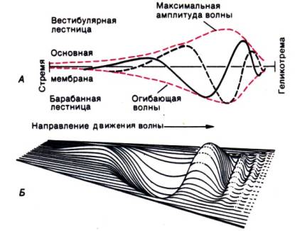
Rice. 12.9. A. Diagram of a traveling wave at two points in time. The envelope shows its maximum amplitude at a constant frequency in different parts of the cochlea. B. 3D wave reconstruction
Transformation processes in hair cells . As discussed in the previous section, due to the mechanical properties of the cochlea, a certain sound frequency causes vibrations of the basilar membrane with an amplitude sufficient to excite sensory cells only in one, strictly limited place. Since the basilar and tectorial membranes move relative to each other a shear force occurs on the cilia, both during their direct contact with the tectorial membrane, and as a result of the movement of subtectorial lymph; in both cases, their bending serves as an adequate stimulus for the auditory receptors (as in the vestibular receptors).
This bending starts conversion process(transduction): microscopic mechanical deformations of the cilia lead to the opening of ion channels in the hair cell membrane and, consequently, to their depolarization. Its prerequisite is the presence endocochlear potential. Microelectrode measurements showed that the endolymphatic space has a positive (approximately +80 mV) charge relative to the scala vestibularis and other extracellular spaces of the body. The stria vascularis and organ of Corti carry a negative charge (~ –70 mV; Fig. 12.10). The potentials recorded in the organ of Corti probably correspond to the intracellular potentials of hair and supporting cells. Positive endocochlear potential is provided by energy-dependent processes in the stria vascularis. The shift of cilia upon stimulation changes the resistance of the hair cell membrane as a result of the opening of ion channels. Because between

Rice. 12.10.Continuous cochlear potentials

Rice. 12.11.Microphonic cochlear potential (MP) and compound action potential (CAP) of the auditory nerve recorded at the round window during the sound of a click
endolymphatic space and their intracellular environment, there is a significant potential difference (at least 150 mV), synchronously with the stimulus local ionic currents arise, changing the membrane potential of the hair cells, i.e. generating a receptor potential (the so-called battery hypothesis). Registering it is difficult, but possible. It is easier, however, to place microelectrodes near the receptors in the scala tympani or on the round window and record cochlea microphone potential(Fig. 12.11).
It is similar to the output voltage of a microphone and fairly accurately reflects changes in sound pressure. A tape recording of speech made by connecting to the microphone potential of an experimental animal is quite intelligible. The origin of this potential is unclear; the initial assumption that it consists of extracellularly recorded components of hair cell receptor potentials is no longer entirely acceptable. As shown by intracellular leads from inner and outer hair cells, although receptor potentials are generated by them, at a high frequency of stimuli only constant pressure: Hair cell membrane potential does not change synchronously with high-frequency sound. Microphone potential:
1) synchronous with the sound stimulus with virtually no latency period;
2) deprived of a refractory period;
3) lacks a measurable threshold;
4) not subject to fatigue; those. differs in all respects from a neural action potential.
Depolarization of hair cells causes the release of a transmitter (possibly glutamate) from their basal part, which excites afferent nerve fibers. When a click (a short pressure pulse) is heard near the ear, the fibers of the auditory nerve are activated synchronously and from the round window, in addition to the microphone, a compound action potential can also be recorded. Longer sounds cause asynchronous impulses that do not sum up into individual action potentials. In Fig. Figure 12.11 shows the cochlear microphonic potential (CMP) and the compound action potential (CAP) evoked by a click. They have been recorded in cats, but they can also be recorded in humans when, for diagnostic purposes, an electrode is passed through the eardrum and brought to the round window.
Coding of sound in auditory nerve fibers .
In the cochlear nerve, 90% of the afferent fibers are myelinated and arise from the inner hair cells. Each contacts only one of them, i.e. with a very small part of the cochlea. These fibers are thick enough to record action potentials with microelectrodes and study the response to sound stimulation (the fibers coming from the outer hair cells are too thin for this). Since each part of the cochlea corresponds to a certain frequency, each of these fibers is most strongly excited by its characteristic frequency sound, but is not activated at all by other frequencies or is activated only when the sound pressure increases. This is reflected in Fig. 12.12, which shows a graph of the dependence of the perception threshold on the stimulus frequency for two different fibers. The criterion for establishing a threshold is a certain increase in activity above its spontaneous level. Each fiber is excited at frequency and intensity values within the shaded area in the figure. At the curve bounding it frequency-threshold characteristics a narrow, pointed low-threshold area and a wide high-threshold area are noticeable. Frequency threshold characteristics reflect

Rice. 12.12.Schematic frequency-threshold curves of two afferent fibers of the auditory nerve (a, b) with different characteristic frequencies (HF). Curve B is typical for fiber with pathological changes caused by damage to the inner ear
distribution of frequency maxima on the main membrane. The response of a single fiber to a stimulus, expressed as such a curve, represents a spectral analysis of sound. If it has several different frequencies, several groups of nerve fibers are activated. Duration sound stimulus is encoded by the duration of neural activity, and intensity–its level. As the sound pressure increases, the frequency of neuron impulses also increases (up to a certain limit, after which saturation occurs). At very high pressure, in addition, neighboring fibers that were previously in a state of rest are activated. This process is shown in Fig. 12.12; both fibers are excited if the sound corresponds to the area of overlap of their frequency-threshold characteristics. So, at the level of primary afferents, the sound stimulus is decomposed into frequency components. Each of them excites the corresponding nerve fibers. At higher levels of the auditory tract, neurons may behave differently.
When the cochlea is damaged, the sensitivity and frequency selectivity of afferent fibers decrease (Fig. 12.12). The receptor potential of the inner hair cells also changes and, as mentioned above, the same happens with the mechanical vibrational properties of the main membrane. The latter, it can be assumed, determine the behavior of these cells and fibers, but they themselves depend on the process of active mechanical reinforcement, for which the outer hair cells may be responsible. According to the current hypothesis, they are stimulated by sound first and generate additional vibrational energy of the same frequency. It is then transmitted to the inner hair cells. If this is correct, we are talking about a kind of hybrid between a sensory cell and a mechanical energy generator.
Many questions here have not yet been answered, but the ability of the cochlea to both produce and analyze sound energy is undeniable. The sound generated in the cochlea can be measured even outside the eardrum. These processes are very often disrupted by various injuries.
Outer hair cells are suitable as active amplifiers because they contain contractile proteins and are served by an extremely developed efferent network. Moreover, their afferents are clearly not essential for transmitting information to the brain.
Coding of sound frequencies based on the principle of receptor localization is discussed above. The second type of information encoding in the auditory nerve is as follows. Tones with frequencies up to 5 kHz usually cause neural firing in the auditory nerve only during certain phases of the cycle sound vibration. As a result, the temporal structure of the stimulus (for example, period T in Fig. 12.5, B) is represented by groups of action potentials transmitted to the central nervous system along the auditory nerve at appropriate times. The brain is apparently capable of assessing the temporal structure of impulses and determining the underlying sound frequency (the so-called periodicity analysis). Particularly clear evidence of this was obtained from direct electrical stimulation of the auditory nerve of patients suffering from deafness; periodic stimuli were processed in such a way that a tone of a certain pitch was perceived, which shows the real importance of periodicity analysis for hearing.
Anatomy of the auditory tract in the form of a highly simplified diagram is shown in Fig. 12.13. For simplicity, only the path from the left ear is shown. Arrows indicate synapses with higher order neurons. In order not to overload the figure, recurrent collaterals and interneurons are omitted, although in the auditory system such connections are very common.
The primary afferent fiber bifurcates, sending one process to ventral, and the other - to dorsal cochlear (cochlear) nuclei. Their fine structure(especially dorsal) is very complex. The ventral tract (from the ventral nucleus) is directed (partially through the trapezius nucleus) to the ipsi- and contralateral olivary complexes, the neurons of which thus receive signals from both ears. It is this neural level that allows us to compare acoustic signals coming from two sides of the body (we will return to this comparison process below). Dorsal tract (from

Rice. 12.13.Highly simplified diagram of the auditory tract (left ear only). To demonstrate binaural interactions in the superior olive, connections are also shown right ventral cochlear nucleus. Centrifugal paths omitted
dorsal nucleus) passes to the opposite side of the body and goes to nucleus of the lateral lemniscus (lateral lemniscus). The ascending processes of the cells of the olivary complex are both ipsilateral and contralateral. After synaptic switching in the nucleus of the lateral lemniscus, the auditory tract passes through lower colliculi quadrigeminal and medial geniculate body to primary auditory cortex, covering the transverse temporal gyri of the upper part of the temporal lobes (Heschl's gyrus). This zone corresponds to Brodmann's field 41; most of it is hidden in the depths of the Sylvian fissure. Neighboring the primary auditory cortex are other projection areas of the auditory system, called the secondary auditory cortex (Brodmann's area 42). Thus, the precortical auditory tract consists of at least five or six neurons, and since the additional synaptic switches and reentrant collaterals in Fig. 12.13 are not shown; longer chains are possible. This is described in more detail in the works. Finally, in addition to afferent pathways, the auditory system also includes centrifugal efferent fibers, also not shown in Fig. 12.13.
Excitation of central neurons of the auditory system . While the primary afferents of the auditory nerve are excited by pure tones, that is, very simple sound stimuli, neurons at higher levels are generally not capable of this. In the ventral cochlear nucleus they still behave like primary neurons. Clean

Rice. 12.14.Activity of four neurons of the dorsal cochlear nucleus in response to a tone of characteristic frequency for 50 ms (as modified). By x-axis – time; By ordinate axes– number of action potentials
tones of suprathreshold intensity always cause their excitement; they have narrow, pointed frequency-threshold curves and short latent periods. However, already in dorsal cochlear nucleus the picture is completely different. Although here again most neurons are excited by pure tones, the types of their responses vary widely. As an example in Fig. Figure 12.14 shows the reactions of various fibers emanating from this nucleus: in each case, a tone lasting 50 ms with a frequency characteristic of a given cell was presented. Neuron in Fig. 12.14, A behaves like the primary afferent, while the behavior of the others is significantly different. In some of them, the sound can cause braking; others are excited by strictly defined frequencies and inhibited by slight deviations from them. There are also neurons that respond in a special way to sounds of variable frequency (so-called frequency-modulated tones), although they also respond to pure tones. The anatomical basis of such complex behavior is collateral connections, some of which are excitatory and others inhibitory.
Functional meaning All this, obviously, is that neurons respond especially clearly to certain features of the sound stimulus, facilitating image recognition already on such lowest level tract. At higher levels, the specificity of their response gradually increases.
The further away from the cochlea along the auditory tract, the more complex sound characteristics are required to activate neurons. Many cells do not respond to pure tones at all. IN lower hills quadrigeminal, for example, there are cells that respond only to frequency-modulated tones with a specific direction and degree of modulation. Other neurons here respond only to amplitude-modulated (i.e., variable intensity) tones. And in this case, the modulation must often have certain characteristics, otherwise it will not cause excitement.
In general, we can say that the information contained in a sound stimulus is repeatedly recoded as it passes through different levels of the auditory tract. During this process, neurons of one type or another release “their” properties of the stimulus, which ensures a fairly specific activation of neurons higher levels.
In everyday life, we practically never encounter pure tones. The sounds around us are made up of different frequency components that change constantly and independently of each other. Their amplitude and duration also vary; they can arise and end suddenly or gradually, repeat or be unique; their source can be located closer or further from us, move, etc. A person, at least with trained hearing, is able to evaluate all these properties. The neural processes underlying this assessment have been identified mainly in auditory cortex. For example, some neurons in the primary auditory cortex respond only to the beginning of a sound stimulus, others only to its end. Some groups of neurons are excited by sounds of a certain duration, others by repeated sounds. There are also cells that are activated only by one or another frequency or amplitude modulation of sound. Many neurons are activated by a wide range of frequencies, i.e. noise, while others have frequency-threshold characteristics that differ in one or more pronounced minima. Most cortical cells are excited by afferents from the contralateral ear, but some respond to ipsilateral stimulation and others only to bilateral stimulation. A significant portion of neurons in the primary auditory cortex are not activated by any experimental influence; perhaps they are highly specific and respond only to stimuli that are too difficult to reproduce in a laboratory setting.
Overall cell responses primary auditory cortex are similar to those known for complex or supercomplex neurons of the visual cortex. Obviously, they are involved in auditory image recognition, a process that is very important, for example, For understanding speech. Even in the auditory cortex of monkeys, cells have been found that respond mainly to sounds associated with intraspecific communication. However, the properties of these neurons often depend on some unknown parameters, and their responses vary unpredictably.
Damage to the temporal lobes of the brain, where the auditory cortex is located, makes it difficult to understand speech, spatially localize the source of a sound (see below), and identify its temporal characteristics. However, such lesions do not affect the ability to distinguish between the frequency and intensity of sound. The central processing of sound information is discussed in more detail in the works.
Recent studies have shown that the tonotopic organization characteristic of the cochlea is preserved at higher levels of the auditory system, including the cortex. The presence of such an organization, i.e. An orderly distribution of regions associated with specific sound frequencies in the primary auditory cortex was previously denied.
Another result that contradicts earlier assumptions was the fact that auditory neurons at higher levels are not characterized by pronounced peaks in frequency-threshold characteristics. In the primary afferents of the auditory nerve, if the experimental animal is in optimal conditions, they are very clear.
Auditory orientation in space . The central auditory system is very important for spatial orientation. As is known from everyday experience, when binaural hearing The direction to the sound source can be determined quite accurately. The physical basis is this directionality is that usually one ear is located further from it than the other. Propagating at a finite speed, sound reaches a more distant ear Later and with less by force, and the auditory system is able to detect its difference in two ears already at the level of 1 dB. In Fig. Figure 12.15 shows a method for calculating the difference in sound travel time. Distance difference Δ S = d sin α , where (d is the distance between the ears, andα – the angle at which the sound source is located relative to the subject. This means that the time delay Δ t = Δs /s, where c is the speed of sound. A person is able to detect a delay of only 3–10–5 s, which corresponds to a deviation of the sound source from the midline by approximately 3°. Under optimal conditions, an angle that is half as small can be discerned.
Both psychophysical and neurophysiological experiments have shown that directional hearing is based on differences in conduction time and sound intensity. When headphones are used to stimulate each ear independently, a delay in the signal or a decrease in its intensity on one side causes the sensation of sound localization in the opposite ear. The delay can be compensated for by increasing the intensity; in this case, the sound source seems to be located in the head. Similar results were obtained in neurophysiological experiments. In the upper olive, first level

Rice. 12.15. Calculation of the difference in time for sound to reach the right and left ears (see text)
auditory system with bilateral afferentation, there are neurons that behave in a similar way with respect to temporal characteristics and signal intensity. Excitation in them is maximum when the sound in one ear is louder than in the other and precedes it. Another type of cell here is most active when the stimuli reaching both ears differ in certain ways in arrival time and intensity. This means that the first type of cells responds maximally to sound localized along the axis of one of the ears, and the other - to sound arriving at a certain angle. In the superior colliculus, auditory and visual afferentations combine to provide a three-dimensional “map” of space. IN auditory cortex some cells are also activated only when the sound source is located at a very specific location relative to the listener. When it is destroyed, spatial orientation also suffers. However, it is still not entirely clear how the central nervous system copes with determining a time difference of less than 10 –4 s.
Differences in conduction time and intensity are not enough to determine whether the sound source is in front or behind, above or below the head relative to the head. To do this, you need an additional device—the auricle. Its structure “distorts” the signal depending on the location of its source in such a way that it can be localized. This can be used in technology by placing microphones in the head of a mannequin in place of the eardrums: the stereophonic recordings obtained with their help will have excellent quality.
Hearing in noisy environments . Binaural hearing also has another, more important function than spatial orientation; it helps analyze acoustic information in the presence of extraneous noise. Interaural differences in the intensity and direction of signals are used by the central nervous system to suppress background noise and highlight useful sounds (for example, when concentrating on a desired conversation in a crowded meeting). This selective filtering process increases audibility by approximately 10 dB. This does not happen in people who are deaf in one ear, as you can easily verify by plugging your ear. Therefore, in case of hearing loss, it is important to restore binaural hearing, for example, with the help of hearing aids.
Adaptation of the auditory system . The auditory system, like other sensory systems, is capable of to adaptation. Both the peripheral ear and central neurons are involved in this process. Adaptation manifests itself in a temporary increase in the auditory threshold. This is useful because it reduces the threshold for loudness discrimination and thus promotes differentiation of auditory sensations. In an adapted ear, the isophones are shifted upward and closer together. More detailed information is contained in the works.
Pathophysiology of hearing impairment
Hearing loss and deafness have a very significant impact on the lives of patients, and therefore attract great attention from clinicians. The causes of these disorders can be divided into three categories.
1. Sound conduction disorders. These include injuries to the middle ear. For example, when it is inflamed, the tympanic-ossicular apparatus does not transmit the normal amount of sound energy to the inner ear. As a result, even if it is healthy, hearing deteriorates. There are microsurgical methods to effectively eliminate such hearing defects.
2. Impaired sound perception. In this case, the hair cells of the organ of Corti are damaged, so that either signal transduction or neurotransmitter release is impaired. As a result, the transmission of information from the cochlea to the central nervous system suffers.
3. Retrocochlear disorders. The inner and middle ear are healthy, but either the central part of the primary afferent fibers or other components of the auditory tract are damaged (for example, with a brain tumor).
Testing patients' hearing is called audiometry. To identify and localize damage hearing aid Numerous tests have been developed (for more details, see).
The most important among them – threshold audiometry. The patient is presented with different tones through one earphone. The doctor starts with a clearly subthreshold sound intensity and gradually increases the sound pressure until. the patient will not report hearing a sound. This sound pressure is plotted on a graph (Fig. 12.16) called audiogram.
On standard audiographic forms, the normal hearing threshold level is represented by a bold line and marked “About dB”. Unlike Fig. 12.18, higher thresholds are plotted below the zero line and characterize the degree of hearing loss - how many decibels they are below the norm. We emphasize that we are not talking about decibels of ultrasound, but about hearing loss by so many dB. For example, if you put your fingers in both ears, the reduction will be approximately 20 dB (when performing this experiment, of course, you must try not to make too much noise with your fingers). Using headphones, the perception of sound is tested when it is air conduction. Bone conduction are tested in a similar way, but instead of headphones, a tuning fork is used, applied from the side being tested to the mastoid process of the temporal bone, so that the vibrations propagate directly through the skull. By comparing threshold curves for bone and air conduction, it is possible to distinguish between deafness caused by damage to the middle or inner ear.
Deafness caused by damage to the middle ear is caused by impaired conduction of sound. The inner ear is healthy. Under these conditions, hearing loss is detected by the air conduction test (cf. Fig. 12.16), and the threshold for bone conduction is normal because the sound energy when tested reaches the hair cells without bypassing the middle ear.
Deafness due to pathology of the inner ear caused by damage to hair cells; the middle ear is great. In this case, the threshold for both types of conduction is increased, since in both cases the signal is converted through the same receptor process. Retrocochlear disorders also increase both thresholds.
Using a tuning fork (usually with a frequency of 256 Hz), conduction disorders are very easily distinguished from damage to the inner ear or retrocochlear pathologies, when it is known which ear hears worse (Weber test). The stem of the oscillating tuning fork is placed on the midline of the skull; if the inner ear is affected, the patient reports that the tone sounds from the healthy side; if average, with the affected one.
This phenomenon is easy to explain in the case of pathology of the inner ear. Damaged receptors excite the auditory nerve weaker, so the tone sounds louder in the healthy ear, and due to this difference, a directional sensation arises. In the case of damage to the middle ear, we are faced with three simultaneous processes. Firstly, the deterioration of the oscillatory properties of the ossicular apparatus weakens the transmission of sound not only from the outside to the inside, but also in the opposite direction. Consequently, the inner ear, excited by sound, when
Information in the brain
Part 2. Analysis of vestibular and sound
The anatomy of the vestibular tract is extremely complex (Fig. 24). Afferent fibers from the crests of the semicircular canals and macules of the sacculus and utriculus are directed to Scarpa's ganglion (vestibular) close to the external auditory canal, where the bodies of neurons are located, and then, after connecting with cochlear fibers, they form vestibulocochlear nerve , going to ipsilateral vestibular complex , located in the ventral part of the medulla oblongata under the fourth cerebral ventricle. The complex consists of four important cores: lateral (Deiters nucleus), medial, superior and descending. There are also many smaller nuclei located here, united by a complex system of afferents and efferents.
This complex of nuclei is innervated by descending fibers from the cerebellum and reticular formation. In addition, each complex receives innervation from the contralateral complex . In some cases, this contralateral innervation underlies the push-pull mechanism. For example, the crest cells of the semicircular canal also receive information from the crest of the contralateral canal. On top of all this, complex receives information from the eyes and proprioceptive fibers ascending along the spinal cord . Thus, the vestibular complex is an extremely important center for the integration of information related to movement and orientation. Rice. 24 shows that in addition to powerful connections with cerebellum And oculomotor nuclei, the vestibular complex sends fibers to the cerebral cortex. They are believed to end in postcentral gyrus near the lower end of the sulcus intraparietalis (intraparietal groove). Epileptic seizures that focus on this area are usually preceded by an aura (one of the components of an epileptic seizure characterized by disturbances of perception), characterized by feelings of dizziness and disorientation.
Vestibular apparatus also tracks the stationary orientation of the head in space (otoliths) And acceleration of its movement (crests of semicircular canals). All this is complemented by information from numerous somesthetic receptors throughout the body. To eliminate the flow of information from these sensors, you need to place the body in water or on an orbital station. Under these conditions, all the work falls on the eyes and vestibular apparatus; if now the object is also blinded, only the information from the membranous vestibule will remain.
The role of information from the semicircular canals can be clearly demonstrated by placing a test subject on a rapidly rotating swivel chair. In this case, the eyes shift to the side opposite to the rotation, in an attempt to fix the gaze on a stationary object, and then (when it is lost from the field of vision) they quickly jump in the direction of the rotation to find another point of fixation of the gaze. Similarly, when rotation suddenly stops, the eyes continue moving in the direction of the previous rotation and then make a leap in the opposite direction. This sudden change occurs as a result of the crests of the semicircular canals being affected by the flow of endolymph, which reverses the direction of flow. These characteristic eye movements are called nystagmus. They are conditioned three neuronal pathways (Fig. 25):
Ø from the semicircular canals to the vestibular nuclei,
Ø to the external eye muscles.
Meaning vestibulo-oculomotor reflex can be clearly demonstrated by comparing the vision of a rotating ocular system with vision when the head is stationary and the environment is rotating. The details of the rotating environment are very quickly lost: at two revolutions per second, the point of fixation of the gaze blurs into a blur. On the contrary, a test subject sitting in a swivel chair loses some visual acuity only at a rotation speed of about 10 revolutions per second.
Finally, it is worth saying a few words about motion sickness. This unpleasant feeling arises mainly due to mismatched touch inputs . In some cases, this mismatch occurs in the vestibular apparatus itself. If the head loses its normal orientation and rotates, signals from the crests of the semicircular canals no longer correlate with signals from the otoliths. Another source of motion sickness is mismatch of signals from the eyes and from the vestibular apparatus. If, in a rough sea in a cabin, the eyes report a lack of relative movement between the head and the walls of the cabin, while the vestibular system, on the contrary, is under strain, symptoms of “sea sickness” are observed. It is also worth mentioning that excessive alcohol consumption also leads to dangerous disorientation. This is due to the fact that ethanol changes the specific density of the endolymph, so that the cupula can now sense gravity and therefore send unusual signals to the central vestibular system.
Motor reactions of the eyes to irritation of the vestibular apparatus (oculocephalic reflex, vestibulo-ocular reflex) are mediated by pathways running through the brain stem from the vestibular nuclei of the medulla oblongata to the nuclei of the abducens and oculomotor nerves. Normally, rotation of the head causes endolymph to move in the semicircular canals in the direction opposite to rotation. In this case, in one labyrinth an endolymph flow occurs towards the ampulla of the horizontal semicircular canal, and in the other labyrinth - in the direction from the ampulla of the canal, while the irritation of the receptors of one channel increases, and the irritation of the opposite one decreases, i.e. an imbalance of impulses arriving to the vestibular nuclei occurs. When the vestibular nuclei are stimulated on one side, information is immediately transmitted to the contralateral nucleus of the abducens nerve in the pons, from where impulses pass through the medial longitudinal beam reach the nucleus of the oculomotor nerve in the midbrain on the side of the irritated vestibular apparatus. This ensures synchronous contraction of the lateral rectus muscle of the eye opposite to the irritated labyrinth and the medial rectus muscle of the eye of the same name, which ultimately leads to a slow friendly deviation of the eyes in the direction opposite to the direction of head rotation. This reflex allows you to stabilize the position of the eyes and fix your gaze on a stationary object, despite the rotation of the head. In a healthy, awake person, it can be voluntarily suppressed due to the influence of the cerebral cortex on the brainstem structures. in a patient who is in clear consciousness, the integrity of the structures responsible for this reflex is determined as follows. They ask the patient to fix their gaze on a centrally located object and quickly (two cycles per second) turn the patient’s head in one direction or the other. If the vestibulo-ocular reflex is preserved, then the movements of the eyeballs are smooth, they are proportional to the speed of head movements and are directed in the opposite direction. To assess this reflex in a comatose patient, the doll eye test is used. It allows you to determine the safety of stem functions. The doctor fixes the patient’s head with his hands and turns it left and right, then tilts it back and lowers it forward; The patient's eyelids should be raised (the test is absolutely contraindicated if a cervical spine injury is suspected).
The test is considered positive if the eyeballs involuntarily deviate in the direction opposite to the rotation (the "doll's eyes" phenomenon). In case of intoxication and dysmetabolic disorders with bilateral damage to the cerebral cortex, the "doll's eyes" test is positive (the patient's eyeballs move in the direction opposite to the direction of head rotation). With lesions of the brain stem, the oculocephalic reflex is absent, that is, the test is negative (when turning, the eyeballs move simultaneously with the head as if they were frozen in place). This test is also negative in case of poisoning with certain medicines(for example, with an overdose of phenytoin, tricyclic antidepressants, barbiturates, sometimes muscle relaxants, diazepam), but the normal sizes pupils and their reaction to light.
Caloric tests are also based on reflex mechanisms. Stimulation of the semicircular canals cold water, which is poured into the outer ear, is accompanied by a slow friendly deviation of the eyeballs towards the irritated labyrinth. A cold caloric test is carried out as follows. First, you need to make sure that the eardrums in both ears are intact. Using a small syringe and a short thin soft plastic tube, 0.2-1 ml is carefully injected into the external auditory canal ice water. In a healthy, awake person, nystagmus will appear, the slow component of which (slow deviation of the eyeballs) is directed towards the irritated ear, and the fast component - in the opposite direction (nystagmus, traditionally determined by the fast component, is directed in the opposite direction). After a few minutes, repeat the procedure for opposite side. This test can serve as an express method for identifying peripheral vestibular hypofunction.
In a comatose patient with the brain stem intact, this test causes a tonic coordinated deviation of the eyeballs towards the cooled labyrinth, but there are no rapid eye movements in the opposite direction (that is, nystagmus itself is not observed). If the structures of the brain stem are damaged in a patient in a coma, the described test does not cause any movements of the eyeballs at all (there is no tonic deviation of the eyeballs).
Vestibular ataxia
Vestibular ataxia is detected using the Romberg test and examining the patient's gait (he is asked to walk in a straight line with his eyes open and then with his eyes closed). With unilateral peripheral vestibular pathology, instability is observed when standing and walking in a straight line with a deviation towards the affected labyrinth. Vestibular ataxia is characterized by changes in the severity of ataxia with sudden changes in head position and turns of gaze. A pointing test is also carried out: the subject is asked to raise his hand above his head and then lower it, trying to get his index finger into the doctor’s index finger. The doctor's finger can move in different directions.
First, the patient performs the test with his eyes open, then he is asked to perform the test with his eyes closed. A patient with vestibular ataxia misses both hands toward the slow component of nystagmus.
Static and statokinetic reflexes. Balance is maintained reflexively, without the fundamental participation of consciousness in this. There are static and statokinetic reflexes. Vestibular receptors and somatosensory afferents, especially from proprioceptors in the cervical region, are associated with both. Static reflexes ensure adequate relative position of the limbs, as well as stable orientation of the body in space, i.e. postural reflexes. Vestibular afferentation comes in this case from the otolith organs. A static reflex, easily observed in a cat due to the vertical shape of its pupil, is a compensatory rotation of the eyeball when turning the head around the long axis of the body (for example, with the left ear down). At the same time, the pupils always maintain a position very close to vertical. This reflex is also observed in humans. Statokinetic reflexes are reactions to motor stimuli that are themselves expressed in movements. They are caused by stimulation of the receptors of the semicircular canals and otolith organs; examples include the rotation of a cat's body in a fall to ensure it lands on all four paws, or the movements of a person regaining his balance after tripping.
One of the statokinetic reflexes is vestibular nystagmus. As discussed above, the vestibular system causes various eye movements; nystagmus as their special form is observed at the beginning of a more intense rotation than ordinary short turns of the head. In this case, the eyes turn against the direction of rotation in order to maintain the original image on the retina, however, without reaching their extreme possible position, they sharply “jump” in the direction of rotation, and another part of space appears in the field of view. Then follows their slow return movement.
The slow phase of nystagmus is triggered by the vestibular system, and the rapid “jump” of gaze is triggered by the prepontine part of the reticular formation.
When the body rotates around a vertical axis, almost only the horizontal semicircular canals are irritated, i.e., the deviation of their cupulae causes horizontal nystagmus. The direction of both its components (fast and slow) depends on the direction of rotation and, thus, on the direction of cupular deformation. If the body is rotated around a horizontal axis (for example, through the ears or sagittally across the forehead), the vertical semicircular canals are stimulated and vertical, or rotational, nystagmus occurs. The direction of nystagmus is usually determined by its fast phase, i.e. with “right nystagmus,” the gaze “jumps” to the right.
With passive rotation of the body, two factors lead to the occurrence of nystagmus: stimulation of the vestibular apparatus and movement of the visual field relative to the person. Optokinetic (caused by visual afferentation) and vestibular nystagmus act synergistically.
Diagnostic value of nystagmus. Nystagmus is used clinically to test vestibular function. The subject sits in a special chair, which rotates for a long time at a constant speed and then suddenly stops. Stopping causes the cupula to deviate in the direction opposite to that in which it deviated at the beginning of the movement; the result is nystagmus. Its direction can be determined by recording the deformation of the cupula; it must be opposite to the direction of the previous movement. The recording of eye movements resembles that obtained in the case of optokinetic nystagmus. It's called a nystagmogram.
After conducting a test for post-rotational nystagmus, it is important to eliminate the possibility of fixation of the gaze at one point, since during oculomotor reactions, visual afferentation dominates over vestibular afferentation and, in some conditions, can suppress nystagmus. Therefore, the subject is put on Frenzel glasses with highly convex lenses and a built-in light source. They make him “shortsighted” and unable to fixate his gaze, while allowing the doctor to easily observe eye movements. Such glasses are also necessary in the test for the presence of spontaneous nystagmus - the first, simplest and most important procedure in the clinical study of vestibular function.
Another clinical way to trigger vestibular nystagmus is thermal stimulation of the horizontal semicircular canals. Its advantage is the ability to test each side of the body separately. The head of a sitting subject is tilted back by approximately 60° (for a person lying on his back, it is raised by 30°) so that the horizontal semicircular canal occupies a strictly vertical direction. Then the external auditory canal is washed with cold or warm water. The outer edge of the semicircular canal is located very close to it, so it immediately cools or heats up. In accordance with Barany's theory, the density of endolymph decreases when heated; consequently, its heated part rises, creating a pressure difference on both sides of the cupula; the resulting deformation causes nystagmus. Based on its nature, this type of nystagmus is called caloric. When heated, it is directed towards the place of thermal impact, and when cooled, it is directed in the opposite direction. In people suffering from vestibular disorders, nystagmus differs from normal qualitatively and quantitatively. Details of its testing are given in the work. It should be noted that caloric nystagmus can occur in spacecraft under conditions of weightlessness, when differences in endolymph density are insignificant. Consequently, at least one more, as yet unknown, mechanism is involved in its triggering, for example, direct thermal effects on the vestibular organ.
The function of the otolithic apparatus can be tested by observing oculomotor reactions when the head is tilted or during back-and-forth movements of the patient located on a special platform.


